Investigation of the effect of freeze-dried human serum albumin on the biocompatibility of cancellous bone
allograft
Ph.D. Thesis
Miklós Weszl
Semmelweis University PhD School of Basic Medicine
Supervisor: Zsombor Lacza, MD, DSc Official Reviewers: József Piffkó, DMD, MD, PhD
Tibor Glasz, MD, PhD Head of the Complex
Examination Committee: István Antal, D.Pharm, PhD Members of the Complex
Examination Committee: Zoltán Benyó, MD, DSc
Emília Madarász, Professor emerita Éva Szökő, D.Pharm, DSc
Budapest 2017
Table of contents
1 LIST OF ABBREVIATIONS ... 5
2 INTRODUCTION ... 7
2.1 Overview of bone replacement ... 9
2.1.1 Aetiology of bone defects ... 10
2.1.2 Categorization of bone defects ... 10
2.1.2.1 Traumatic and long bone defects ... 11
2.1.2.2 Maxillary and mandibular alveolar bone defects ... 11
2.1.2.3 Vertebral compression fracture ... 12
2.1.2.4 Nonunions ... 13
2.1.3 Management strategy of bone defects ... 13
2.1.3.1 Replacement of traumatic bone losses and long bone defects ... 14
2.1.3.2 Fixation of orthopaedic implants ... 14
2.1.3.3 Replacement of maxillary and mandibular alveolar bone defects ... 15
2.1.3.4 Compression fracture repair ... 15
2.1.3.5 Clinical trial of bone grafts ... 17
2.2 Bone remodelling and fracture healing ... 17
2.3 Bone grafts ... 22
2.3.1 Bone graft incorporation ... 22
2.3.1.1 Osteoinduction ... 23
2.3.1.2 Osteoconduction ... 23
2.3.1.3 Osteogenesis ... 24
2.3.1.4 Mechanical environment ... 24
2.3.1.5 Complications associated with bone grafts ... 24
2.3.2 Bone graft types ... 27
2.3.2.1 Autogenic bone grafts ... 27
2.3.2.2 Allogeneic bone grafts ... 28
2.3.2.3 Xenogeneic bone grafts ... 29
2.3.2.4 Synthetic bone graft materials ... 29
2.3.2.5 Presentations of bone grafts ... 30
2.3.3 Growth factors ... 31
2.3.3.1 Bone morphogenetic proteins ... 31
2.3.3.2 Vascular endothelial growth factor (VEGF) ... 33
2.3.3.3 Platelet-derived growth factor (PDGF) ... 34
2.3.3.4 Complications associated with growth factors ... 35
2.3.4 Trends in bone graft development ... 35
2.3.4.1 Nanotechnology ... 35
2.3.4.2 Coatings ... 37
2.3.4.3 Tissue engineering ... 38
2.3.4.4 Bioreactors ... 39
2.4 Evaluation of the pre-clinical performance of bone grafts ... 39
2.4.1 Biomechanics of bone ... 39
2.4.2 Mechanotransduction ... 42
2.4.2.1 Chemical composition ... 45
2.4.2.2 Porosity ... 46
2.4.2.3 Hardness ... 47
2.4.2.4 Topography ... 48
2.4.3 Biological assessment of bone graft materials ... 50
2.4.3.1 In vitro biocompatibility studies ... 51
3.1 Investigation of the in vivo biocompatibility of chemically sterilized, antigen-
extracted freeze-dried human bone grafts ... 58
3.2 Identification of a coating substance to improve the biocompatibility of the chemically sterilized, antigen-extracted freeze-dried cancellous allogeneic bone grafts 58 3.3 Investigation of the in vitro and in vivo biocompatibility coated freeze-dried cancellous allogeneic bone grafts ... 59
4 MATERIALS AND METHODS ... 60
4.1 In vivo investigation of the biocompatibility of chemically sterilized, antigen extracted freeze-dried human bone graft ... 60
4.1.1 Surgical procedure ... 60
4.1.2 In vivo multimodal imaging ... 61
4.1.3 Ex-vivo µCT analysis ... 61
4.1.4 Histology ... 61
4.2 In vitro experimental design ... 62
4.2.1 Bone graft types ... 63
4.2.2 In vitro experimental groups ... 64
4.2.3 Coating substances ... 64
4.2.4 Coating methods ... 64
4.2.4.1 Aqueous coating ... 65
4.2.4.2 Freeze-dried coating ... 65
4.2.5 Physical characterization of the bone grafts ... 65
4.2.5.1 Micro-hardness measurement ... 65
4.2.5.2 Scanning Electron Microscopy ... 66
4.3 In vitro biocompatibility study of coated human bone grafts ... 66
4.3.1 Isolation and cultivation of bone marrow derived MSCs ... 66
4.3.2 Isolation and cultivation of dental pulp derived MSCs ... 67
4.3.3 Characterization of the stem cells ... 67
4.3.4 Labelling of MSCs ... 67
4.3.5 Seeding of MSCs under standard conditions ... 68
4.3.6 Seeding of cells under dynamic conditions ... 68
4.3.7 Assessment of the proliferation of BMSCs ... 68
4.3.8 Assessment of the proliferation of DPSCs ... 69
4.4 In vivo biocompatibility study of coated human bone grafts ... 69
4.4.1 Nonunion model and study design ... 70
4.4.2 Statistical analysis ... 71
5 RESULTS ... 73
5.1 In vivo biocompatibility of chemically sterilized, antigen extracted freeze-dried human bone allografts ... 73
5.1.1 NanoSPECT-CT analysis ... 73
5.1.2 Ex-vivo µCT analysis ... 74
5.1.3 Results of histology ... 76
5.2 Physical characteristics of bone grafts ... 76
5.2.1 Optical characteristics of bone grafts ... 76
5.2.2 Microhardness of bone grafts ... 79
5.3 In vitro biocompatibility of bone grafts ... 79
5.3.1 Characterization of stem cells ... 79
5.3.2 Adherence and survival of MSCs on bone grafts ... 80
5.3.2.1 Control group ... 80
5.3.2.2 Test group A ... 80
5.3.2.3 Test group B ... 81
5.3.2.4 Effect of dynamic seeding on MSC adherence ... 83
5.4 In vivo biocompatibility of freeze-dried albumin coated bone grafts ... 83
6 DISCUSSION ... 85
7 CONCLUSION ... 92
7.1 Investigation of the in vivo biocompatibility of chemically sterilized, antigen- extracted freeze-dried human bone grafts ... 92
7.2 Identification of a coating substance to improve the biocompatibility of the chemically sterilized, antigen-extracted freeze-dried cancellous allogeneic bone grafts 92 7.3 Investigation of the in vitro and in vivo biocompatibility coated freeze-dried cancellous allogeneic bone grafts ... 92
8 SUMMARY (English, Hungarian) ... 93
9 REFERENCES ... 95
10 PUBLICATION LIST ... 116
11 ACKNOWLEDGMENTS ... 118
1 LIST OF ABBREVIATIONS
α-MEM alpha modification of Eagle’s medium β-cat β-catenin
BA Bone area
BMSC Bone marrow derived mesenchymal stem cell BMP Bone morphogenetic protein
rhBMP Recombinant human bone morphogenetic protein BMU Bone multicellular unit
BRC Bone remodelling compartment
CD Cluster of differentiation
CT Computed tomography
CPC Calcium phosphate cement
Dvl Dishevelled
DPSC Dental pulp derived stem cell
DMEM Dulbecco’s Modified Eagle’s Medium ECM Extracellular matrix
FA Focal adhesion
FAK Focal adhesion kinase
FCS Foetal calf serum
FGF Fibroblast growth factor
FYN Src family tyrosine-protein kinase Grb2 Growth factor receptor-bound protein 2
HAP Hydroxyapatite
HV Numeric value of Vickers hardness LEF Lymphoid enhancer factor
LTPB Latent TGF-β-binding protein MAKP Mitogen-activated kinase
MDP Methylene bisphosphonate
MEK MAPK/Erk kinase.
MLC Myosin light chain
MSC Mesenchymal stem cell
NMMII Non-muscle myosin II
OB Osteoblast
OC Osteoclast
Pax Paxillin
pCPC Premixed calcium phosphate cement
PCL Polycaprolactone
PDGF Platelet-derived growth factor PI3K Phosphatidylinositol 3-kinase
PLA Polylactide
PMMA Poly(methyl methacrylate)
PPAR-γ Peroxisome proliferator-activated receptor γ
PPF Polypropylenefumarate
RANKL Receptor activator of nuclear factor kappa-B ligand Rho GEFs Rho guanine nucleotide exchange factors
ROCK Rho-associated coiled-coil-containing protein kinase
ROI Region of interest
Runx2 Runt-related transcription factor 2
RPM Rotation per minute
SEM Scanning electron microscopy
Shc SH2-containing collagen-related proteins Src Rous sarcoma oncogene cellular homolog SrCPS Sr-doped calcium phosphate composite spheres
SUN Sad1p and UNC-84 homology
VASP Vasodilator-stimulated phosphoprotein
Vin Vinculin
VOI Volume of interest
VEGF Vascular endothelial growth factor
TA Tissue area
TAZ Transcriptional co-activator with PDZ-binding motif Tb.Th. Trabecular thickness
Tb.Sp. Trabecular separation Tb.Pf. Trabecular pattern factor
99mTc-MDP Technetium 99m-methyl diphosphonate YAP Yes-associated protein
2 INTRODUCTION
A serious limitation of the clinical performance of bone grafts is their unreliable incorporation1. Bone grafts have the capability of turning into live bone, however sometimes the graft fails to coalesce with the host bone with no foreseeable reason. The pathophysiologic variability does not allow the simplification and regard bone defects as a uniform condition, but depending on their aetiology and anatomical location they must be evaluated and treated on a personalized manner. Age, sex, metabolism and physical activity are factors that may significantly influence the innate regenerative potential of the bone after fracture or bone replacement. Unfortunately, the biology of bone development and fracture healing is not fully understood yet, which is one of the major limitations of the development of bone replacement technologies that ensure reliable bone graft incorporation. Another limitation is the lack of potent biomaterials that may become alternatives of autogenic bone grafts. However, the biomimetic approach of tissue engineering (i.e. the development of artificial materials that mimic natural bone) and the discovery of the role of biophysical cues in bone remodelling may open a new chapter in the development of the next generation of bone graft materials.
Pursuing biomimetic approach, the individual characteristics of the host bone and its environment should be taken into consideration, e.g. bone morphometric indices, elasticity, defect size and morphology, in order to develop personalized bone grafts with good clinical performance. However, the implementation of this approach presumes the existence of highly developed integrated technologies, such as diagnostic imaging and additive manufacturing. The development of diagnostic image processing that is able to provide data on the mechanical characteristics of host bone would be the prerequisite of personalized treatment. In the meantime, the development of additive manufacturing technologies would also be required because mass produced bone graft materials will always have biological weaknesses that limit their clinical performance2. However, the capability of producing artificial bone grafts that corresponds at least the physical environment (elasticity, mineral content, resorption rate, shape, etc.) of the host in order to activate biophysical cues may increase the reliability of graft incorporation.
At the current technology level the best biomimetic materials are of human origin, such as allogeneic bone graft. Allogeneic bone graft (allograft) is usually the
second choice for bone replacement after autogenic bone. Fresh, frozen, and freeze- dried allografts are most often used at load-bearing sites, however there are no exclusively applied protocols for their preparation. For patient safety the cadaveric and donor grafts are supposed to be subjected to disinfection to avoid the transfer of contagious agents from the donor to the recipient. Freeze-drying technique allows the disinfection of allogeneic bone grafts using volatile agents, such as ethylene-oxide and acids that form non-toxic residual salts3. As the disadvantageous effect of harsh disinfection the osteogenic cells are killed and most of the cell adhesive proteins become denatured, which impairs the biological value of the allograft. Thus, in such a way manufactured freeze-dried allograft can be characterized with good mechanical resistance and low biological value, which may be responsible for its unreliable incorporation. The re-establishment of the original or just quasi-original biological properties of the allografts would provide an unlimited source of potent bone grafts that could be an alternative to autogenic bone, which was the objective of the present doctoral work.
This thesis follows a bottom-up approach, meaning that the current achievements and challenges of bone replacement with bone grafts will be outlined first in order to put into context the objectives of the experimental work. The aim of the thematic introduction is to highlight the versatility of bone defects and clinical challenges that need to be solved by different management strategies. The different clinical challenges call for alternative approaches both at acute treatment and regenerative therapy levels. This is the underlying reason of the need for different bone grafts with various characteristics that could challenge the myth of the existence of an ultimate or ‘ideal’ bone graft. Each graft type has limitations – even the autogenic bone – in clinical applications, whereas there is poor extrapolation between the results of pre- clinical studies and clinical outcomes with bone grafts4. Therefore, the biological performance of a bone graft should be verified in each intended indication in controlled human studies, while the underlying biological mechanism should be evaluated in
orthopaedic use, which biological performance has been verified in subsequent animal and human studies by other members of our research group5,6,7.
2.1 Overview of bone replacement
Bone possesses the intrinsic capability of regenerating itself after injuries through a well-orchestrated process involving multiple molecular signalling pathways that induce series of cellular and intercellular biological events. In the clinical setting, bone healing after fractures is the most common form of bone regeneration. In contrast to other tissues, most of the bone injuries heal without the formation of scar tissue, which is resulted in the total recovery of the pre-existing bone function, while the newly formed bone is undistinguishable from uninjured bone tissue. However, there are cases when the self-healing property of the bone is impaired or insufficient that results either in persistent bone defects or nonunion.
Bone defects may develop by various reasons at different anatomical locations in the human body that makes it difficult to conduct an exhaustive discussion that covers every aspect of the problem. The magnitude of the clinical problem of bone defects is highlighted by the fact that the human bone is the second most common transplant tissue after blood, which means ~2.2 million bone grafts used just in orthopaedic procedures annually worldwide8,9. The growing demand for a higher quality of life after bone and joint replacement has become an essential requirement from the patient side and the volume of research and development activities in the domain of bone tissue replacement has dramatically increased in the last decade. The driving force of the intense research of bone graft materials is to find alternative(s) of human bone tissue transplants (autografts), which has limited availability in general. In this endeavour several approaches have been published in the scientific literature concerning the construction and evaluation of artificial bone graft materials. However, there is consensus in the art concerning the desired biological properties of an ideal bone graft, it will be detailed later in paragraph 2.3, but there is controversy about how to achieve and measure those properties.
The underlying basis of the arguments may stem form that sometimes underemphasized fact that bone defects have different pathophysiology depending on
their aetiology. The diverse pathophysiology may require various approaches concerning the management of the bone defects in order to achieve the intended clinical outcome. There could be considerable differences in the clinical pictures when bone replacement is indicated, if we consider the different ends of the spectrum, for instance, critical size bone defects when the bone fails to bridge an oversized gap in the healing process, in contrast with the compression fracture of a vertebrae when a relatively small bone defect can result in serious complications. The different clinical need driven approaches are manifested in the development and evaluation strategies of the various bone graft materials.
2.1.1 Aetiology of bone defects
Bone defects may develop in several different ways: bone fracture can end up in the occurrence of a pseudoarthrosis in 5 – 10% of the cases, high energy trauma, prosthesis revision arthroplasty, treatment of musculoskeletal infections or excision of bone tumours can also result in large bone deficiencies. Depending on the aetiology, anatomical location and associated complications bone defects may be characterized with different pathophysiologic properties.
2.1.2 Categorization of bone defects
The importance of bone defect analysis and classification is to determine the best regenerative treatment for each specific defect. The categorization of bone defects can be performed according to various considerations, however the management-based approach may offer a clinically reasonable overview. Concerning clinical setting, perhaps the greatest need for enhanced bone repair is in the treatment of segmental bone defects and chronic nonunion of fractures. Even within these subcategories there are significant differences concerning the properties of bone defects depending on their aetiology and anatomical location of bone defects.
2.1.2.1 Traumatic and long bone defects
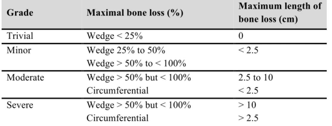
The overall incidence of long bone fractures in the Western world is estimated to be between 300 and 400 individuals per 100,000 per year10. The majority of trauma- induced fractures in adults will heal within nine months11. Apparently, 5–30% of the patients develop complications during the healing process, leading to delayed union or even nonunion of the fracture 12. Traumatic bone loss implies a wide spectrum ranging from a small butterfly fragment through to complete loss of large sections of bone. The most commonly employed system for the classification of traumatic injuries was given by Gustilo and Anderson, which has been later modified by Gustilo, Mendoza and Williams; however, traumatic bone loss is not part of this classification13,14. Robinson and his colleagues proposed a classification of bone loss in tibial fractures that could be applied to all long bone diaphyseal fractures (Table 1). It must be noted that there is no widely recognized classification of traumatic bone loss.
Table 1. Classification of tibial bone loss according to Robinson and his colleagues15.
Grade Maximal bone loss (%) Maximum length of
bone loss (cm)
Trivial Wedge < 25% 0
Minor Wedge 25% to 50%
Wedge > 50% to < 100%
< 2.5 Moderate Wedge > 50% but < 100%
Circumferential
2.5 to 10
< 2.5 Severe Wedge > 50% but < 100%
Circumferential
> 10
> 2.5
2.1.2.2 Maxillary and mandibular alveolar bone defects
Parameters that can describe alveolar bony defects are the anatomic position of defect in the jaws, dimensions and morphology of the defect (vertical, horizontal, ridge contour, etc.), defect base width and number of residual bony walls surrounding the defect16. Various classifications have been developed to describe alveolar ridge defects.
Dimension based classification divided the alveolar ridge defects into 3 classes, such as horizontal defects (Class I), vertical defects (Class II) and a common variant of them, i.e. horizontal and vertical defects (Class 3)17. These classes can be subdivided based on
the amount of the deficiency18. The recently proposed more quantitative classification that takes into consideration the size and orientation is more descriptive and applicable in the diagnostic work-up of alveolar bone defects19. Perhaps, Khojasteh et al. proposed the most comprehensive classification that takes into consideration the recipient site characteristic, as well; (A: Two-wall defects, B: One-wall defects, C: A defect with no surrounding walls) and width of defect base (I: A bony defect with a base width of 5 mm or more, II: A bony defect with a base width of 3 mm or more, but less than 5 mm, III: A bony defect with a base width less than 3 mm)16.
2.1.2.3 Vertebral compression fracture
Spine fractures are the most frequent fragility fractures and the second ones for morbidity and mortality in the elderly group after hip fractures20. The prevalence of vertebral fractures is increasing in the aging global population that is a consequence of the modern lifestyle that requires less movement, thus many elderly today have weak bone structure. Vertebral fractures are indicators for osteoporosis, which incidence will continue to rise and so will the incidence of osteoporotic vertebral fractures21,22. More than 25% of women 50 year of age and older will have one or more vertebral fractures by 202523. The most frequent site is the lumbar spine, and the primary and major symptom is localized back pain that can be debilitating. Osteoporotic vertebral fractures happen for axial compression that is not always associated with trauma, but especially in old patients, a simple lateral bending or weight lifting can be the cause (Figure 1).
Spinal deformity index has been introduced for the morphometric characterization of the fractures, which is important for fracture classification and treatment24,25. The risk of mortality is 2-fold higher in patients with osteoporotic vertebral fractures, while osteoporotic men are at higher risk for mortality than women. Compared to hip fractures, there is a 25% higher mortality risk after osteoporotic vertebral fractures23.
Figure 1. Side views of a normal spine and a spine with a compression fracture. An osteoporotic compression fracture causes the front of the vertebral body to collapse in a wedge- shape (red lines). The figure and legend were reprinted from the webpage of Mayfield Clinic26.
2.1.2.4 Nonunions
The clinical symptoms and physical findings of nonunion include pain and motion at the fracture site as well as radiographic evidence of failure of union27. The incidence of nonunion can be as high as 5% to 20%, but varies by fracture site and is influenced by a number of factors27. Nonunions are classified as hypertrophic, and atrophic (oligotrophic). Hypertrophic nonunions have adequate vascularity and exuberant callus formation, and generally only require appropriate mechanical stabilization with fixation devices to support healing (this condition is also referred as pseudoarthrosis). In contrast, in oligotrophic or atrophic nonunions, there is minimal or no callus formation with diminished or absent vascularity. These types of nonunions may benefit most from bone grafting28.
2.1.3 Management strategy of bone defects
The standard clinical approaches to stimulate or enhance bone regeneration include distraction osteogenesis, bone transport and various bone-grafting methods29. This thesis focuses on the discussion of bone-grafting methods from clinical point of
view and highlights the variety of the clinical approaches that need to comply with different clinical needs.
2.1.3.1 Replacement of traumatic bone losses and long bone defects
The goal in the management of any fracture with bone loss is to achieve solid bone union, adequate alignment, equal limb length and restoration of function. For fracture stabilization of traumatic bone losses i) plate fixation, ii) intramedullary fixation and iii) external fixation have been conventionally used30,31. Although, when severe traumatic bone loss is involved bone grafting can be applied as an alternative surgical technique. Vascularized free fibular graft may be used to bridge a bone defect >
12 cm in size; however, it is a technically demanding procedure that may cause undue burden to the patient32,33. Allografts can avoid the complication of donor-site morbidity but their use may be associated with lengthy recovery period, fracture and nonunion34,35,36. An alternative method of the reconstruction of long bone defects is a two-stage procedure (Masquelet technique) where the formation of a biological membrane is induced by the application of a cement spacer (first stage) that acts as a chamber for the insertion of a non-vascularized autogenic graft (second stage)37,38,39.
2.1.3.2 Fixation of orthopaedic implants
Bone grafts may be used to fill bone voids in total joint (hip and knee) arthroplasty when the autologous bone tissue supply is limited, albeit there is still a debate on the cemented and cementless (biological) fixation40. There are pros and cons concerning each fixation techniques, however in some cases the biological fixation (using bone graft instead of cement) may provide more beneficial clinical outcome than cementation. For instance, in younger patients the cement-implant interface may alter in time due to the growth of the bone that may cause micro-fractures in the cement texture leading to osteolysis and implant loosening41. In contrast, biological fixation may allow tissue remodelling around the implant during the development and avoid the emergence
extent of bone remodelling and the quality of the biological fixation. Unfortunately, the scarcity of clinical data from controlled, randomized human studies makes it difficult to carry out the objective evaluation of the short-, and long-term clinical outcomes of cemented and cementless fixations techniques and even less the efficiency of (various) bone grafts in this application41,42.
2.1.3.3 Replacement of maxillary and mandibular alveolar bone defects
The availability of adequate alveolar bone in terms of quality and quantity in all spatial dimensions is the prerequisite of successful dental implant placement. There are several surgical techniques for the vertical and horizontal augmentation of the alveolar bone, however guided bone regeneration and block graft techniques have become prevalent over distraction osteogenesis and osteoperiosteal flaps45. The block augmentation technique utilizes an autologous bone block that is fixed to the recipient ridge. The remodelling and histological performance of the bone block is affected primarily by its revascularization and the invasion rate by osteogenic cells. On the other hand, the principle of guided bone regeneration is to exclude encleftation (i.e. the proliferation of connective tissue into the sinus cavity) that would detrimentally affect the remodelling of particulate bone grafts, which occurs at a slower rate than soft tissue ingrowth43. In spite of the technical differences in the surgical techniques of block bone grafting and guided bone regeneration there are intrinsic similarities in their methodical principles, such as the exclusion of the epithelium and connective tissues, space maintenance, stability of fibrin clot and primary wound closure44,45.
2.1.3.4 Compression fracture repair
The most frequent treatment of vertebral fractures is conservative because the majority of these fractures are stable and do not have radicular or medullar involvement.
The indication of surgery depends on age, general conditions, fracture pattern and stability, involvement of medullary canal, bone quality, time elapsed from fracture20. The surgical options are vertebroplasty or kyphoplasty, vertebral stabilization and/or fusion with eventual decompression of the medullary canal. Vertebroplasty and kyphoplasty are minimal invasive interventions, which are performed through a hollow
needle that is driven directly into the fractured vertebra. In vertebroplasty, injectable bone filler is pressed through the hollow needle into the fractured bone. In kyphoplasty, a balloon is inserted first and inflated to expand the compressed vertebra to its normal height before filling the space with the bone filler46. The cemented bone filler in the vertebra allows to regain upright position, reduces pain, and prevents further fractures (Figure 2). Historically, poly(methyl methacrylate) (PMMA) cement has been used in these procedures first. However there have been complications associated with the use of PMMA, such as cement leakage and increased risk of adjacent vertebral fractures.
Calcium phosphate, calcium sulphate and hydroxyapatite pastes have become the alternatives to PMMA because of their chemical resemblance to bone that renders good biocompatibility and biodegradability allowing both the biological and mechanical repair of the fracture. In clinical setting, there are two major obstacles in conjunction with calcium-based and other injectable bone fillers in spinal applications: handling and radiopacity47. Furthermore, the calcium-based injectable fillers are prone to syneresis and contraction as well as for brittleness after cementation48. However, most of these weaknesses of those injectable bone fillers can be eliminated by using additives, such as polymers that improve the mechanical properties and oxides that enhance the radiopacity47,48,49.
Figure 2. Illustration of compression fracture repair by kyphoplasty. The balloon is inserted into the working channel inside the vertebra, and then inflated to raise the vertebra to the appropriate height. The balloon is removed and bone cement is injected into the cavity.
2.1.3.5 Clinical trial of bone grafts
There are a large number of reported clinical trials concerning bone grafts but most of them have never been completed50. Furthermore, only a small fraction of the completed clinical studies have been published in peer-reviewed scientific journals.
Hence, it may be concluded that the clinical safety and/or efficacy of the vast majority of bone grafts have not been established in controlled clinical studies. As a consequence, there are a lot of bone graft brands in clinical application, which have the risk of severe complications that may be associated with their use, e.g. rejection, early resorption or fracture. Concerning clinical study design, controlled, randomized, blinded, parallel assignment and multicentre arrangement would be appropriate to investigate the clinical performance of a bone substitute when the test graft is compared to a widely accepted comparator in selected patient groups. Interestingly, there are considerable amount of reliable and credible clinical data available for bone grafts that are intended for alveolar bone replacement, but concerning other indications and bone grafts the availability of such high quality clinical data is relatively low50,51,52,53. However, the extrapolation to the clinical performance of a bone graft based on pre- clinical experiments is questionable mainly due the lack of adequate animal models.
Furthermore, even the results of a registered clinical study should be critically appraised and special emphasis should be taken to the evaluation of the correct study design and patient selection criteria54,55. It should also be kept in mind that the clinical performance of a bone graft in a selected indication may not be predictive for its performance in other indications (e.g. alveolar bone versus long bone replacement)56.
2.2 Bone remodelling and fracture healing
Bone is constantly being remodelled throughout life in a sequence characterized by removal of old bone by osteoclasts and its replacement by osteoblasts. The main reason for this physiologic process is likely the removal of fatigue microcracks that occur in the skeleton as the result of daily physiological load. The remodelling process is driven in the basic multicellular unit (BMU) that comprises osteoclasts, osteoblasts, osteocytes, and lining cells (Figure 3). Under two-dimensional light microscopic
evaluation, bone remodelling compartments appear as narrow cleavages between trabecular bone and the bone marrow, which are linked by a layer of flattened lining cells on the marrow side and by the bone remodelling surface on the trabecular side.
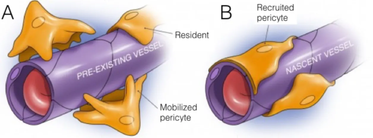
Given their tent-like appearance, the cells separating the bone remodelling compartment (BRC) from the bone marrow are thus termed the BRC canopies57. Osteoclasts are observed directly underneath canopies at the edges of the bone remodelling compartment (Figure 3)58. These canopy cells are connected to lining cells on the quiescent bone surface that are connected to osteocytes that reside in the bone matrix via gap junctions and cannaliculi59. The osteocytes have the capability of sensing biomechanical signals, such as mechanical strain and microcracks and initiate bone remodelling in respond to these signals presumably via its communication with lining cells60. In turn, the bone lining cells begins to form the BRC canopy and regulate osteoclast recruitment and differentiation by expressing receptor activator of nuclear factor κ B ligand (RANKL). Some recent studies show that activated osteoclasts may stimulate angiogenesis by secreting matrix metalloproteinase-9 that is able to release extracellular matrix (ECM)-bound vascular endothelial growth factor (VEGF)61,62. The ingrowth of a marrow capillary by penetrating the canopy of lining cells may serve as a conduit for the cells needed for the remodelling. At tissue level, mesenchymal stem cells (MSCs) reside in perivascular location close to sheets of osteoblast, as a cellular component of the hematopoietic niche or as an inactive marrow stromal cell63. When angiogenic stimuli occur the MSCs of bone marrow and fat has the capability of becoming pericytes on newly forming blood vessels (Figure 4). In the BRC, the pericytes are detached and act as MSCs that is driven by chemotactic factors released by inflammatory and other cells in the callus (Figure 5). There is emerging evidence that perivascular cells within the bone marrow exhibit mesenchymal lineage specific characteristics64. These mesenchymal cell-like perivascular cells form a unique niche, which possess self-renewing potential and the ability to commit to osteogenic, chondrogenic and adipogenic lineages (Figure 5)65,66. Thus, recent evidence indicates that the presence of blood vessels associated with the BRC may be a prerequisite for the
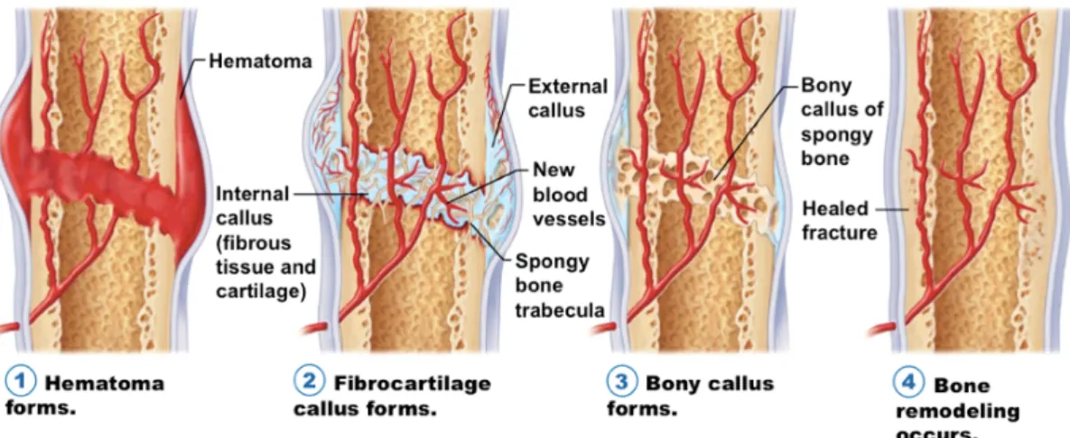
called secondary bone healing. Secondary bone healing occurs in the vast majority of bony injuries, involving both intramembranous and endochondral ossification that lead to callus formation. Callus is a physiological reaction to inter-fragmentary movement and requires the existence of residual cell vitality and adequate blood flow (Figure 6)68. The fracture hematoma has been proven to be a source of signalling molecules, such as interleukins, tumour necrosis factor-α, fibroblast growth factor, insulin-like growth factor, platelet-derived growth factor, vascular endothelial growth factor, and the transforming growth factor β superfamily members that are supposed to induce a cascade of cellular events that initiate healing69,71. Along with these biological cues the progressive union of a fracture requires the presence of four factors combined in the so- called diamond concept: an adequate cellular environment, sufficient growth factors, a bone matrix and mechanical stability68. Intriguingly, it appears that the initial cartilaginous callus forms even in the absence of a blood vessel, but the replacement of cartilage by bone only occurs following the penetration of blood vessels into the callus70. It is worth to note that oligotrophic and atrophic nonunions are characterized by the absence of blood vessels, which contain calcified cartilage that has not made the conversion step to bone, presumably due to the failure of the ingrowth of blood vessels and associated lack of appropriate osteoblast progenitor cells71.
20
Figure 3. Schematic drawing of the basic multicellular unit within bone remodelling compartment (BRC). The figure shows the key cells involved in normal bone remodelling, including the osteocytes embedded within bone, osteoclasts (OC), osteoblasts (OB), bone lining cells, and, at least in mice, osteal macrophages. As depicted, normal bone remodelling may largely serve to repair fatigue microcracks in bone. Note also the close relationship between the BRC and the blood vessel, which likely carries the perivascular stem cells destined to become osteoblasts on the bone surface. Image and legend were reprinted from reference 28.
Figure 4. Mobilization and transfer of pericytes72. The residing MSCs have the capability of associating with bone pre-existing marrow vessels as pericytes for angiogenic stimuli (A). The pericytes are transferred to the BRC on the surface a newly forming marrow capillary (B).
interest, a recent study using human mesenchymal stem cells for healing segmental bone defects in rats found that a combi- nation of the stem cells with bone morphogenetic protein (BMP)-7, which is known to induce osteoblastic differentia- tion, resulted in a better osteoinductive graft than either the mesenchymal stem cells or BMP-7 alone, [22] suggesting that combining mesenchymal stem cells with specific growth fac- tors may represent a fruitful approach to pursue. Along these lines, a number of studies have successfully used mesenchy- mal cells expressing vectors encoding factors, such as BMPs, to enhance bone healing [23, 24].
In terms of human studies, the use of culture-expanded osteoprogenitor cells in conjunction with porous hydroxyapa- tite scaffolds was reported in the treatment of four patients with diaphyseal segmental defects ranging in size from 3.0 to 28.3 cm3in a tibia, a humerus, and two separate ulnar fractures [25, 26]. Autologous bone marrow-derived pluripotent mesen- chymal stem cells were expanded in vitro and loaded on to 100% hydroxyapatite macroporous ceramic scaffolds. The grafts were seeded with the mesenchymal cells and the fracture defects were stabilized with an external fixator. There was pro- gressive integration of the implants with the surrounding bone, new bone formation inside the bioceramic pores, and vascular ingrowth. A good integration of the implants with the pre- existing bone was maintained during all the follow-up periods and no major adverse reactions were observed. Radiography and tomography showed that bone formation was far more prominent over the external surface and within the inner canal of the implants. This could be due to a higher density of loaded cells and/or a better survival of cells within the outer- most portions of the bioceramics. The patients all recovered limb function. With time, the implants revealed a progressive appearance of cracks and fissures indicative of some biocer- amic disintegration, whereas bone formation progressed and the implants were completely integrated into the existing bone.
ume of mineralized callus at 4 months and the number and concentration of fibroblast colony-forming units in the graft.
In the seven patients, who did not achieve union, both the concentration and the total number of stem cells injected were significantly lower than in the patients with osseous union.
One potential weakness of the study was the absence of a cohort with a placebo treatment. However, the success of the treatment of fracture nonunion with percutaneous bone mar- row grafting did appear to be dependent on the number and concentration of stem cells available for injection.
Despite the emerging evidence that bone marrow mesen- chymal stem cells may have utility in animal models and in humans for skeletal repair, the precise mechanism(s) by which these cells enhance tissue repair and regeneration remain unclear, not only for bone but also for other tissues. For exam- ple, Arthur et al. [19] found that BrdU-labeled human mesen- chymal stem cells that survived and contributed to ectopic bone formation when transplanted subcutaneously into immunocom- promised mice exhibited little or no proliferation in vivo, sug- gesting that expansion of these cells and their subsequent differ- entiation into osteoblastic cells may have had only a limited contribution to the bone that was formed. In analogous studies examining the use of mesenchymal stem cells in islet cell regeneration, Lee et al. [28] demonstrated that intracardiac infusion of human mesenchymal stem cells into diabetic non- obese diabetes/severe combined immune deficiency (NOD/
SCID) mice resulted in a reduction in blood glucose levels, but this was due to the production of mouse (and not human) insu- lin resulting from the human mesenchymal stem cell-induced regeneration of endogenous mouse b cells. This has led to the general concept, summarized by Prockop, [29] that although tissue repair by mesenchymal stem cells may be mediated to some extent by differentiation and/or transdifferentiation of these cells into specific functional cells (e.g., osteoblasts), a sig- nificant (and perhaps major) mechanism by which these cells Figure 1. Schematic of the basic multicellu- lar unit within the bone remodeling compart- ment (BRC) showing the key cells involved in normal bone remodeling, including the osteo- cytes embedded within bone, osteoclasts, osteoblasts, bone lining cells, and, at least in mice, osteal macrophages. As depicted, normal bone remodeling may largely serve to repair fatigue microcracks in bone. Note also the inti- mate relationship between the BRC and the blood vessel, which likely carries with it the perivascular stem cells destined to become osteoblasts on the bone surface. Abbreviations:
OB, osteoblast; OC, osteoclast.
Figure 5. Pericyte - MSC transitions. MSCs reside in situ as perivascular cells, which can be released to enter an osteoblastic differentiation program and develop into secretory osteoblasts/embedded osteocytes. Alternatively, the released perivascular cells can become activated to exert trophic and immunomodulatory effects. Image is an adaptation from reference 63.
Figure 6. The stages of fracture repair. (1) Hematoma formation: following injury, fracture disrupts bony blood supply leading to hematoma formation in and around the bony defect; (2) Fibrocartilage (soft) callus formation: fracture hematoma is rich in VEGF, which promotes blood vessel ingrowth from surrounding vessels (angiogenesis) along with the formation of a cartilage intermediate by endochondral ossification (internal callus) and the external callus (intramembranous ossification); (3) Bony (hard) callus formation: the callus is mineralized as hypertrophic chondrocytes undergo apoptosis (partially regulated by VEGF) and woven bone is formed and eventually replaced by lamellar bone; (4) Bone remodelling: the fracture callus composed of primary lamellar bone is remodelled to secondary lamellar bone, and the vascular supply returns to normal. Image was reprinted from the internet73 and legend is an adaptation from reference 62.
2.3 Bone grafts
A bone graft can be defined as either an inorganic or an organic 3-dimensional structure, or the combinations thereof that is intended for the replacement of a bone defect. Bone grafts can be used: i) to fill bone cavities and defects that emerged after cyst or tumour resection; ii) to bridge joints by creating arthrodesis; iii) to replace major defects and establish the continuity of a long bone; iv) to create bone block in order to limit joint motion; v) to establish union in pseudoarthrosis; vi) to promote union or fill defects in delayed union, malunion, fresh fractures and osteotomies; vii) to fix implants.
2.3.1 Bone graft incorporation
The successful incorporation of a bone graft depends on new bone formation that is driven by adaptive remodelling in response to mechanical stress. This process takes places in sequential phases that is supposed to be similar to those in fracture healing (Figure 6)74,80. The diagnostic follow-up of bone graft by computed tomography revealed that the discrete boundary between host and graft is initially identifiable;
however, as union progresses, the graft-host junction is obliterated as a result of trabecular ingrowth, and the medullary canal is replaced by fibrous tissue, which may be attributed to fibrocartilage (soft) callus formation80. Morone and his co-workers gave a general description concerning the incorporation process of a bone graft into a host, e.g. the ‘‘process of envelopment and interdigitation of the donor bone tissue with new bone deposited by the recipient’’75. Campana and his colleagues gave an explanatory overview on the multiple stage process of bone graft incorporation76:
i) “Initially, the bone graft induces a response that leads to the accumulation of inflammatory cells that is followed by the chemotaxis of host mesenchymal cells to the graft site;
ii) Thereafter, the primitive host cells differentiate into chondroblasts and osteoblasts in a process that is directed by the cohort of osteoinductive factors;
iii) In the subsequent processes bone graft revascularization and necrotic graft
In order to stimulate this process a bone graft is supposed to possess particular biological and mechanical features, e.g. osteoinductive, osteoconductive and osteogenic properties similar to native bone, however this terms should be critically appraised or maybe overruled in this context.
2.3.1.1 Osteoinduction
Osteoinduction refers to the recruitment and stimulation of undifferentiated cell types to develop into osteogenic cell lineages77. This is a basic biological mechanism that occurs regularly during bone remodelling, fracture healing and bone graft incorporation. Even if pre-existing osteoblasts may help to form new bone, it is getting generally agreed that such pre-existing cells only contribute to a minor portion of the new bone formation after bone graft placement. The instant bony injury induces intense inflammatory response at the fracture site, where inflammatory and other cells release signalling molecules, such as growth factors that attract cell types needed for bone repair. The fate of the recruited stem and other cell types is modulated locally by both soluble biological and insoluble biophysical cues. In summary, the initial part of the healing response includes osteoinduction, a process that starts immediately after the injury and is very active during the first week thereafter, even though the action of the newly recruited pre-osteoblasts is not obvious until several weeks later, in the callus stage77.
2.3.1.2 Osteoconduction
When bone graft placement is indicated either the size of the bone defect or the insufficient local supply of osteogenic cells compromise the physiologic fracture healing. In such cases, due to the lack of native bone structure the recruited bone forming need a matrix or scaffold to adhere, migrate, proliferate and differentiate.
Hence, an osteoconductive bone graft may be defined as a scaffold that permits bone growth on its surface, including down into pores, channels or pipes77.
2.3.1.3 Osteogenesis
It is often said that the osteogenic property of a bone graft derives from dwelling cells that synthetize bone at the recipient site. This interpretation assumes the pre- existence of dwelling stem cells or other osteogenic cells on the surface of a bone graft that are supposed to contribute to the new bone formation. In contrast, compelling studies support that pre-existing cells cannot survive on a bone graft in vivo because of the lack of proper blood supply78. There is growing evidence that the major source of bone forming cells in physiological bone remodelling and fracture healing process is delivered by blood vessels to the repair site days or weeks after the bone graft placement. Therefore, the osteogenic property of a bone graft may be interpreted as the consequence of its osteoconductive property and biophysical cues that are mediated by the mechanical features of the graft, such as hardness and topography that support the adherence, proliferation and differentiation of stem cells.
2.3.1.4 Mechanical environment
Mechanical stability is of primary importance to support the vascularisation and angiogenesis during bone regeneration63. In many cases, the bone defects are mechanically unstable that requires additional fixation by using metallic devices. The metallic fixation should cause minimal destruction in the local blood supply, supplement and protect the implanted bone graft from undue mechanical load.
However, the optimal instrumentation allows small intramedullary movements that do not compromise the formation and integrity of marrow capillary and vessels within the callus. In such a mechanically stable environment fragile blood vessels are able to span distances and form anastomoses, while the MSCs both stabilize the blood vessels and form sheets of osteoblasts that generate osteoid, which becomes calcified into trabecular bone68. This newly formed bone is restructured as controlled by its loading dynamics.
2.3.1.5 Complications associated with bone grafts
metabolic diseases (e.g. osteoporosis), under medication or treatment that suppresses bone metabolism (bisphosphonate, radiation treatment), and septic conditions, like osteomyelitis and other generic infections. Relative contraindications of bone graft placement may defer depending on the aetiology and the anatomical location of the bone defect that need to be taken into consideration on case-by-case basis by the medical team. Adverse events may emerge even if absolute and relative contraindications do not compromise the clinical outcome of the bone graft placement.
The most common complications that may be associated with bone graft placement include early graft resorption, nonunion or delayed union of bone fragments, graft fracture, graft extrusion, and infection79,80.
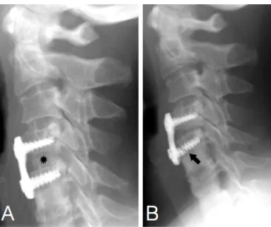
In surgical reconstruction, bone grafts are often placed along with implants and fixation devices at load-bearing sites. Early graft resorption and graft fracture allow excessive mechanical load on the hardware that may lead to consequential hardware failure (Figure 7). The extrusion of allografts or synthetic bone grafts may also be a major failure mode as it is shown on Figure 8. The early resorption of human and synthetic bone grafts are shown on Figure 9 and Figure 10, respectively.
Figure 7. Cancellous bone allograft resorption with hardware loosening and failure in a 46-year old woman. Panel A: Lateral radiograph obtained on postoperative day 1 shows the allograft (*) as an area of high opacity in the C4-C5 interspace and C4-C5 anterior cervical plate (Atlantis Vision; Medtronic Sofamor Danek, Memphis, Tenn). The graft was coated with injectable bone paste (Osteofil; Regeneration Technologies. Panel B: One-year follow-up radiograph shows focal allograft resorption, hardware loosening, and failure of the inferior screw (arrow). Figure and legend are adaptations from reference 80.
26
Figure 8. Extrusion of a cortical allograft and failure of fusion in a 61-year-old woman.
Panel A: Lateral radiograph of the right foot, obtained 9 months after graft placement, shows the bone graft (*) and a screw bridging the subtalar joint fragments. Panel B: Sagittal reconstruction CT image, obtained 4 days after ‘A’, shows extrusion of the bone graft (*) into the sinus tarsi and persistence of the subtalar joint fracture, with no osseous union. Figure and legend are adaptation from reference 80.
Figure 9. Failure of an autograft in the wrist of a 24 year old man. Anteroposterior radiograph shows a Herbert screw that bridges an old nonunited scaphoid fracture deformity (arrow), accompanied by evidence of scapholunate advanced collapse. The autograft that was initially placed to aid in fracture union has failed and cannot be seen. The proximal pole of the scaphoid is diminutive and not well defined, and there is marked cystic change of the capitate (*) and distal radius, with ulnar positive variance. Figure and legend are adaptations from reference 80.
Complications
The possible complications of bone graft place- ment include nonunion or delayed union of bone fragments, graft fracture, graft extrusion, and in-
sory loss (2,6). Possible complications related to allografts include disease transmission and mild rejection (4).
Figure 18. Failure of an autograft in the wrist of a 24- year-old man. Anteroposterior radiograph shows a Her- bert screw that bridges an old nonunited scaphoid fracture deformity (arrow), accompanied by evidence of scapholu- nate advanced collapse. The autograft that was initially placed to aid in fracture union has failed and cannot be seen. The proximal pole of the scaphoid is diminutive and not well defined, and there is marked cystic change of the capitate (*) and distal radius, with ulnar positive variance.
Figure 19. Failure of a vascularized fibular autograft in a 49-year-old man. Anteroposterior radiograph(a)and coronal reconstruction CT image(b)show a subtrochanteric transverse fracture of the right femur and an associated fracture of the vascularized fibular autograft (*). The linear area of opacity inais a K-wire placed for graft fixation.
386 March-April 2006 RG f Volume 26 ● Number 2
RadioGraphics
Teaching Point
Figure 10. Calcium sulphate ceramic bone graft substitute used for joint repair in a 42- year-old man. Panel A: Preprocedural axial CT image shows a unicameral bone cyst (*) in the right posterior ilium at the level of the superior sacroiliac joint. Panel B: Axial CT image, obtained 1 month after graft placement, shows partial resorption of the graft material (arrow).
Panel C: Axial CT image, obtained 2 years after graft placement, shows complete resorption of the graft material and minimal ingrowth of bone (arrows). Figure and legend are adaptations from reference 80.
2.3.2 Bone graft types
Various classifications of bone grafts exist but none of them is comprehensive enough to outline the whole spectrum of the available products in detail. Perhaps the most plausible categorization is based on the origin of the bone grafts because this approach provides an illustrative description of their clinical performance and limitations.
2.3.2.1 Autogenic bone grafts
The first use of autogenic bone grafts in large volume is dated back to World War II when massive cancellous bone grafts constituted the mainstay of treatment of bone loss in war injuries81. Based on the experience of these early procedures the development of grafting techniques has been commenced and volume of treatment methods in the surgeon’s armamentarium is still growing82. Autogenic bone grafts (autografts) are harvested from a patient who is in need of bone replacement, thus it is more likely to be incorporated than a foreign graft material. Autograft can be harvested from non-essential bones, like iliac crest, fibula, chin, rib, mandible or the skull depending on the type of the surgery and the size of the bone defect. The unique bone healing potential of the autologous bone graft derives from its good osteoconductive,
osteoinductive and osteogenic potential, while the risk of immune reaction or the transmission of diseases is not implicated. The osteogenic property of an autograft is provided by the osteoblasts, osteoclasts, and stem cells residing between the lamellae and trabeculae of the freshly harvested autologous bone grafts. Albeit, the complications associated with the harvest of autologous bone, like chronic pain at the donor site and its limited availability, especially in paediatric and elder patients often impede their use83. The incidence of autograft donor site morbidity and associated complications can be as high as 25.3% even exceeding the complication rate of the grafting procedure in the second surgery84.
2.3.2.2 Allogeneic bone grafts
Allogeneic bone graft is supposed to be a good alternative of autografts because of its human origin85. The off-the-shelf availability and the lack of donor site morbidity due to the elimination of need for a second surgery (bone harvesting) are the undisputed advantages of the allograft compared to autograft86,87. Allografts88 may be collected either from patients who are subjected to total joint replacement surgery or from cadavers. The allografts must be processed in a certified bone tissue bank before distribution. Fresh, frozen and freeze-dried allografts are seemed to be the most popular, however there are not exclusively applied protocols for their preparation87,89.For the patient safety the allografts should be subjected to disinfection so as to avoid the transfer of contagious agents from the donor to the host90,91. Freeze-drying technique allows the preliminary disinfection of allogeneic bone grafts with chemicals, like acids and ethylene-oxide because these agents eliminate from the allograft in the rinsing and freeze-drying process. An unwanted effect of such disinfection is that, the osteogenic cells are killed on the allograft and most of the osteoinductive proteins become denatured, which impairs the biological value of the allograft91,92. On the other hand, allografts are often subjected to gamma-sterilization that may detrimentally affect the mechanical property of the bone, while deactivates proteins that are normally found in the bone tissue. Thus, the 3-dimensional (structural) freeze-dried and irradiated
incorporation, which is the greatest disadvantage of bone allografts compared to autografts.
2.3.2.3 Xenogeneic bone grafts
Xenogeneic bone grafts (xenografts) are collected from non-human species, such as bovine bone, porcine bone or coralline grafts93. The advantage of xenografts is their easy availability, good osteoconductivity, mechanical property and low cost. In spite of these advantages xenografts are rarely used in clinical practice. The reason of the neglect may be the contradictory results, however favourable data have also been published with these kinds of grafts94,95. Perhaps bovine bone has achieved the highest penetration in oral surgery and has become an alternative of autogenic block bone grafts , but scarce validation in orthopaedics93. There are continuous arguments over the safety of xenogeneic grafts because the transmission of diseases (“zoonosis”) may not be excluded for sure96.
2.3.2.4 Synthetic bone graft materials
Synthetic bone grafts are supposed to be the alternatives of the human and xenogeneic bone grafts by taking the advantage of their unlimited availability and the complete elimination of the risk associated with the transfer of diseases. Such bone grafts are synthetized in chemical reactions with the goal to create artificial materials that support the replacement of specific types of bone defects. Although, the ultimate goal is to create an artificial bone graft material that possesses autograft-like properties, but in reality the engineering projects are driven by a specific clinical problem, which solution relies on only a few specific features of a synthetic bone graft. From practical point of view, either the mechanical or the biological features of a synthetic bone graft material can be enhanced depending on the type of the bone defect. For instance, at load-bearing sites the mechanical strength is the most important feature of a bone graft, while its biodegradability is a secondary attribute. In contrast, at non-load-bearing sites the biodegradability is the paramount feature of a bone graft, while its poor mechanical resistance, such as brittleness has lower clinical relevance. This kind of appraisal concerning the biological and mechanical properties of a synthetic bone graft is
unavoidable because the currently known bone graft materials have their own specific strengths and weaknesses compared to an autograft, which is still the gold standard to replace a bone defect. Based on their chemical compositions the synthetic bone graft materials can be categorized into three main groups, e.g. ceramics, polymers and composites. Many other subgroups are possible within the main categories of synthetic bone graft materials and further characterized according to their biological, chemical and mechanical properties, however its relevance is questionable. The mechanical and biological properties of a synthetic bone graft strongly depends on multiple factors, such as the raw material, the method and process parameters of manufacturing, the presence of additives and so on and forth. This means that depending on the manufacturing parameters, for instance, a ceramic bone graft material may show remarkably different mechanical properties that will influence its biological behaviour.
Therefore, it cannot be concluded that there are well-defined sets of physical properties that are distinctive concerning the biological behaviour of the ceramic, polymer and composite bone graft materials. On the other hand, the composition-based categorization of the synthetic bone graft materials is still predominant in the scientific publications. However, an indication-based categorization system of the synthetic bone graft materials may also be useful in order to support the decision making of the surgeons.
2.3.2.5 Presentations of bone grafts
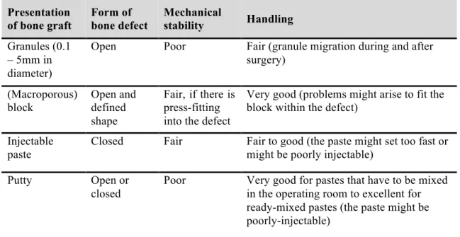
The availability of bone grafts in various forms may be necessary depending on the characteristic of the bone defect to be replaced. Bohner gave a practical overview about the specific features of the four most common forms of bone grafts, such as granules, blocks, cements and putties (Table 2)174. The forms of bone defects are categorized based on their dimensions in Table 2. ’Open’ refers to open cancellous bone defects; ‘defined shape’ refers to osteotomy site; ‘closed’ refers for cavital defect that is surrounded by bone substance.