R E S E A R C H A R T I C L E
Serum albumin ‐ coated bone allograft (BoneAlbumin) results in faster bone formation and mechanically stronger bone in aging rats
Dénes B. Horváthy
1,2 |Károly Schandl
1 |Charlotte M. Schwarz
1 |Károly Renner
3 |István Hornyák
1 |Bence T. Szabó
4 |Eugenia Niculescu ‐ Morzsa
5 |Stefan Nehrer
5 |Csaba Dobó ‐ Nagy
4 |Attila Doros
2 |Zsombor Lacza
1,61Institute of Clinical Experimental Research, Semmelweis University, Budapest, Hungary
2Department of Transplantation and Surgery, Semmelweis University, Budapest, Hungary
3Department of Physical Chemistry and Material Science, Budapest University of Technology and Economics, Budapest, Hungary
4Department of Oral Diagnostics, Semmelweis University, Budapest, Hungary
5Center for Regenerative Medicine and Orthopedics, Department for Health Sciences and Biomedicine, Danube University Krems, Krems an der Donau, Austria
6Research Center for Sport Physiology, University of Physical Education, Budapest, Hungary
Correspondence
Stefan Nehrer Center for Regenerative Medicine and Orthopedics, Department for Health Sciences and Biomedicine, Danube University Krems, Dr.‐Karl‐Dorrek‐Straße 30 3500 Krems an der Donau, Austria Email: stefan.nehrer@donau‐uni.ac.at Funding information
Lower Austria Research Promotion Agency (NfB); OrthoSera GmbH
Abstract
Serum albumin
‐coated bone allografts (BoneAlbumin) have successfully supported bone regeneration in various experimental models by activating endogenous progenitors.
However, the effect of tissue aging, linked to declining stem cell function, has yet to be explicitly examined within the context of BoneAlbumin's regenerative capacity. Stem cell function was tested with an in vitro attachment assay, which showed that albumin coating increases stem cell attachment on demineralized bone surfaces in an aging cell population. Bone regeneration was investigated in vivo by creating critical size bone defects on the parietal bones of aging female rats. Demineralized bone matrices with and without serum albumin coating were used to fill the defects. Bone regeneration was determined by measuring the density and the size of the remaining bone defect with computed tomography (CT). Microcomputed tomography (MicroCT) and mechan- ical testing were performed on the parietal bone explants. In vivo CT and ex vivo microCT measurements showed better regeneration with albumin
‐coated grafts. Addi- tionally, the albumin
‐coated group showed a twofold increase in peak fracture force compared with uncoated allografts. In the present study, serum albumin
‐coated demineralized bone matrices successfully supported faster and functionally superior bone regeneration in aging rats. Because stem cell function, a key contributor of bone remodelling, decreases with age and serum albumin is an effective activator of endog- enous progenitor cells, this method could be an effective and safe adjuvant in bone regeneration of aging adult and osteo
‐compromised populations.
K E Y W O R D S
aging, bone, bone substitute, BoneAlbumin, serum albumin, stem cells
1
|I N T R O D U C T I O N
Endogenous stem cell function is one of the most important factors contributing to proper tissue healing. Mesenchymal stem cells (MSCs)
are essential to the bone regenerative cascade, with the localization of these cells appearing paramount (Gibon, Lu, & Goodman, 2016). Peri- osteal and endosteal tissue is the most abundant source of MSCs after bone injury, highlighting the role of local progenitors in bone - - - - This is an open access article under the terms of the Creative Commons Attribution‐NonCommercial License, which permits use, distribution and reproduction in any medium, provided the original work is properly cited and is not used for commercial purposes.
© 2019 The Authors Journal of Tissue Engineering and Regenerative Medicine Published by John Wiley & Sons Ltd DOI: 10.1002/term.2803
416 wileyonlinelibrary.com/journal/term J Tissue Eng Regen Med. 2019;13:416–422.
regeneration (Colnot, 2009). However, aging tissue, linked to dimin- ished stem cell function, can result in inferior and slower tissue remod- elling, including compromised bone healing (Liu, Xia, & Li, 2015).
Osteoporosis, a diseased bone state that generally develops with increasing age, also affects the regenerative potential of bone tissue and increases the likelihood of traumatic fracture injuries. The medical cost of osteoporosis and fractures in older adults was estimated at $22 billion in the United States alone in 2008 (Blume & Curtis, 2011). Con- sequently, aging adults are susceptible to prolonged hospitalization and dramatically affected functional status as a result of compromised bone quality and regenerative capacity (Office of the Surgeon General [US], 2004). Additionally, about 15% of all fractures in the United States require bone grafting to support proper tissue healing (Khanzada, Holy, Jerry Volenec, & Bruder, 2008). For these reasons, adequate bone sub- stitution and subsequent treatment modalities remain a dire challenge in aging adult and osteo‐compromised populations.
Serum albumin‐coated bone allograft (BoneAlbumin) has been inten- sively investigated lately, with outcomes data demonstrating an ability of BoneAlbumin to facilitate new bone formation in non‐union and critical size defect models in rats (Horvathy, Vacz, et al., 2016; Skaliczki et al., 2013; Weszl et al., 2012). BoneAlbumin was also shown to decrease donor site morbidity and enhance bone formation after anterior cruciate ligament reconstruction with bone‐patellar tendon‐bone autografts (Schandl et al., 2016). Moreover, in a thorough review article, the albumin molecule was shown to have an important and potentially active but not yet completely understood role in bone regeneration (Horvathy, Simon, et al., 2016). Additionally, stem cell function was tested on various serum albumin‐coated surfaces. It was shown that the serum albumin coating supports stem cell attachment and proliferation, suggesting that serum albumin‐coated biomaterials can be used as vehicles for cell transplanta- tion (Bernards, Qin, & Jiang, 2008; Horvathy et al., 2013; Liu et al., 2014; Weszl et al., 2012). From these studies, the authors concluded that the BoneAlbumin technique increases local albumin concentration, lead- ing to endogenous stem cell activation. As a result, a higher number of progenitors are present at the injury site, permitting almost complete bone healing even in critical size defects.
Accordingly, given BoneAlbumin's robust facilitation of bone for- mation and stem cell function, the aim of the present study was to investigate whether such a regenerative modality could yield faster and mechanically superior ossification in an aging rat model. If such findings are substantiated here within, the implications for treatment in the aforementioned aging adult and osteo‐compromised demo- graphics would be clinically advantageous.
2
|M A T E R I A L S A N D M E T H O D S 2.1
|Animals
Because female rats enter menopause between the ages of 15 and 20 months, female Wistar rats (animal) older than 20 months (and younger than 24 months) were used for all experiments (Sengupta, 2013). The animals were maintained on lab chow and tap water ad libitum with 12‐hr day/night cycle in the animal facility of the Institute of Clinical Experimental Research in Budapest. The investigation was
approved by the local animal research committee according to the guidelines for animal experimentation (date of issue: 2009.10.07; reg- istration number: 22.1/2960/003/2009)
2.2
|Stem cell harvest and culture
Animals were euthanized with urethane (5 mg/ml). Both tibiae and femora were cleaned of soft tissue and placed in 70% ethanol. Under a sterile hood, both ends of the bones were cut, and the medulla was flushed out slowly with 10‐ml culture medium (Dulbecco's Modified Eagle's medium supplemented with 1‐g/L glutamine, 1‐g/L glucose, 10% fetal bovine serum, 100‐U/ml penicillin, and 100‐μg/ml strepto- mycin) into 15‐ml tubes. Bone marrow was centrifuged at 274gfor 8 min at room temperature and was placed in 100‐mm Petri dishes.
After harvesting, Bone marrow‐derived mesenchymal stromal cells (BMSCs) were maintained at 5% CO2level at 37°C. Two days later, the culture was washed in Dulbecco's phosphate‐buffered saline, and fresh culture medium was given every 48 hr. Cell cultures were used when they reached 80% of confluency, before reaching the fourth passage.
2.3
|Technology background: BoneAlbumin preparation
Animals were euthanized with urethane (5 mg/ml). Parietal bone was identified, and cortical bone pieces were harvested with a 4‐mm inter- nal diameter trephine bur. The demineralized bone matrix (DBM;
Graft) was prepared by following the classical method originally described by Urist, Mikulski, and Boyd (1975). In the case of albumin‐treated grafts (Graft + Albumin; BoneAlbumin), protocol was followed by the guidelines of OrthoSera GmbH: graft soaking in 20%
human albumin solution (Biotest, Hungary) was performed prior to standard freeze drying (frozen at−80°C and freeze dried at−50°C overnight). The albumin content in the BoneAlbumin group was deter- mined previously (Horvathy, Vacz, et al., 2016). All chemicals were purchased from Sigma‐Aldrich Co., Budapest, Hungary.
2.4
|In vitro attachment
Prepared DBM grafts with and without albumin were put into 96 well ultra‐low attachment wells (Costar, Corning Inc.). A 200‐μl cell suspen- sion containing 50,000 rat BMSCs and stem cell media were co‐ cultured with the grafts for 6 and 12 hr. Cellular attachment was mea- sured with methylthiazole tetrazolium assay (MTT, Sigma‐Aldrich Co.;
Holst Hansen, 1998). Briefly, after 6 and 12 hr of co‐culture, grafts were allocated in a new 96‐well plate, and fresh media (200μl) con- taining 5‐mg/ml MTT (1:9) were added (37°C, 1 hr). After 1 hr of incu- bation, MTT‐containing media were removed, and 200‐μl propanol was added for 1 hr under gentle shaking. The absorbance of MTT was measured using a spectrophotometer at the characteristic wave- length of 570 nm. The background was measured at 690 nm. Because absorbance values reflect on metabolic activity, increased optical den- sity suggests better cell attachment.
2.5
|Experimental protocol in vivo
In the in vivo experiment, critical size bone defects were created on the parietal bones of the aging animals. Bone formation was moni- tored with computed tomography (CT) on the 5th and 11th post‐ operative week. On the 11th week, parietal bones were harvested for microcomputed tomography (microCT) scans and mechanical test- ing. Three experimental groups were established: albumin‐coated DBMs (Graft + Albumin; BoneAlbumin), uncoated DBMs (Graft), and unfilled control (sham). Thirteen aging animals were involved in the present experiment, each with two randomly filled bone defects. Nine rats with 18 bone defects were examined with CT, microCT, and mechanical testing; accordingly, each experimental group occupied a total of six defects. Four animals with randomly filled defects were used for histologic purpose only.
2.6
|Surgical procedure
The surgical procedure was performed as described by Spicer et al.
(2012) inNature Protocols. Under ketamine–xylazine (100–10 mg/kg, Richter Gedeon Plc., Budapest, Hungary–Sigma‐Aldrich Co.) anaes- thesia, and after proper disinfection, the skin was incised over the parietal bone of each animal. Periosteum was carefully elevated from the bone. Bone defects were created using a 4‐mm trephine bur (external diameter) on both parietal bones. DBMs (Graft) with and without albumin were used to fill the defects, and the periosteum was united in the midline with a thin absorbable suture (6–0 Vicryl, Johnson & Johnson, Janssen‐Cilag Ltd., Hungary).
2.7
|Computed tomography
Under ketamine–xylazine anaesthesia, CT was performed with a Phil- lips Brilliance 16 Slice CT machine (Philips International B.V. Amster- dam, The Netherlands). Axial slices were obtained with 120 kV and 300 mA (slice thickness: 0.8 mm, increment: 0.4 mm, collimation:
16 × 0.75 mm, rotation time: 0.75 s, and pitch: 0.4). Images from the CT scan were analysed to determine bone density. Density was reported using the Hounsfield unit (HU) scale. Circular region of inter- est (ROI) was set on the fifth post‐operative week for every defect.
Reference points were used to set the same ROI at each time point.
Area of the remaining bone defect was measured from the CT scans.
Windows were set to visualize animal bone without soft tissue back- ground (2572/1595 Window/Center). The remaining bone defect was calculated on an enlarged reconstructed image, with freehand technique. The area of the bone defect (mm2) was compared between the albumin‐coated, uncoated allografts, and empty defects.
2.8
|Ex vivo microCT
The harvested parietal bones were scanned using a microCT scanner (Skyscan 1172 X‐ray microtomograph, Kontich, Belgium) at 59 kV, 167μA with a 23μm3isovolumetric pixel size. The centre of the bone defect was determined via horizontal and coronal images utilizing CTAn analysis software. On coronal images, a circular ROI with 150
pixels of radius was drawn, and 100 slices were selected cranial and caudal from the centre. As described previously, bone volume per tissue volume (BV/TV) was calculated to determine the relative bone content (%) in the defect area and in uninjured parts (Kallai et al., 2011).
2.9
|Histology
Four animals with randomly filled defects were sacrificed after the 11th post‐operative week. Paraffin sections from the formalin‐fixed parietal bones were stained with haematoxylin and eosin.
2.10
|Mechanical push ‐ out testing
Mechanical push‐out testing was performed with an Instron 5566 device (Instron, Norwood, MA, USA) according to Kretlow's method (Kretlow et al., 2010). A custom‐made holder was created to hold the parietal bone. The tissue samples were gently fixed between two flat surfaces with a 5‐mm hole in the middle. The graft was guided to the centre of the holder. To determine the breaking force, the bone tissue was loaded with a 2.5‐mm diameter rod with a flat bottom and rounded edges at 10 mm/min cross‐head speed, whereas load– displacement data was registered every 100 ms. The force was regis- tered as the rod was pressed through the bone. Peak breaking force (N) of the samples was identified by the highest force measured. The slope of the triangular shaped load–displacement curves represented material stiffness (N/mm2).
2.11
|Statistical analysis
All of the values are reported as the means ± standard error of the mean. The statistical analysis was performed with t test and one‐ way analysis of variance with Bonferroni multiple comparison tests using the GraphPad Prism statistical software. Probability values of p< 0.05 were considered significant.
3
|R E S U L T S
3.1
|In vitro attachment
Decreased cell function was apparent in the aging tissue population, with attached cell number post‐harvest definitively lower than with cells originating from young animals. Furthermore, it took approxi- mately 3 weeks to reach the desired cell number (~800,000 cells/Petri dish). Once this was reached, the attachment assay was started. After 6 hr of incubation, the albumin‐coated grafts attached more than twice as many cells as the uncoated grafts (Graft + Albumin:
0.12 ± 0.02 AU; Graft: 0.06 ± 0.01 AU;p< 0.01). After 12 hr of incu- bation, cell number did not change on the albumin‐coated surface.
There was a slight increase in cell number on the surface of uncoated grafts after 12 hr, but it was still significantly lower compared with the albumin‐coated grafts (Graft + Albumin: 0.11 ± 0.01 AU; Graft:
0.07 ± 0.01 AU;p< 0.5; Figure 1).
3.2
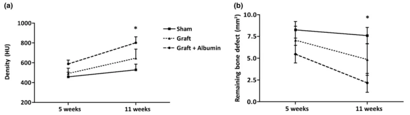
|In vivo imaging
Bone formation was investigated with CT on the 5th and 11th post‐ operative week by calculating the remaining bone defect and measur- ing the density (Figure 2). After 5 weeks, no statistically significant dif- ferences in bone defect density were found between the experimental groups; however, the albumin‐coated group did show the smallest remaining bone defect (Graft + Albumin: 5.47 ± 1 mm2; Graft:
7.07 ± 1.6 mm2; sham: 8.27 ± 0.95 mm2). After 11 weeks, the differ- ences between the study groups became more evident. At this time point, the albumin‐coated group showed significantly lower remaining bone defect compared with the empty defects, although the bone gap was not completely closed. By contrast, the defects left empty showed very low healing with a relatively large remaining bone defect on the 11th week (Graft + Albumin: 2.2 ± 1.08 mm2; Graft:
4.85 ± 1.83 mm2; sham: 7.6 ± 0.93 mm2;p< 0.05).
The same findings were found in the density measurements. After 5 weeks, no significant differences were found, but the albumin‐ coated group showed the highest value (Graft + Albumin:
588 ± 38 HU; Graft: 492 ± 54 HU; sham: 458 ± 47 HU). By 11 weeks, the albumin treated group showed significantly higher density values compared with the unfilled controls (Graft + Albumin: 802 ± 60 HU;
Graft: 646 ± 91 HU; sham: 529 ± 58 HU;p< 0.05; Figure 2).
3.3
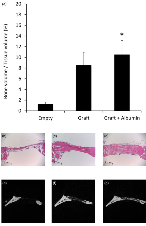
|Ex vivo microCT and histology
The BV/TV values of uninjured parietal bones were 20 ± 0.2%, which represents 100% (normalized) on Figure 3. MicroCT scans of injured parietal bones were compared with the uninjured bone. As expected, the measurements showed open bone defects and low BV/TV values in the unfilled (sham) group. Increased, but not significant, bone healing was seen with uncoated allografts. However, in the albumin enhanced group, significantly higher BV/TV values were observed compared with empty control group (Graft + Albumin: 11 ± 2.6%;
Graft: 9 ± 2.5%; sham: 1 ± 0.4% HU;p< 0.05; Figure 3). Microscopic analysis also showed better bone regeneration in the albumin‐coated group. In the sham operated defects, only connective tissue was observed. Bone edges were round shaped, indicating the termination of the regenerative process. Uncoated allografts, on the other hand, resulted in more intense bone formation, whereas albumin‐coated allografts showed even better osteogenesis and tissue remodelling after the 11th post‐operative week (Figure 3).
3.4
|Mechanical push ‐ out testing
Peak breaking force and material stiffness were measured on samples from the uncoated and albumin‐coated groups (Figure 3). Failure of the bone tissue occurred at the graft/host interface in all cases. Frac- ture force of the albumin‐coated group was two times higher com- pared with the uncoated bone grafts. (Graft + Albumin: 26.8 ± 4.5;
Graft: 14.3 ± 1.8 N;p< 0.05). The albumin‐coated grafts also showed higher but not significant stiffness values (Graft + Albumin:
34 ± 3.5 N/mm2; Graft: 25.8 ± 4.9 N/mm2;p= 0.2; Figure 4).
4
|D I S C U S S I O N
In the present study, the authors showed that stem cells originating from aging rats do not readily attach to DBM surfaces, but with the use of albumin coating stem cell, adherence increases significantly.
Additionally, albumin‐coated DBM produced significantly faster bone formation and stronger new bone in critical size calvaria defects of the aging rats.
FIGURE 1 Cell attachment on the allograft surface in vitro. The panel shows absorbance values (arbitrary unit) after 6 and 12 hr after cell seeding. Albumin‐coated group presented significantly better cell adherence after each time point (n= 6, error bars:
mean ± standard error of the mean, one‐way analysis of variance,
**p< 0.01, *p< 0.05)
FIGURE 2 Computed tomography measurements of new bone formation in vivo. Panel (a) shows density values of the defect area (Hounsfield unit [HU]). Albumin‐coated allograft showed significantly higher density values after the 11th week. Panel (b) shows the remaining bone defect (mm2). Albumin‐coated allograft group shows significantly lower remaining bone defect after the 11th week (n= 6, error bars: mean ± standard error of the mean, one‐way analysis of variance,*p< 0.05)
FIGURE 3 Ex vivo microcomputed tomography analysis and histology of calvaria defects at the 11th post‐operative week. Panel (a) shows qualitative analysis of regenerated bone in the defect site, showing significant difference between the albumin‐coated allograft and unfilled control group (n= 6, error bars: mean ± standard error of the mean, one‐way analysis of variance, *p< 0.05). Panels (b, c, and d) show representative histologic images of empty, uncoated, and albumin‐coated filling, respectively. Panels (e, f, and g) shows representative microcomputed tomography images of empty, uncoated, and albumin‐coated filling, respectively
As mentioned previously, stem cell function plays an essential role in bone regeneration. In order to increase the number of stem cells at the injury site, Hernigou, Poignard, Zilber, and Rouard (2009) transplanted autologous bone marrow into necrotic femoral heads and found better outcomes in patients receiving higher number of progenitors. Goodman and Hwang (2015) found similar results with the same technique treating young individuals with secondary osteonecrosis of the knee. However, stem cell functionality decreases with age, thereby increasing the need for high multipotent cell number at the injury site (Quarto, Thomas, & Liang, 1995; Shigeno & Ashton, 1995). According to these results, stem cell therapy could be a useful tool for clinicians, but a population of young donor cells is necessary to reach optimal regeneration in the elderly. Allogenic cell delivery, on the other hand, raises regulatory, immunologic, and ethical ques- tions that hinder this therapeutic option (Yim, 2005). Therefore, targeting the endogenous stem cell population seems to be a reason- able strategy. With this in mind, the authors showed that serum albumin‐coated bone allograft (BoneAlbumin) successfully supports bone regeneration in various experimental models. The idea behind the phenomenon is that locally increased albumin concentration induces endogenous progenitor recruitment, resulting in the presence of a higher cell number to support the tissue remodelling phase (Horvathy, Simon, et al., 2016). Evidence of stem cell activation was supported by a first‐in‐human investigation, in which large albumin‐ coated structural allografts were used to support recovery after total joint revision arthroplasty, with single photon emission computed tomography (SPECT) analysis showing increased osteoblast activation even 1 year after operation (Klara, Csonge, Janositz, Csernatony, &
Lacza, 2014). This also suggests that albumin could be a successful adjuvant in the aging and/or elderly population to overcome the diminished healing potential caused by decreasing stem cell function.
The idea is strongly supported by the current experiment, where albu- min coating was shown to attach twice as many BMSCs compared with uncoated DBMs. This ratio was nearly the same in previous work, in which stem cells originating from young donors were used (Horvathy, Vacz, et al., 2016). Those results showed the same attach- ment pattern, but the cell number was twofold higher at every time point, indicating the decreased functionality of aging multipotent cells.
Additionally, albumin‐coated DBM was a successful bone substitute in young specimens, resulting in nearly complete regeneration on the seventh post‐operative week (Horvathy, Vacz, et al., 2016). In the present study, the albumin‐coated DBMs also resulted in significantly better bone healing compared with the uncoated material in remaining
bone defect, density, BV/TV, and peak fracture force, even though the values showed less intensive bone regeneration, because this popula- tion did not reach full consolidation even after the 11th post‐operative week. One limitation of the present work is the size of the study groups, but even with this element number, we observed both the reduced healing potential and the beneficial effects of albumin, sug- gesting that the population was adequately chosen for the experiment.
5
|C O N C L U S I O N
Serum albumin‐coated bone allograft (BoneAlbumin) showed effec- tiveness in bone substitution in several ways by activating the endog- enous stem cell population. Because stem cell function generally declines with age, aging and osteo‐compromised patients may benefit from this technique. This method achieves a faster and more complete regeneration, facilitating recovery and potentially decreasing the risk of refracture. The authors emphasize that implication for clinical prac- tice remains that of extrapolation; however, the scalability of such a technology presents as a high potential downstream orthopaedic modality.
A C K N O W L E D G E M E N T S
The authors thank Daniel Katics for figure preparation. The authors thank Dr. Joel Batts for his invaluable input in finalizing the manu- script. The authors would like to acknowledge the Lower Austria Research Promotion Agency (NfB) and OrthoSera GmbH for supporting this study.
C O N F L I C T O F I N T E R E S T
The principal investigator Z. L. is inventor in a granted patent for albu- min coating of bone and holds stock in a startup company OrthoSera GmbH that owns the patent.
O R C I D
Dénes B. Horváthy https://orcid.org/0000-0001-8136-7100
R E F E R E N C E S
Bernards, M. T., Qin, C., & Jiang, S. (2008). MC3T3‐E1 cell adhesion to hydroxyapatite with adsorbed bone sialoprotein, bone osteopontin, and bovine serum albumin. Colloids and Surfaces. B, Biointerfaces, 64(2), 236–247. https://doi.org/10.1016/j.colsurfb.2008.01.025 FIGURE 4 Mechanical testing. Panel (a)
shows peak breaking force (N) of the implanted material after 11 weeks. Panel (b) shows the stiffness (N/mm2) of the implanted material after 11 weeks. Significantly higher breaking force can be seen in the albumin‐ coated group (n= 6, error bars:
mean ± standard error of the mean,ttest,
*p< 0.05)
Blume, S. W., & Curtis, J. R. (2011). Medical costs of osteoporosis in the elderly Medicare population. Osteoporosis International, 22(6), 1835–1844. https://doi.org/10.1007/s00198‐010‐1419‐7
Colnot, C. (2009). Skeletal cell fate decisions within periosteum and bone marrow during bone regeneration. Journal of Bone and Mineral Research: the Official Journal of the American Society for Bone and Min- eral Research,24(2), 274–282. https://doi.org/10.1359/jbmr.081003 Gibon, E., Lu, L., & Goodman, S. B. (2016). Aging, inflammation, stem cells,
and bone healing.Stem Cell Research & Therapy,7, 44. https://doi.org/
10.1186/s13287‐016‐0300‐9
Goodman, S. B., & Hwang, K. L. (2015). Treatment of secondary osteonecrosis of the knee with local debridement and osteoprogenitor cell grafting.The Journal of Arthroplasty,30(11), 1892–1896. https://
doi.org/10.1016/j.arth.2015.05.013
Hernigou, P., Poignard, A., Zilber, S., & Rouard, H. (2009). Cell therapy of hip osteonecrosis with autologous bone marrow grafting.Indian Journal of Orthopaedics, 43(1), 40–45. https://doi.org/10.4103/0019‐ 5413.45322
Holst Hansen, C. B. (1998). MTT‐cell proliferation assay. In J. E. Celis (Ed.), Cell biology: A laboratory handbook(2nd ed., Vol. 1) (pp. 16–18). San Diego: Academic Press.
Horvathy, D. B., Simon, M., Schwarz, C. M., Masteling, M., Vacz, G., Hornyak, I., & Lacza, Z. (2016). Serum albumin as a local therapeutic agent in cell therapy and tissue engineering. BioFactorshttps://doi.
org/10.1002/biof.1337,43, 315–330.
Horvathy, D. B., Vacz, G., Cselenyak, A., Weszl, M., Kiss, L., & Lacza, Z.
(2013). Albumin‐coated bioactive suture for cell transplantation.Surgi- cal Innovation, 20(3), 249–255. https://doi.org/10.1177/
1553350612451353
Horvathy, D. B., Vacz, G., Szabo, T., Szigyarto, I. C., Toro, I., Vamos, B.,… Lacza, Z. (2016). Serum albumin coating of demineralized bone matrix results in stronger new bone formation.Journal of Biomedical Materials Research. Part B, Applied Biomaterials, 104(1), 126–132. https://doi.
org/10.1002/jbm.b.33359
Kallai, I., Mizrahi, O., Tawackoli, W., Gazit, Z., Pelled, G., & Gazit, D. (2011).
Microcomputed tomography‐based structural analysis of various bone tissue regeneration models.Nature Protocols,6(1), 105–110. https://
doi.org/10.1038/nprot.2010.180
Klara, T., Csonge, L., Janositz, G., Csernatony, Z., & Lacza, Z. (2014). Albu- min‐coated structural lyophilized bone allografts: A clinical report of 10 cases.Cell and Tissue Banking,15(1), 89–97. https://doi.org/10.1007/
s10561‐013‐9379‐8
Kretlow, J. D., Spicer, P. P., Jansen, J. A., Vacanti, C. A., Kasper, F. K., &
Mikos, A. G. (2010). Uncultured marrow mononuclear cells delivered within fibrin glue hydrogels to porous scaffolds enhance bone regener- ation within critical‐sized rat cranial defects.Tissue Engineering. Part A, 16(12), 3555–3568. https://doi.org/10.1089/ten.TEA.2010.0471 Liu, H., Xia, X., & Li, B. (2015). Mesenchymal stem cell aging: Mechanisms
and influences on skeletal and non‐skeletal tissues.Experimental Biol- ogy and Medicine, 240(8), 1099–1106. https://doi.org/10.1177/
1535370215591828
Liu, X., Zhou, X., Li, S., Lai, R., Zhou, Z., Zhang, Y., & Zhou, L. (2014). Effects of titania nanotubes with or without bovine serum albumin loaded on
human gingival fibroblasts. International Journal of Nanomedicine, 9, 1185–1198. https://doi.org/10.2147/IJN.S55514
Office of the Surgeon General (US) (2004). The burden of bone disease. In Bone health and osteoporosis: A report of the surgeon general. Rockville (MD): Office of the Surgeon General (US).
Quarto, R., Thomas, D., & Liang, C. T. (1995). Bone progenitor cell deficits and the age‐associated decline in bone repair capacity.Calcified Tissue International,56(2), 123–129. https://doi.org/10.1007/BF00296343 Khanzada RN., Holy C. E., Jerry Volenec F., Bruder S. P. (2008). Cell thera-
pies for bone regeneration. In A. Atala (Ed.),Principles of regenerative medicine(pp. 868–887): Burlington, USA: Elsevier Inc.
Schandl, K., Horvathy, D. B., Doros, A., Majzik, E., Schwarz, C. M., Csonge, L.,…Lacza, Z. (2016). Bone‐Albumin filling decreases donor site mor- bidity and enhances bone formation after anterior cruciate ligament reconstruction with bone‐patellar tendon‐bone autografts. Interna- tional Orthopaedics, 40(10), 2097–2104. https://doi.org/10.1007/
s00264‐016‐3246‐8
Sengupta, P. (2013). The laboratory rat: Relating its age with human's.Inter- national Journal of Preventive Medicine,4(6), 624–630.
Shigeno, Y., & Ashton, B. A. (1995). Human bone‐cell proliferation in vitro decreases with human donor age.The Journal of Bone and Joint Surgery.
British Volume,77(1), 139–142.
Skaliczki, G., Schandl, K., Weszl, M., Major, T., Kovacs, M., Skaliczki, J.,… Lacza, Z. (2013). Serum albumin enhances bone healing in a nonunion femoral defect model in rats: A computer tomography micromorphometry study.International Orthopaedics,37(4), 741–745.
https://doi.org/10.1007/s00264‐012‐1770‐8
Spicer, P. P., Kretlow, J. D., Young, S., Jansen, J. A., Kasper, F. K., & Mikos, A. G. (2012). Evaluation of bone regeneration using the rat critical size calvarial defect.Nature Protocols,7(10), 1918–1929. https://doi.org/
10.1038/nprot.2012.113
Urist, M. R., Mikulski, A., & Boyd, S. D. (1975). A chemosterilized antigen‐extracted autodigested alloimplant for bone banks. Archives of Surgery, 110(4), 416–428. https://doi.org/10.1001/archsurg.1975.
01360100058011
Weszl, M., Skaliczki, G., Cselenyak, A., Kiss, L., Major, T., Schandl, K.,… Lacza, Z. (2012). Freeze‐dried human serum albumin improves the adherence and proliferation of mesenchymal stem cells on mineralized human bone allografts. Journal of Orthopaedic Research, 30(3), 489–496. https://doi.org/10.1002/jor.21527
Yim, R. (2005). Administrative and research policies required to bring cellu- lar therapies from the research laboratory to the patient's bedside.
Transfusion,45(4 Suppl), 144S–158S. https://doi.org/10.1111/j.1537‐ 2995.2005.00616.x
How to cite this article: Horváthy DB, Schandl K, Schwarz CM, et al. Serum albumin‐coated bone allograft (BoneAlbumin) results in faster bone formation and mechanically stronger bone in aging rats. J Tissue Eng Regen Med. 2019;13:
416–422.https://doi.org/10.1002/term.2803