The regenerative roles of serum albumin in the musculoskeletal system
PhD thesis
Dénes Balázs Horváthy
Doctoral School of Clinical Medicine Semmelweis University
Supervisor: Zsombor Lacza, M.D., D.Sc.
Official reviewers: Sándor Mester, M.D., Ph.D.
Gergely Holnapy, M.D., Ph.D.
Head of the Final Examination Committee:
György Szőke, M.D., D.Sc.
Members of the Final Examination Committee:
Zoltán Bejek, M.D., Ph.D.
István Kádas, M.D., Ph.D.
Budapest
2017
1. Introduction
Regenerative medicine already appeared in Greek mythology due the story of Prometheus. As a punishment, Zeus prisonned him on the Caucasian Mountin, while an eagle ate his liver, which completely regenerated by the next morning. The story highlights two important aspects of regenerative medicie: the immortality and functional healing of the tissues. Immortal stem cells are capable to renew themselves by creating identical daughter cells, while functional healing is the main goal of regenerative medicine, which through the differentiation of daughter cells, eventually leads to the functionally remodelled cells, tissues and organs. The most human tissues, however react to injury with scar tissue formation. Scar tissue is a strong structure, replacing the damaged tissue, but it is also functionally inactive. From the patients prospective on the other hand, fully functional tissue healing would be the best outcome. For this reason, there is an increasing demand towards laboratory techniques to reach clinical application as fast as possible. In order to understand these processes, one has to be aware of the normal path of regeneration, the potential stem cell populations, growth factors and 3D scaffolds. After injury, a complex process is activated, which is called healing. The most tissues initial reaction to injury is inflammation, which is followed by the proliferation of local progenitors cells.
Remodelling only starts a few days after the injury. Remodellation is the phase aiming at completing functional healing, but it is not always succesful. The result of this phase differentiates between regeneration and reparation. Regeneration provides fully functional tissues, while reparation results in functionally inferior scar tissue. Through the augmentation of physiologic healing processe, the goal of regenerative medicine is to regenerate the injured tissue as fast as possible. The phases of regeneration are controlled by growth factors, for this reason growth factors are one of the most important participants in regenerative medicine. Growth factors can also be used in combination and the most favourite form of these molecule coctails are autologous serum fractions. Stem cells also have an important role in regenerative medicine because with the help of in vitro techniques even larger tissue defects can be remodelled. The third important participants in regenerative medicine are three dimensional scaffolds. Without these strucutres tissue substituation and in vitro tissue engineering cannot be succesfull. The clinical application of growth factors and serum
fractions have already become common therapeutic strategies, while stem cells therapy is still hindered by regulatory hurdles for obvious reasons. Even so, there is a great enthusiasm towards stem cell therapies, which is due to the good clinical results of bone marrow transplantation. Ex vivo expansion of multipotent cells is also possible, through which autologous kerationcyte and chondrocyte transplantation can be achieved. Even though the clinical customs favors growth factors and serum fractions, stem cell therapy is gaining attention in the field of musculoskeletal regenartion as well. Nevertheless, cell therapies are promising and also recognized by the scientific community. In 2007 Evans received the Nobel prize for discovering embrional stem cells, why in 2012 the prize was given to Gurdon and Yamanaka for the development of induced pluripotent cells. Even though we have to wait a little more for stem cell therapy to become a common therapeutic strategy, functional healing is needed in the everyday clinical practice. For this reason the present work is dedicated to investigate stem cell therapy as well as develope simple and safe solutions to supporting functional tissue healing.
2. Aims
Albumin is a well known molecule with several important physiologic functions and it is also investigated in the field of biotechnology and regenerative recently. Data shows that albumin supports in vitro cell proliferation, it also has krioprotective function, and also useful in in vitro fertilization techniques. As a transzport protein, it carries several important molecules and it can also be used for controlled drug delivery. The molecule can also be used as a replellent surface for bacteria, which can be useful in several biotechnologic approach. Albumin is also helping the attachment and proliferation of stem cells, which is a useful feature in tissue engineering or in developping cell transplantation techniques. Additionally, albumin also supports bone regeneration, probably in an active way. In the present work I aimed to investigate the following questions:
1. Can serum albumin support the attachment of mesenchymal stem cells on the surface of previously not investigated biomaterials like demineralised bone matrix and poly- glycolate suture material?
2. Can serum albumin coated sutures function as stem cell delivery vehicles into injured soft tissues?
3. Can serum albumin coated demineralised bone matrices enhance bone formation in critical size bone defects?
3. Methods 3.1. Animals
Male Wistar rats weighing 250-300g were used in the experiments. The animals were maintained on lab chow and tap water ad libitum with 12 h day/night cycle in the animal facility of the Institute of Clinical Experimental Research in Budapest.
3.2. Mesenchymal stem cells
Rat bone marrow derived mesenchymal stem cells were harvested from the femur and tibia. Human barrow was donated by the Orthopedic Clinic of Semmelweis University.
Bone marrow was diluted with stem cell media and kept under standard cell culture conditions.
3.3. Statistics
The results were evaluated as mean ± SEM. For the statistical analysis Graphpad Prism software was used and t-tests, one and two way ANOVA was performed with Bonferroni and Dunett post hoc tests. p < 0.05, p < 0.01, és p < 0.001 significance values were applied.
3.4. Sutures
Absorbable suture materials were coated with albumin, poly-L-lizine and fibronection in order to increas cell attachment, which was investigated 6, 12 and 24 hours after seeding. It is important whether some original features of the sutures change throughout the preparation process, so we investigated the absorbtion and a tensile strength of the cell coated sutures as well.
The purpose of the cell coated sutures is whether it can deliver stem cells into soft tissues. It is a question, whether the cells endure the suturing process at all, or will they be able to migrate into deeper layer of the injured tissue. For this reason we investigated cells delivered with suturesin skeletal muscle after 48, 168 hours and 5 weeks post injury.
3.5. Mesenchymal stem cell attachment on demineralized bone surfaces
We have manufactured demineralised bone matrices acccording to well known literature procedures. Thereafter, freeze-drying with serum albumin solution was performed. We investigated mesenchymal stem cell attachment after 6, 12, and 24 hours of the seeding.
3.6. Bone regeneration
According to literature suggestions, critical size bone defect were created on the parietal bone of the rat calvarium. Bone defects were randomly filled with albumin coated and uncoated DBM's. Defects filled with blood clot only served as negative controls. IN vivo bone formation was monitored with computed tomoraphy bi-weekly, 1,3,5,7,9,a nd 11 weeks postoperatively. Remaining bone defect, bone density was measured. After the 11th postoperative week animals were sacrifized and microCT measurements and mechanical testing was performed.
4. Results
4.1. Mesenchymal stem cell attachment on suture surfaces
After 24 hours of the initial seeding, all surgical sutures had stem cells attached to the surface. There was no significant difference between the different protein coatings. The results was the same for human and for rat mesenchymal stem cells. After 48 hours, the cell number increased in all sutures, but the albumin coated sutures attached significantly higher cell number of both human and rat mesenchymal stem cells. After 1 week the cell number further increased on the albumin coated surface and almost completely covered the suture material.
4.2. Absorption of the sutures
We investigated macroscopically the remains of the sututres in the muscle tissue. It was obvious The sutures treated for 48 hours are clearly visible, while sutures treated for 168 hours are hard to find. We investigated microscopically the diameter and the fiber number of the sutures. Three weeks postoperatively, we found no significant differences between the treatment groups. Five weeks after the operation, on the other hand, the 168 hours incubated sutures presented significantly lower cross-sectional diameter and fiber
number. These differences further increased by the seventh week. Albumin treatment did not interfere with the absorbtion of the sutures at any time.
4.3. Biomechanical measurements
We investigated the effects of freeze-drying and albumin treatment by measureing the tensile strength of the sutures. After 48 hours of incubation, we found no difference compared to untreated control sutures. After 168 hours of incubation the tensile strenght decreased with 16-19% compared to original sutures. We also investigated whether cell attachment changes to tensile strenghth. We found similar results, namely that 48 hours of treatment does not make any difference, while 168 hours of incubation with attached cells significantly decreases the tensile properties of the prepared sutures.
4.4. Stem cell transplantation in vivo
We investigated the fate of the cells delivered on the surface of surgical sutures. We found that after 48 hours the cells can be found on the sutures, but also in the sourrounding tissue. The sam results were found after 168 hours of in vivo incubation.
We found transplanted cells after 5 weeks as well, but their number was very low.
4.5. Mesenchymal stem cell attachment on demineralised bone surfaces
In this experiment, we investigated the cell attachement in serum albumin coated demineralised bone surfaces. We found that mesenchymal stem cells adhere on the albumin coated surface better, their number is twice in every investigated time point (6, 12, 24 hours).
4.6. Remaining bone defect in vivo
Bone regeneration was investigated with bi-weekly performed computed tomography.
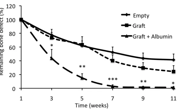
The first CT scans were performed on the first post-operative week, at this time point we found no significant differneces between the study groups. On the third post- operative week bone regeneration was underway in ervery group, since all bone defects were smaller. Additionally, the albumin group showed significantly smaller defects already on the third week. From the fifth post-operative week the albumin groups showed further decrease in bone defecet size, while the other groups showed over 60%
remaining bone defecet. At the seventh post.opertive week, the albumin coated group was almost completely closed, while the other bone defects still showed significant bone defects.
Figure 1. New bone formation. The albumin coated (Graft + Albumin) showed significantly faster bone fomration, by the 7th week bone defects were completely healed.. (n=6, two-way
ANOVA, Bonferroni post test, *: p < 0.05, **: p < 0.1, ***: p < 0.01)
4.7. Densitometry in vivo
We measured the density of bone defects from reconstructed images. We found definite bone defects on the first post-operatve week. According to the measurements at three and five weeks, bone formation was underway, the density values increased. Albumin coated group showed bigger density values, which became significant from the seveth week. At this time empty defects showed 550 ± 69 HU, uncoated defects showed 582 ± 108 HU, while albumin treated group showed 835 ± 64 HU. These differences further increased by th 11th week.
4.8. Ex vivo microCT
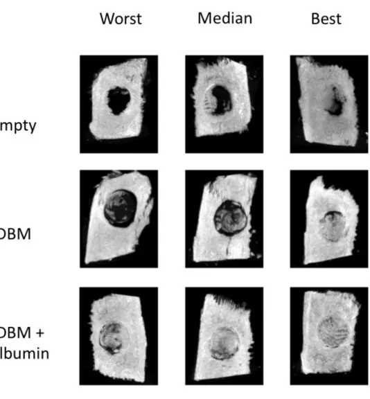
MicroCt was performed to more closely visualize the regenerated bone tissue, whihc we assesed with Bone volume/Tissue volume % measurements. After 11 weeks of regeneration the empty defects showed 4.2 ± 2.7%-ot, uncoated defects showed 8.2 ± 3.2% of BV/TV. These values were significanty lower from the bone defects treated with albumin coated grafts. The albumin coated group presented 24.1 ± 2.4 %.
Figure 2. Ex vivo MicroCT after 11 weeks. The figure is showing representative images from every study group. It also shows median pictures next between the worst and best pictures.The
albumin coated group shows significantly better bone formation.
4.9. Biomechanical testing
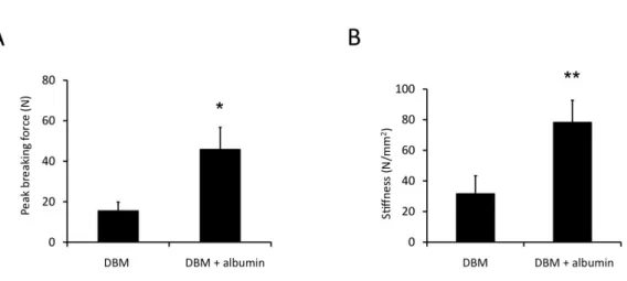
In the biomechanical testing experiement, we wanted to see how stable the regenerated bone tissue is. in the uncoated group 15.7 ± 4 N force was needed to breake the graft- host interface. In the albumin coated group a significantly higher peak breaking force was needed (46.1 ±11 N). We also gained data from the bone stiffness and realised the the stiffness of uncoated bone grafts was 32 ± 11 N/mm2, while the albumin coated group was significants harder (78.6 ± 14 N/mm2).
Figure 3. Ex vivo mechanical testing after 11 weeks. Panel A shows peak breaking force, while Panle B shows stiffness of the regenerated grafts. In both measurments the albumin coated
groups showed significantly higher value. (n=6, unpaired t-test, *: p < 0.05)
5. Conclusions
According to the result from the above mentioned experiments, the following thesises can be concluded:
1. Mesenchymal stem cells are willing to attach different biocompatible, albumin coated surfaces, like poly-esther surgical sutures and demineralised bone matrices. This process is probably due to the convenient physico-chemical and biological parameters
of the serum albumin molecule. For this reason it createds a convenietn milliue for stem cell attachment, survival and proliferation.
2. We have shown, that serum albumin coated surgical sutures are potent vehicles of stem cell delivery into soft tissues. After transplantation, transplanted cells leave the surface of the sutures and migrate into deeper layer in the injured. For this reason sutures are capable to deliver stem cells in soft tissues in a concentrated fashion.
3. Serum albumin molecule also supported bone regeneration, probably in an active way, since it enhanced to healing potential of demineralised bone matrices in critical size bone defects. Defects filled with albumin coated bone graft also produced mechanically more stable bone. The activ role of albumin is that it recrutes endogenous progenitors and therefore supports bone formation.
6. Summary
In the present work we investigated the role of serum albumin in the regeneration of the musculoskeletal system. Stem cell therapy is a promising adjuvant supporting the remodelling of musculoskeletal soft tissues, and since serum albumin provides a convenient milieu for stem cell attachment, survival and proliferation, application of coated sutures seems to be an ideal tool for this purpose. From a clinical point of view, this new cell transplantation technique can be used in the everyday surgical practice without complicating the surgical procedure itself. Exogenous stem cell delivery regarding bone tissue, on the other hand, is questionable, since both bone marrow and periosteum possess a large number of progenitors, ready to act after bone injury. In critical size bone defects this activation is still not enough, therefore various bone substitutes and osteoinductive agents are used to repair the damaged tissue. In the present work, serum albumin coated demineralized bone matrix successfully enhanced the healing of critical size bone defects without exogenous stem cell delivery. The regenerated bone tissue showed significantly better results both radiographically and mechanically, compared to uncoated demineralized bone matrix. Our conclusion was that serum albumin recruits endogenous progenitors and supports their proliferation, as a result a higher cell number is present at the defect site initiating the tissue
remodellation phase. Considering the strong osteoinductive capacity of serum albumin, stem cell therapy can be achieved without exogenous stem cell transplantation. Serum albumin is also easily accessible and, due to its purified nature and well investigated properties, it is safe to use. There is no question that highly developed tissue engineering techniques will provide better or maybe perfect solution in the future, but until then, serum albumin based products could be important adjuvants in musculoskeletal tissue regeneration.
List of publications
Publications of the current dissertation:
Denes B. Horvathy, Gabriella Vacz, Tamas Szabo, Imola C. Szigyarto, Ildiko Toro, Boglarka Vamos, Istvan Hornyak, Karoly Renner, Tamas Klara, Bence T. Szabo, Csaba Dobo-Nagy, Attila Doros, Zsombor Lacza 2015. Serum albumin coating of demineralized bone matrix results in stronger new bone formation. J Biomed Mater Res Part B 2016 Jan;104(1):126-32. doi: 10.1002/jbm.b.33359. IF: 2.881
Horvathy, D.B., Vacz, G., Cselenyak, A., Weszl, M., Kiss, L., Lacza, Z., Albumin-Coated Bioactive Suture for Cell Transplantation. Surg Innov. 2013 Jun;20(3):249-55. doi:
10.1177/1553350612451353. IF: 1.338
Dénes B. Horváthy, Gabriella Vácz, Tamás Szabó, Károly Renner, Kinga Vajda, Balázs Sándor and Zsombor Lacza Communication: Absorption and Tensility of Bioactive Sutures Prepared for Cell Transplantation Materials 2013, 6(2), 544-550; doi:10.3390/ma6020544 IF:
1.879
Horváthy DB, Simon M, Schwarz CM, Masteling M, Vácz G, Hornyák I, Lacza Z. Serum albumin as a local therapeutic agent in cell therapy and tissue engineering. Biofactors. 2016 Nov 11. doi: 10.1002/biof.1337. IF:4.504
Schandl K, Horváthy DB, Doros A, Majzik E, Schwarz CM, Csönge L, Abkarovits G, Bucsi L, Lacza Z. Bone-Albumin filling decreases donor site morbidity and enhances bone formation after anterior cruciate ligament reconstruction with bone-patellar tendon-bone autografts. Int Orthop. 2016 Jun 29. DOI:10.1007/s00264-016-3246-8 IF: 2.387
Horvathy DB, Vacz G, Toro I, Szabo T, May Z, Duarte M, Hornyak I, Szabo BT, Dobo- Nagy C, Doros A, Lacza Z. 2015. Remineralization of demineralized bone matrix in critical size cranial defects in rats: A 6-month follow-up study. J Biomed Mater Res Part B 2015:00B:000–000 IF: 2.881
Hornyák I, Madácsi E, Kalugyer P, Vácz G, Horváthy DB, Szendrői M, Han W, Lacza Z.
Increased release time of antibiotics from bone allografts through a novel biodegradable coating. Biomed Res Int. 2014;2014:459867. doi: 10.1155/2014/459867. IF: 1.579
D. B. Horvathy, P. P. Nardai, T. Major, K. Schandl, A. Cselenyak, G. Vacz, L. Kiss, M.
Szendroi, Z. Lacza: Muscle regeneration is undisturbed by repeated polytraumatic injury, Accepted for publication in Eur J Trauma Emerg Surg on May 31, 2010 IF: 0.328
Independent publications:
Vácz G, Cselenyák A, Cserép Z, Benkő R, Kovács E, Pankotai E, Lindenmair A, Wolbank S, Schwarz CM, Horváthy DB, Kiss L, Hornyák I, Lacza Z. Effects of amniotic epithelial cell transplantation in endothelial injury. Interv Med Appl Sci. 2016 Dec;8(4):164-171. doi:
10.1556/1646.8.2016.4.6. IF: 0
Marianna Király, Kristóf Kádár, Dénes B. Horváthy, Péter Nardai, Gábor Z. Rácz, Zsombor Lacza, Gábor Varga, Gábor Gerber: Integration of neuronally predifferentiated human dental pulp stem cells into rat brain in vivo. Neurochem Int. 2011 Sep;59(3):371-81. doi:
10.1016/j.neuint.2011.01.006. IF: 2.857
Horvathy DB, Hauck EF, Ogilvy CS, Hopkins LN , Levy EI, Siddiqui AH: Complete Preoperative Embolization of Hemangioblastoma Vessels with Onyx: Technical Note.
Accepted for publication in J Clin Neurosci on June 14, 2010. IF: 1.247
Hauck EF, Natarajan SK, Horvathy DB, Hopkins LN , Siddiqui AH, Levy EI: Stent- assisted Basilar Reconstruction for a Traumatic Vertebral Dissection with a Large Basilar Artery Thrombosis. Accepted for publication in J NeuroIntervent Surg 2010 IF: 0.923
Xantus G, Kovacs A, Horvathy DB: Hiedelmek és Hagyományok a sebkezelésben Háziorvos továbbképző szemle 2008 IF: 0