R E S E A R C H A R T I C L E
Rare manifestations of spinal tuberculosis in the Robert J. Terry Anatomical Skeletal Collection (National Museum of Natural History, Smithsonian Institution, Washington, DC, USA)
O. Spekker
1 |D.R. Hunt
2 |O.A. Váradi
1,3 |W. Berthon
1,4,5 |E. Molnár
1*
|G. Pálfi
1*
1Department of Biological Anthropology, University of Szeged, Szeged, Hungary
2Department of Anthropology, National Museum of Natural History, Smithsonian Institution, Washington, DC, USA
3Department of Microbiology, University of Szeged, Szeged, Hungary
4École Pratique des Hautes Études, PSL Research University Paris, Paris, France
5UMR 5199 PACEA, CNRS & University of Bordeaux, Pessac cedex, France
Correspondence
Olga Spekker, Department of Biological Anthropology, University of Szeged, Közép fasor 52, H‐6726 Szeged, Hungary.
Email: olga.spekker@gmail.com Funding information
National Research, Development and Innovation Office (Hungary), Grant/Award Number: K 125561; Tempus Public Foundation, Hungarian State Eötvös Fellow- ship; Hungarian Ministry of Human Capacities, Human Capacities Grant Management Office, Grant/Award Number: NTP‐NFTÖ‐16
Abstract
Today, osteoarticular tuberculosis (TB) is a rare extrapulmonary manifestation of TB, developing in about 1
–3% of patients. Although any part of the skeleton can be affected by tuberculosis, the spine represents the most common site of involvement (~50%). Spinal TB or tuberculous spondylitis typically affects 2 or more contiguous vertebrae, predominantly in the lower thoracic (40
–50%) and upper lumbar (35
–40%) regions. In the minority of patients (less than 10%), multilevel noncontigu- ous involvement occurs. By the location of lesions in the vertebra, 5 main types of spinal TB can be distinguished: paradiscal, central, anterior subligamentous, posterior, and articular forms, with the first being the most common type (90
–95%). In the palaeopathological literature, the majority of the described spinal TB cases represent the paradiscal form affecting 2 or more contiguous vertebrae in the thoracolumbar region. The aim of our paper is to provide 3 example cases of known age at death, sex, and cause of death from the Terry Collection that show vertebral alterations indicative of rare manifestations of tuberculous spondylitis, and to interpret the observed lesions with regard to their diagnostic value in the palaeopathological prac- tice. Terry No. 468 (23
‐year
‐old, male, died of pulmonary and spinal TB) exhibited bony changes presumably attributed to the anterior subligamentous form, whereas Terry No. 902 (c. 36
‐year
‐old, male, died of pulmonary TB) very likely represents the articular and anterior subligamentous types. The vertebral lesions recorded in the skeleton of Terry No. 1124R (49
‐year
‐old, female, died of pulmonary TB) are suggestive of the paradiscal form with multilevel noncontiguous involvement. Our paper provides palaeopathologists with a stronger basis for identifying tuberculosis in ancient human remains that reveal unusual vertebral alterations resembling that of our cases and, therefore, with a more sensitive means of assessing TB prevalence in past populations.
K E Y W O R D S
palaeopathology, rare spinal TB types, Terry Collection, tuberculosis
1
|I N T R O D U C T I O N
Today, skeletal tuberculosis (TB) occurs in 1–3% of TB patients and accounts for 10–35% of extrapulmonary TB cases (Garg & Somvanshi,
*Shared last authors.
DOI: 10.1002/oa.2658
Int J Osteoarchaeol. 2018;28:343–353. wileyonlinelibrary.com/journal/oa Copyright © 2018 John Wiley & Sons, Ltd. 343
2011; Kumar, 2005; Pigrau‐Serrallach & Rodríguez‐Pardo, 2013;
Vanhoenacker, Sanghvi, & De Backer, 2009). It is usually secondary to haematogenous spread of TB bacteria from a primary location out- side the skeleton into the bone and/or synovial tissue (Esteves, Catarino, Lopes, & Sousa, 2017; Garg & Somvanshi, 2011; Spiegel, Singh, & Banskota, 2005). Any part of the skeleton can be affected by tuberculosis, but its most preferential skeletal localisation is the spine, followed by the hip and knee joints (Garg & Somvanshi, 2011;
Vanhoenacker et al., 2009). The deposition of TB bacteria into the skeleton can lead to the development of three main forms of osteoarticular tuberculosis: spinal TB or tuberculous spondylitis (i.e., combination of tuberculous vertebral osteomyelitis and arthritis), tuberculous osteomyelitis of the extra‐spinal bones, and tuberculous arthritis of the extra‐spinal joints, with the first being the most common type (~50%; Golden & Vikram, 2005; Spiegel et al., 2005;
Vanhoenacker et al., 2009).
Tuberculous spondylitis is also known as Pott's disease in honour of English surgeon Sir Percivall Pott (1779) who first described this clinical entity in the modern era (De la Garza Ramos et al., 2017; Fuentes Ferrer, Gutiérrez Torres, Ayala Ramírez, Rumayor Zarzuelo, & del Prado González, 2012; Garg & Somvanshi, 2011). Any part of the vertebral column can be involved by tubercu- losis, but the most frequently affected regions are the lower thoracic (40–50%) and upper lumbar (35–40%) spine (De la Garza Ramos et al., 2017; Garg & Somvanshi, 2011; Kumar, 2005). The cervical region is involved in only the minority of spinal TB patients (~10%; De la Garza Ramos et al., 2017; Fuentes Ferrer et al., 2012; Pigrau‐Serrallach & Rodríguez‐Pardo, 2013). Although tuber- culous spondylitis typically affects two or more contiguous vertebrae, multilevel noncontiguous (less than 10%) or solitary verte- bral involvement can also occur in a few cases (Garg & Somvanshi, 2011; Kumar, 2005; Pigrau‐Serrallach & Rodríguez‐Pardo, 2013;
Spiegel et al., 2005). Based on the location of tuberculous lesions in the vertebra, five main types of spinal TB can be distinguished:
paradiscal, central, anterior subligamentous, posterior, and articular forms (Esteves et al., 2017; Garg & Somvanshi, 2011; Kumar, 2005; Sridhar & Krishnan, 2009).
The most common type of tuberculous spondylitis (90–95%) is the paradiscal form that arises from the anterior subchondral (paradiscal) region of the vertebral body (Esteves et al., 2017; Rivas‐Garcia, Sarria‐Estrada, Torrents‐Odin, Casas‐Gomila, &
Franquet, 2013; Spiegel et al., 2005). The presence of TB bacteria triggers the formation of osteolytic lesions in the paradiscal area, with subsequent involvement of the entire vertebral body and occa- sionally of the posterior elements (Esteves et al., 2017; Rivas‐Garcia et al., 2013). Later, the infection may spread vertically beneath the anterior longitudinal ligament from the primarily affected vertebra into a similar location at one or more contiguous vertebrae or beyond (Aufderheide & Rodríguez‐Martín, 1998; Esteves et al., 2017). The progressive destruction of the subchondral region results in the weakening of the affected vertebral bodies, with their conse- quent collapse under the weight of the trunk (Aufderheide &
Rodríguez‐Martín, 1998). Because the cavitation of the infected vertebrae predominantly involves their anterior part, the collapse is characterised by a wedge‐shaped appearance (Aufderheide &
Rodríguez‐Martín, 1998; Garg & Somvanshi, 2011). Depending on its localisation in the spine, the collapse of one or more contigu- ous vertebrae may lead to the development of different spinal deformity types, most frequently of a sharply angular kyphosis in the thoracic region (i.e., Pott's gibbus; Aufderheide & Rodríguez‐ Martín, 1998; Garg & Somvanshi, 2011). The progressive destruction of the affected vertebrae may be associated with mechanical instability of the spine (Esteves et al., 2017). Therefore, subsequent bony fusion of the remnants of the collapsed vertebral bodies, bony ankylosis of the facet joints, and formation of bony extensions interconnecting the adjacent vertebrae (i.e., bony bridges) may occur to restabilise the spine (Aufderheide & Rodríguez‐Martín, 1998;
Ortner, 2003).
Other types of tuberculous spondylitis (e.g., central, anterior subligamentous, posterior, and articular forms) all together constitute approximately 5–10% of spinal TB cases (Esteves et al., 2017;
Sridhar & Krishnan, 2009). The central type arises from the midsec- tion of one or more distant or adjacent vertebral bodies (Aufderheide & Rodríguez‐Martín, 1998; Garg & Somvanshi, 2011).
Later, the infection may extend centrifugally to involve the entire body (Garg & Somvanshi, 2011; Rivas‐Garcia et al., 2013). Destruc- tion of the vertebral body may lead to its subsequent ballooning and concentric collapse (Esteves et al., 2017; Rivas‐Garcia et al., 2013). It resemblesvertebra planaand indicates complete compres- sion of the body under the weight of the trunk, with consequent development of spinal deformity (Esteves et al., 2017; Garg &
Somvanshi, 2011).
The anterior subligamentous form of tuberculous spondylitis is characterised by erosive cortical bone destruction mainly affecting the anterior and lateral surfaces of the involved vertebral bodies (Aufderheide & Rodríguez‐Martín, 1998; Sorrel & Sorrel‐ Dejerine, 1932). It may be secondary to the development of an extravertebral TB abscess between the vertebral body and the anterior longitudinal ligament or periosteum that can extend verti- cally over several contiguous vertebrae beneath the aforemen- tioned fibrous structures (Aufderheide & Rodríguez‐Martín, 1998;
Kumar, 2005; Rivas‐Garcia et al., 2013; Spiegel et al., 2005). The subligamentous spread of infection results in shallow erosion of the anterior and lateral surfaces of the affected vertebral bodies that produces a scalloped appearance; later, the anterior and lateral body portions can also be involved (Aufderheide &
Rodríguez‐Martín, 1998; Kumar, 2005; Rivas‐Garcia et al., 2013;
Spiegel et al., 2005). Interconnecting, stabilising bony extensions (i.e., bony bridges) may also occur on the anterior and/or lateral aspects of the involved adjoining bodies that contribute to the pres- ervation of the intervertebral disc spaces and to the prevention of the development of vertebral collapse and subsequent spinal defor- mity even in cases with severe bone loss (Ortner, 2003; Sorrel &
Sorrel‐Dejerine, 1932).
In one of the less frequent spinal TB types (2–3%), namely, in the posterior or appendiceal form, the tuberculous process is isolated to the posterior elements, without or with secondary involvement of the vertebral body (Garg & Somvanshi, 2011; Kumar, 2017; Sridhar
& Krishnan, 2009). The osteolytic lesions mainly affect only one vertebra, with pedicles and laminae representing the most common
sites of involvement (Kumar, 2017; Sridhar & Krishnan, 2009).
Destruction of the posterior elements only is rarely accompanied by spinal deformity, because it barely affects the stability of the spine (Sridhar & Krishnan, 2009).
The articular or synovial form of tuberculous spondylitis, affecting the atlanto‐occipital, atlanto‐axial, or facet joints, occurs in only the minority of spinal TB patients (e.g., the involvement of the atlanto‐ occipital joint can be observed in less than 1% of cases; Garg &
Somvanshi, 2011; Kumar, 2017; Qureshi, Afzal, Khalique, Pasha, &
Aebi, 2013). The progressive destruction of the affected joint with consequent instability may be associated with the development of subluxation or dislocation (Aufderheide & Rodríguez‐Martín, 1998;
Qureshi et al., 2013).
Tuberculosis is one of the oldest known infectious diseases that has been plaguing humans for thousands of years (Galagan, 2014;
Pálfi, Dutour, Deák, & Hutás, 1999; Pálfi, Dutour, Perrin, Sola, &
Zink, 2015; Roberts & Buikstra, 2003): The earliest recognised and verified TB cases (e.g., Baker et al., 2015; Köhler et al., 2014;
Masson et al., 2015; Sparacello, Roberts, Kerudin, & Müller, 2017) come from the Neolithic period. Traditionally, the diagnosis of TB in human remains from archaeological sites relies on the identification of pathological lesions in the skeleton that can be related to different forms of osteoarticular TB (Aufderheide & Rodríguez‐Martín, 1998;
Ortner, 2003; Pálfi, Bereczki, Ortner, & Dutour, 2012). Although a number of studies on spinal TB have been published in the palaeopathological literature since the beginning of the 20th century, the vast majority of the described cases (e.g., Baker et al., 2015; Balázs et al., 2015; Hajdu et al., 2012; Sorrel & Sorrel‐Dejerine, 1932) represent the most common type of the disease by location of lesions in the vertebra, that is, the paradiscal form with involvement of two or more contiguous thoracic and/or lumbar vertebrae. The aim of our paper is to provide three example cases of known cause of death from the Terry Collection that show vertebral changes indicative of rare manifestations of tuberculous spondylitis, namely, the anterior subligamentous and articular types, and the paradiscal form with mul- tilevel noncontiguous involvement, and to interpret the observed alter- ations with regard to their diagnostic value in the palaeopathological practice.
2
|M A T E R I A L S A N D M E T H O D S
The Robert J. Terry Anatomical Skeletal Collection—currently curated in the Department of Anthropology at the National Museum of Natural History (NMNH; Smithsonian Institution, Washington, DC, USA)—consists of 1,728 human skeletons (Hunt & Albanese, 2005).
For each individual, a series of documentary forms providing various biographical information (e.g., age at death, sex, and cause of death) and basic anthropological data is available at the NMNH (Hunt &
Albanese, 2005). Therefore, the Terry Collection serves as an invalu- able resource for anthropological and medical research, including developing new criteria for the diagnosis of specific infectious diseases in osteoarchaeological series from the preantibiotic era (Hunt & Albanese, 2005). In the last three decades, it has been used to define and refine diagnostic criteria for TB in several studies
(e.g., Pálfi et al., 2012; Roberts, Lucy, & Manchester, 1994; Winland, Rothschild, & Rothschild, 1997).
As part of a comprehensive research project, a detailed investiga- tion was performed on all specimens (N= 302) with TB as the cause of death from the Terry Collection and on a control group consisting of randomly selected individuals (N= 302) from the remaining specimens of the Collection with nontuberculous causes of death. The macro- scopic evaluation of the 604 selected skeletons focused on the macromorphological characteristics, frequencies, and co‐occurrences of pathological alterations probably related to different forms of TB.
These are the following:
• bony changes indicative of osteoarticular TB (osteolytic or erosive vertebral lesions, collapse or fusion of the vertebral bodies, vertebral hypervascularisation, cortical remodelling and reactive new bone formation on the vertebral surfaces, signs of osteomyelitis of the extra‐spinal bones, and destruction, subluxation, or dislocation of the intervertebral or extra‐spinal joints);
• lesions suggestive of pulmonary TB and/or TB pleurisy (periosteal new bone formation (PNBF) on the visceral surface of ribs, erosive costal changes, and signs of diffuse, symmetrical periostitis on the diaphysis of short and long tubular bones); and
• endocranial alterations likely associated with tuberculous meningi- tis (TBM; abnormally pronounced digital impressions [APDIs], periosteal appositions, abnormal blood vessel impressions [ABVIs], and granular impressions).
All bones of specimens were macromophologically evaluated.
For each surveyed individual, detailed written and pictorial descriptions of all observed probable TB‐related alterations were made on a data collection sheet. In a few cases, CT examination was also performed, using a Siemens SOMATOM Emotion 6 CT scanner.
Among the 604 evaluated skeletons from the Terry Collection, there were only three specimens recorded to have died of tubercu- lous spondylitis; nonetheless, bony changes probably associated with spinal TB were identified in the skeleton of more than 30 individ- uals. In our paper, the following three skeletons from the Terry Collection were selected for discussion in detail as they exhibit vertebral lesions very likely related to rare forms of tuberculous spondylitis:
• Terry No. 468: a 23‐year‐old (1902–1926) male recorded to have died of pulmonary and spinal TB. The skeleton is well preserved and complete;
• Terry No. 902: a c. 36‐year‐old (1895?–1931) male whose morgue record states pulmonary TB as the cause of death. The skeleton is well preserved and almost complete (only the left femur and the two 12th ribs are missing); and
• Terry No. 1124R: a 49‐year‐old (1899–1949) female registered to have died of pulmonary TB. The skeleton is well preserved and almost complete (only the manubrium sterni, the left fourth meta- carpal, and three phalanges are missing).
3
|R E S U L T S A N D D I S C U S S I O N
3.1
|Terry No. 468
3.1.1
|Vertebral bony changes
In the vertebral column (Table 1), bony changes presumably resulted from the anterior subligamentous form of spinal TB were observed in the thoracic (T6–12) and lumbar (L1–5) regions. The anterior and lateral cortical bone layers of the bodies showed slight erosion and reactive new bone formation in the thoracic (T6–12) spine (Figure 1) and severe destructive remodelling in the lumbar (L1–4) spine (Figure 2). Moreover, small, shallow erosive lesions were noted on the anterior and lateral aspects of the lower thoracic (T8–12) bodies (Figures 1 and S1a,b), and multiple, tunnel‐like or groove‐like erosive lesions, extensive new bone formation, and bone bridges interconnecting the anterior aspects of the adjacent bodies were detected in the lumbar (L1–4) region (Figure 2). The L5 body (Figure S1c,d) exhibited a shallow erosive lesion, reactive new bone formation, and signs of hypervascularisation.
3.1.2
|Extra ‐ spinal bony changes
In the extra‐spinal skeleton (Table 1), PNBF was registered on the visceral surface of two left side (11th–12th) ribs (Figure S2),
TABLE 1 Registered bony changes in the three specimens described in detail by anatomical regions (+ = present,−= not present)
Registered bony changes Terry No. 468 Terry No. 902 Terry No. 1124R
Skull
Abnormally pronounced digital impressions + (skullcap and base) − −
Abnormal blood vessel impressions − + (frontal and parietals) −
Spine
Cortical remodelling and/or reactive new bone formation
+ (T6–12 and L1–5) + (T5–6, T12, and L4–5) + (T7, T12, and L3)
Erosive vertebral lesions + (T8–12 and L1–5) + (T4, T7, T12, and L5) + (T7 and L3) Bone bridges interconnecting the adjacent
vertebral bodies
+ (L1–4) − −
Signs of vertebral hypervascularisation + (L5) + (T5–6, T10–12, and L1–5) + (L5)
Osteolytic vertebral lesions − − + (T12)
Wedge‐shaped collapse and fusion of vertebral bodies
− − + (C6–7 and L1–2)
Destruction of intervertebral joints − + (left atlanto‐occipital and left T4–5)
+ (left T12–L1 and right L2–3)
Ankylosis of intervertebral joints − − + (C6–7, C7–T1, and L1–2)
Thorax (excluding vertebrae)
Osteolytic lesions − + (sternum) −
Erosive lesions − + (left 3rd–7th and right 4th ribs) −
Cortical remodelling − + (sternum) −
Periosteal new bone formation + (left 11th–12th ribs) + (left 4th–5th and right 3rd–8th ribs) − Pelvis
Erosive lesions + (os coxae) + (right os coxa) −
Periosteal new bone formation + (os coxae) + (right os coxa) −
Sacroiliitis + (bilateral) + (unilateral–left) −
Symphysitis + (bilateral) − −
Diaphysis of short and long tubular bones
Signs of diffuse, symmetrical periostitis − + (tibiae, fibulae, and metacarpals) −
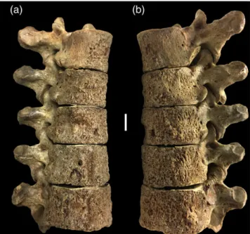
FIGURE 1 Slight cortical erosion accompanied by periosteal new bone formation on the anterior and lateral aspects of the T6–10 vertebral bodies: (a) right antero‐lateral view and (b) left antero‐lateral view (Terry No. 468, 23‐year‐old, male; scale bar = 10 mm) [Colour figure can be viewed at wileyonlinelibrary.com]
predominantly affecting their vertebral end. PNBF on the visceral surface of ribs was associated with pulmonary TB and/or TB pleurisy following examination of skeletons of known cause of death from documented skeletal collections, as they occurred more frequently in individuals recorded to have died of TB than in specimens identified to have died of nontuberculous causes (e.g., Kelley & Micozzi, 1984;
Matos & Santos, 2006; Santos & Roberts, 2001, 2006).
Probably in response to an overlying cold abscess that is a common complication in tuberculous spondylitis (Aufderheide &
Rodríguez‐Martín, 1998; Ortner, 2003), an erosive lesion accompanied by PNBF occurred on the anterior surface of both os coxae (Figure 3a, b). The iliac and sacral articular surfaces of both sacroiliac joints (Figure 3a,b) showed a slightly porous appearance that is indicative of bilateral TB sacroiliitis (Ortner, 2003; Pálfi et al., 2012). Neverthe- less, tuberculous involvement of the sacroiliac joint is rather uncom- mon (affecting approximately 3–10% of osteoarticular TB cases) and usually results from direct extension of a spinal TB focus (Gupta, Bienenstock, Morano, & Gupta, 2005; Papagelopoulos et al., 2005).
The adjoining surfaces of the two pubic symphyses (Figure 3c,d) revealed severe destruction that is suggestive of TB symphysitis, representing an unusual site of the disease (Lal, Jain, & Kannan, 2013; Meena & Gangary, 2015; Ortner, 2003).
In the skull, APDIs were registered all over the inner surface of the skullcap (Figure S3) and skull base. The presence of APDIs is indicative of elevated intracranial pressure probably due to hydrocephalus that might be related to tuberculous meningitis (Schultz, 1993, 1999, 2001, 2003).
3.2
|Terry No. 902
3.2.1
|Vertebral bony changes
In the vertebral column (Table 1), four noncontiguous levels revealed bony changes very likely associated with the articular and anterior subligamentous forms of spinal TB: the left atlanto‐occipital joint,
the middle thoracic (T4–7) region, the T12 vertebra, and the lower lumbar (L4–5) region. The articular surfaces of the left atlanto‐occipital joint (Figure 4a,b) and of the left facet joint between the T4–5 verte- brae (Figure 4c,d) showed a rough porous appearance and destruction.
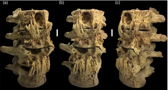
Reactive new bone formation was noted posteriorly to the left occip- ital condyle, adjacent to the foramen magnum (Figure 4a). The anterior aspect of the T5–6 (Figure 5a), T12, and L4–5 bodies exhibited slight FIGURE 2 Deep, spherical, tunnel‐or groove‐like erosive lesions and bone bridges interconnecting the adjacent vertebral bodies in the lumbar region (L1–4): (a) right antero‐lateral view, (b) anterior view, and (c) left antero‐lateral view (Terry No. 468, 23‐year‐old, male; scale bar = 10 mm) [Colour figure can be viewed at wileyonlinelibrary.com]
FIGURE 3 Shallow, roundish erosive lesions and patches of periosteal new bone formation along the iliac crest on the anterior surface of the (a) left and (b) right os coxae, with slight erosion on the iliac articular surface of the sacroiliac joint. Severe destruction of the adjoining surfaces in the (c) left and (d) right pubic symphyses (Terry No. 468, 23‐year‐old, male; scale bar = 10 mm) [Colour figure can be viewed at wileyonlinelibrary.com]
remodelling of the cortical bone layers. Erosive lesions occasionally accompanied by reactive new bone formation were detected on the left lamina and the adjoining part of the spinous process of T4
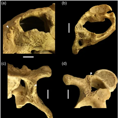
(Figure 5b), the right lateral aspect of the T7 and T12 bodies (Figure 5c), and the left lateral aspect of the L5 body (Figure 5d). Signs of hypervascularisation, indicative of early‐stage spinal TB (e.g., Baker, FIGURE 4 Erosion of the adjoining articular surfaces in the left atlanto‐occipital joint: (a) occipital bone (with new bone formation adjacent to the foramen magnum) and (b) atlas; as well as in the left facet joint between the T4 and T5 vertebrae: (c) T4 and (d) T5 (Terry No. 902, c. 36‐year‐old, male; scale bar = 10 mm) [Colour figure can be viewed at wileyonlinelibrary.com]
FIGURE 5 Superficial vertebral changes: (a) slight cortical remodelling and small, horizontal abnormal blood vessel impressions on the anterior aspect of T6; and erosive lesions accompanied by reactive new bone formation on the (b) left lamina and spinous process of T4, (c) right lateral aspect of T12, and (d) left lateral aspect of L5 (Terry No. 902, c. 36‐year‐old, male; scale bar = 10 mm) [Colour figure can be viewed at wileyonlinelibrary.com]
1999; Mariotti et al., 2015; Ménard, 1888), were recorded on the anterior (T5–6; Figure 5a) and lateral (T10–12 and L1–5) aspects of the thoracic and lumbar bodies.
3.2.2
|Extra ‐ spinal bony changes
In the extra‐spinal skeleton (Table 1), both the external and internal surfaces of the sternal body (Figure 6a,b) revealed multiple perforating lytic lesions and remodelling and destruction of the cortical bone layers that are suggestive of TB osteomyelitis of the sternum.
Nonetheless, tuberculous involvement of the sternum is fairly uncom- mon, accounting for less than 2% of cases with extra‐spinal TB osteo- myelitis (Ortner, 2003; Sachdeva, Sachdeva, & Arora, 2013; Vasa, Ohikhuare, & Brickner, 2009).
Very slight PNBF was registered on the vertebral end of six right‐ side (3rd–8th) and two left‐side (4th–5th) ribs, exclusively affecting the visceral surfaces. Moreover, solitary erosive lesions were observed on the body (3rd left and 4th right) and/or sternal end (4th–7th left) of six ribs (Figure 6c). The above‐mentioned rib lesions were frequently described as not pathognomonic but probable signs of pulmonary TB and/or TB pleurisy in the palaeopathological literature (e.g., Giacon, 2008; Maczel, 2003; Mariotti et al., 2015; Roberts et al., 1994). All surfaces of both tibiae (Figure S4a) and fibulae, and the palmar surfaces of four left‐side (2nd–5th; Figure S4b) and four right‐side (1st–4th) metacarpals also showed slight PNBF, possibly due to hyper- trophic pulmonary osteopathy that can be associated with pulmonary TB (Assis, Santos, & Roberts, 2011; Kelly, Manning, Corcoran, &
Clancy, 1991; Rothschild & Rothschild, 1998; Winland et al., 1997).
A small erosive lesion accompanied by PNBF was observed on the anterior surface of the right os coxa (Figure S5a) that probably devel- oped in response to an overlying TB cold abscess (Aufderheide &
Rodríguez‐Martín, 1998; Ortner, 2003; Pálfi et al., 2012). Both the iliac and sacral articular surfaces of the left sacroiliac joint (Figure S5b,c) exhibited a rough porous appearance that is suggestive of unilateral TB sacroiliitis (Gupta et al., 2005; Ortner, 2003; Papagelopoulos et al., 2005).
In the skull, multifocal, small, serpentine branching ABVIs— described by Schultz (e.g., Schultz, 1993, 1999, 2001, 2003) as non‐specific vestiges of inflammatory‐haemorrhagic processes of the meninges—were registered on the endocranial surface of the frontal and both parietal bones (Figure S6). Although ABVIs are not pathognomonic features of TB, they might be resulted from TBM (Hershkovitz et al., 2002; Lewis, 2004; Schultz, 1993, 1999, 2001, 2003).
3.3
|Terry No. 1124R
3.3.1
|Vertebral bony changes
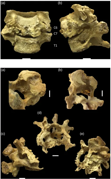
In the spine (Table 1), vertebral lesions indicative of the paradiscal form of tuberculous spondylitis with multilevel, noncontiguous involvement were observed between the C6−T1 and T12−L3 verte- brae. The C6–7 bodies (Figure 7) revealed considerable bone destruc- tion with subsequent wedge‐shaped collapse and fusion of their remnants, and with consequent loss of the C6–7 intervertebral disc space. The facet joints between the C6−7 and C7−T1 vertebrae were ankylosed (Figure 7); however, the T1 body remained intact. In the T7 body, a small erosive lesion accompanied by slight remodelling of the cortical bone layers was detected. The T12 body (Figure 8a,b) exhibited reactive new bone formation on its lateral aspects and small, multiple, confluent, porous‐walled lytic lesions with no evidence of bone repair on its inferior aspect. The posterior part of the T12 body (Figure 8a,b) was almost completely destroyed; however, its height was maintained. The L1–2 bodies (Figure 8c,d,e) were almost completely destroyed and exhibited substantial loss of the vertebral height. The fusion of their small, wedge‐shaped remnants resulted in the complete loss of the intervening disc space and the development of a severe angular kyphosis (Figure 9). The CT images (Movie S1) revealed no narrowing of the spinal canal. The superior and lateral aspects of the L3 body (Figure S7) displayed slight reactive new bone formation, erosive lesions, and signs of hypervascularisation, with maintenance of the vertebral height. The facet joints between the L1–2 vertebrae were ankylosed, and the articular surfaces of the left facet joint between the T12–L1 and of the right facet joint between the L2–3 vertebrae showed a rough porous appearance.
3.3.2
|Extra ‐ spinal bony changes
There were no extra‐spinal bony changes probably related to TB in the skeleton (Table 1).
3.4
|Differential diagnosis
In our paper, three skeletons of known cause of death from the Terry Collection were selected for discussion in detail, as they exhibit FIGURE 6 Severe destruction and remodelling of the (a) external
and (b) internal surfaces of the body of the sternum, with multiple perforating lytic lesions. Small erosive lesion (c) in the costal groove of the fourth left rib (Terry No. 902, c. 36‐year‐old, male; scale bar = 10 mm) [Colour figure can be viewed at wileyonlinelibrary.com]
vertebral lesions very likely related to rare types of spinal TB. Terry No. 468 displayed bony changes that are indicative of the anterior subligamentous form, whereas Terry No. 902 presumably represents the articular and anterior subligamentous types. The alterations detected in the spine of Terry No. 1124R are suggestive of the paradiscal form with multilevel noncontiguous involvement (including the cervical region). The recorded cause of death of the three speci- mens from the Terry Collection supports the tuberculous origin of the observed vertebral lesions. However, it must be noted that even though the morgue record of the above‐mentioned individuals states pulmonary and/or spinal TB as the cause of death, tuberculosis may not have been the only medical condition present in the specimens that could contribute to the development of bony changes in their skeleton. Moreover, there is always the possibility that an inaccurate
cause of death was registered on the morgue record and/or death cer- tificate of individuals from the Terry Collection. Therefore, differential diagnosis is necessary for reinforcing the diagnostic value of the observed vertebral lesions. In addition to spinal TB, it includes pyogenic spondylitis, brucellar spondylitis, fungal spondylitis, and compression fractures (Aufderheide & Rodríguez‐Martín, 1998;
Esteves et al., 2017; Garg & Somvanshi, 2011; Ortner, 2003; Pigrau‐ Serrallach & Rodríguez‐Pardo, 2013; Rivas‐Garcia et al., 2013; Spiegel et al., 2005).
In Terry No. 1124R (49‐year‐old, female, died of pulmonary TB), vertebral compression fractures represent a possible diagnostic option. Similar to spinal TB, compression fractures tend to affect the thoracic and thoracolumbar regions of the vertebral column, but they usually involve a single vertebra, and the kyphosis of the spine
FIGURE 7 Wedge‐shaped collapse and fusion of the C6–7 vertebral bodies, with subsequent ankylosis of the facet joints between the C6–7 and C7–T1 vertebrae:
(a) anterior view and (b) left lateral view (Terry No. 1124R, 49‐year‐old, female; scale bar = 10 mm) [Colour figure can be viewed at wileyonlinelibrary.com]
FIGURE 8 Multiple, confluent, porous‐ walled lytic lesions affecting the inferior and posterior parts of the T12 vertebral body, with severe bone loss but maintained vertebral height: (a) left lateral postero‐inferior view and (b) right lateral postero‐inferior view. Severe destruction of the L1–2 vertebral bodies and fusion of their remnants, with ankylosis of the interconnecting facet joints and gibbus formation: (c) right lateral view, (d) superior view, and (e) left antero‐lateral view (Terry No. 1124R, 49‐year‐old, female; scale bar = 10 mm) [Colour figure can be viewed at wileyonlinelibrary.com]
secondary to the vertebral lesions is not angular. Thus, compression fractures can be ruled out on the above basis along with the presence of osteolytic lesions, cortical remodelling, and reactive new bone formation in the cervical and thoracolumbar spine of Terry No.
1124R that are indicative of an infectious process. Pyogenic and brucellar spondylitis are prone to involve the lumbar region of the spine. They also tend to produce sclerotic bone that is absent in Terry No. 1124R. Moreover, the involvement of more than two vertebrae and the development of spinal deformity are not characteristic of pyogenic or brucellar spondylitis. Therefore, these conditions are unlikely diagnostic options in this specimen. In fungal spondylitis, posterior element involvement is common; however, it was absent in Terry No. 1124R.
In Terry No. 468 (23‐year‐old, male, died of pulmonary and spinal TB) and 902 (c. 36‐year‐old, male, died of pulmonary TB), the presence of anterior scalloping, erosive lesions, and reactive new bone forma- tion in the spine is suggestive of subligamentous spread of extra‐ vertebral cold abscesses along the anterior longitudinal ligament. In Terry No. 468, the infection might extend upwards and downwards from the L1 body, whereas in Terry No. 902, it might start in the inter- vertebral joints, and then spread into other parts of the vertebral column. In spinal TB, the formation of extra‐vertebral cold abscesses is a common complication, but there is no or minimal soft tissue involvement in pyogenic or brucellar spondylitis. Thus, the two latter conditions can be rejected as diagnostic options in both cases.
In summary, based on the recorded cause of death of specimens, the macromorphological characteristics of the observed lesions affecting the vertebral column, and their co‐occurrence with probable
TB‐related extra‐spinal bony changes, the most likely aetiology of alterations detected in the spine of Terry Nos 468, 902, and 1124R is tuberculous spondylitis.
4
|C O N C L U S I O N S
In 1993, tuberculosis has been declared a global health threat by the World Health Organization. Despite significant advances in the global fight against TB in the last few decades, it still presents a health emer- gency, especially in developing countries. Therefore, a renewed inter- est and funding to the research of the disease and of its aetiological agents have sparked since the late 20th century in order to eliminate or at least control TB in the future. The palaeopathological research of tuberculosis (essentially based upon the macromorphological diagnosis of the disease in ancient human remains) provides invaluable novel data on the different manifestations of TB and on the effects of the disease upon human mortality and morbidity in past populations.
In the last three decades, a number of studies (e.g., Matos &
Santos, 2006; Roberts et al., 1994; Santos & Roberts, 2001, 2006) were performed on documented skeletal collections that have revealed a positive correlation between different forms of tuberculosis and bony changes, but only the minority of them (e.g., Ortner, 2003;
Pálfi et al., 2012) described vertebral lesions that are very likely associated with rare types of tuberculous spondylitis. Nevertheless, the establishment of a more reliable and accurate palaeopathological diagnosis of TB and the assessment of a more relevant disease preva- lence in human osteoarchaeological series require excessive scientific FIGURE 9 Severe angular kyphosis (Pott's
gibbus) involving the T12–L3 vertebrae:
(a) right lateral view, (b) left lateral view, (c) right antero‐lateral view, and (d) left antero‐lateral view (Terry No. 1124R, 49‐year‐old, female; scale bar = 10 mm) [Colour figure can be viewed at wileyonlinelibrary.com]
knowledge on the macromorphological diagnostics of tuberculosis, including its rare spinal manifestations, that underlines the importance of the case studies presented in our paper. The three example cases provide palaeopathologists with a stronger basis for identifying tuberculous spondylitis in ancient human remains that reveal unusual vertebral alterations resembling that of our cases and, therefore, with a more sensitive means of assessing the prevalence of TB in past populations. Two of our three specimens revealed vertebral lesions in association with extra‐spinal probable TB‐related bony changes, thus constituting evidence that the aforementioned extra‐spinal lesions do have a diagnostic value in the palaeopathological identifica- tion of TB. Although they cannot be considered as pathognomonic vestiges of the disease, palaeopathologists can use them to diagnose TB in osteoarchaeological series from the preantibiotic era, especially when they occur simultaneously with each other or in association with very likely TB‐related vertebral alterations.
Finally, the three example cases presented in our paper give insight into rare and serious manifestations of spinal TB associated with potentially debilitating physical symptoms and, therefore, into the substantial impact the disease could have on the patients' overall well‐being and quality of life in the recent and ancient past.
A C K N O W L E D G E M E N T S
The support of the Tempus Public Foundation, Hungarian State Eötvös Fellowship, of the Hungarian Ministry of Human Capacities, Human Capacities Grant Management Office, NTP‐NFTÖ‐16, and of the National Research, Development and Innovation Office (Hungary), K 125561, is greatly acknowledged.
O R C I D
O. Spekker http://orcid.org/0000-0002-0350-1906
R E F E R E N C E S
Assis, S., Santos, A. L., & Roberts, C. A. (2011). Evidence of hypertrophic osteoarthropathy in individuals from the Coimbra Skeletal Identified Collection (Portugal). International Journal of Paleopathology, 1(3–4), 155–163. https://doi.org/10.1016/j.ijpp.2012.02.009
Aufderheide, A. C., & Rodríguez‐Martín, C. (1998).The Cambridge encyclo- pedia of human paleopathology. ( pp. 118–141). Cambridge, UK:
Cambridge University Press.
Baker, B. J. (1999). Early manifestations of tuberculosis in the skeleton. In G. Pálfi, O. Dutour, J. Deák, & I. Hutás (Eds.),Tuberculosis: Past and present (pp. 301–307). Budapest, Hungary: TB Foundation: Szeged, Hungary & Golden Book Publisher.
Baker, O., Lee, O. Y.‐C., Wu, H. H. T., Besra, G. S., Minnikin, D. E., Llewellyn, G., … Dutour, O. (2015). Human tuberculosis predates domestication in ancient Syria. Tuberculosis, 95(Suppl. 1), S4–S12.
https://doi.org/10.1016/j.tube.2015.02.001
Balázs, J., Zádori, P. G., Vandulek, C., Molnár, E.,Ősz, B., Bereczki, Z.,… Pálfi, G. (2015). Morphological and paleoradiological studies of Pott's disease cases.Acta Biologica Szegediensis,59(2), 211–216.
De la Garza Ramos, R., Goodwin, C. R., Abu‐Bonsrah, N., Bydon, A., Witham, T. F., Wolinsky, J.‐P., & Sciubba, D. M. (2017). The epidemiol- ogy of spinal tuberculosis in the United States: An analysis of 2002–2011 data.Journal of Neurosurgery: Spine,26(4), 507–512.
Esteves, S., Catarino, I., Lopes, D., & Sousa, C. (2017). Spinal tuberculosis:
Rethinking an old disease.Journal of Spine,6(1), 358. https://doi.org/
10.4172/2165‐7939.1000358
Fuentes Ferrer, M., Gutiérrez Torres, L., Ayala Ramírez, O., Rumayor Zarzuelo, M., & del Prado González, N. (2012). Tuberculosis of the spine. A systematic review of case series.International Orthopaedics, 36(2), 221–231. https://doi.org/10.1007/s00264‐011‐1414‐4 Galagan, J. E. (2014). Genomic insights into tuberculosis.Nature Reviews
Genetics,15(5), 307–320. https://doi.org/10.1038/nrg3664
Garg, R. K., & Somvanshi, D. S. (2011). Spinal tuberculosis: A review.The Journal of Spinal Cord Medicine, 34(5), 440–454. https://doi.org/
10.1179/2045772311Y.0000000023
Giacon, M. (2008). Paleopathology in the osteological collection of Anthro- pological Museum in Padova University: The cases of tuberculosis (PhD Dissertation). Padova University: Padova, Italy.
Golden, M. P., & Vikram, H. R. (2005). Extrapulmonary tuberculosis: An overview.American Family Physician,72(9), 1761–1768.
Gupta, R., Bienenstock, H., Morano, P., & Gupta, A. (2005). Tuberculosis of sacroiliac joint: An unusual presentation.Journal of the National Medical Association,97(8), 1174–1176.
Pálfi, G., Dutour, O., Deák, J., & Hutás, I. (Eds.) (1999).Tuberculosis: Past and present. Budapest, Hungary: TB Foundation: Szeged, Hungary &
Golden Book Publisher.
Pálfi, G., Bereczki, Z., Ortner, D. J., & Dutour, O. (2012). Juvenile cases of skeletal tuberculosis from the Terry Anatomical Collection (Smithsonian Institution, Washington, DC, USA). Acta Biologica Szegediensis,56(1), 1–12.
Hajdu, T., Donoghue, H. D., Bernert, Z., Fóthi, E., Kővári, I., & Marcsik, A.
(2012). A case of spinal tuberculosis from the middle ages in Transylva- nia (Romania).Spine,37(25), e1598–e1601. https://doi.org/10.1097/
BRS.0b013e31827300dc
Hershkovitz, I., Greenwald, C. M., Latimer, B., Jellema, L. M., Wish‐Baratz, S., Eshed, V.,…Rotschild, B. M. (2002). Serpens endocrania symmetrica (SES): A new term and a possible clue for identifying intrathoracic dis- ease in skeletal populations.American Journal of Physical Anthropology, 118(3), 201–216. https://doi.org/10.1002/ajpa.10077
Hunt, D. R., & Albanese, J. (2005). History and demographic composition of the Robert J. Terry Anatomical Collection.American Journal of Physical Anthropology,127(4), 406–417. https://doi.org/10.1002/ajpa.20135 Kelley, M. A., & Micozzi, M. S. (1984). Rib lesions in chronic pulmonary
tuberculosis. American Journal of Physical Anthropology, 65(4), 381–386. https://doi.org/10.1002/ajpa.1330650407
Kelly, P., Manning, P., Corcoran, P., & Clancy, L. (1991). Hypertrophic osteoarthropathy in association with pulmonary tuberculosis.Chest, 99(3), 769–770. https://doi.org/10.1378/chest.99.3.769
Kumar, K. (2017). Posterior spinal tuberculosis: A review.Mycobacterial Diseases,7(2), 243. https://doi.org/10.4172/2161‐1068.1000243 Kumar, R. (2005). Spinal tuberculosis: With reference to the children in
northern India.Child's Nervous System,21(1), 19–26. https://doi.org/
10.1007/s00381‐004‐1029‐9
Lal, H., Jain, V. K., & Kannan, S. (2013). Tuberculosis of the pubic symphy- sis: Four unusual cases and literature review.Clinical Orthopaedics and Related Research, 471(10), 3372–3380. https://doi.org/10.1007/
s11999‐013‐3037‐0
Lewis, M. E. (2004). Endocranial lesions in non‐adult skeletons: Under- standing their aetiology. International Journal of Osteoarchaeology, 14(2), 82–97. https://doi.org/10.1002/oa.713
Maczel, M. (2003).“On the traces of tuberculosis”Diagnostic criteria of tuberculosis affection of the human skeleton and their application in Hungarian and French anthropological series (PhD Dissertation). Uni- versity of La Méditerranée – Aix Marseille II Faculty of Medicine:
Marseille, France & University of Szeged, Faculty of Science: Szeged, Hungary.
Mariotti, V., Zuppello, M., Pedrosi, M. E., Bettuzzi, M., Brancaccio, R., Peccenini, E.,…Belcastro, M. G. (2015). Skeletal evidence of tuberculo- sis in a modern identified human skeletal collection (Certosa cemetery, Bologna, Italy). American Journal of Physical Anthropology, 157(3), 389–401. https://doi.org/10.1002/ajpa.22727
Masson, M., Bereczki, Z., Molnár, E., Donoghue, H. D., Minnikin, D. E., Lee, O. Y.‐C., … Pálfi, G. (2015). 7000‐year‐old tuberculosis cases from Hungary—Osteological and biomolecular evidence. Tuberculosis, 95(Suppl. 1), S13–S17. https://doi.org/10.1016/j.tube.2015.02.007 Matos, V., & Santos, A. L. (2006). On the trail of pulmonary tuberculosis
based on rib lesions: Results from the human identified skeletal collec- tion from the Museu Bocage (Lisbon, Portugal). American Journal of Physical Anthropology, 130(2), 190–200. https://doi.org/10.1002/
ajpa.20309
Meena, S., & Gangary, S. K. (2015). Tuberculosis of symphysis pubis: A case report.Journal of Research in Medical Sciences,20(1), 100–102.
Ménard, V. (1888). Tuberculose vertébrale. Paris, France: Asselin et Houzeau.
Köhler, K., Pálfi, G., Molnár, E., Zalai‐Gaál, I., Osztás, A., Bánffy, E., … Mende, B. G. (2014). A Late Neolithic case of Pott's disease from Hungary. International Journal of Osteoarchaeology, 24(6), 697–703.
https://doi.org/10.1002/oa.2254
Ortner, D. J. (2003). Infectious diseases: Tuberculosis and leprosy. In D. J.
Ortner (Ed.), Identification of pathological conditions in human skeletal remains(pp. 227–271). San Diego, CA, USA: Academic Press.
Pálfi, G., Dutour, O., Perrin, P., Sola, C., & Zink, A. (2015). Tuberculosis in evolution. Tuberculosis, 95(Suppl. 1). https://doi.org/10.1016/j.
tube.2015.04.007
Papagelopoulos, P. J., Papadopoulos, E. C., Mavrogenis, A. F., Themistocleous, G. S., Korres, D. S., & Soucacos, P. N. (2005). Tubercu- lous sacroiliitis. A case report and review of the literature. European Spine Journal, 14(7), 683–688. https://doi.org/10.1007/s00586‐004‐ 0831‐0
Pigrau‐Serrallach, C., & Rodríguez‐Pardo, D. (2013). Bone and joint tuber- culosis.European Spine Journal,22(Suppl. 4), S556–S566. https://doi.
org/10.1007/s00586‐012‐2331‐y
Pott, P. (1779). Remarks on that kind of palsy of the lower limbs which is frequently found to accompany a curvature of the spine, and is supposed to be caused by it, together with its method of cure. To which are added, observations on the necessity and propriety of ampu- tation, in certain cases, and under certain circumstances. J. Johnson:
London, UK.
Qureshi, M. A., Afzal, W., Khalique, A. B., Pasha, I. F., & Aebi, M. (2013).
Tuberculosis of the craniovertebral junction. European Spine Journal, 22(Suppl. 4), S612–S617. https://doi.org/10.1007/s00586‐ 012‐2497‐3
Rivas‐Garcia, A., Sarria‐Estrada, S., Torrents‐Odin, C., Casas‐Gomila, L., &
Franquet, E. (2013). Imaging findings of Pott's disease.European Spine Journal, 22(Suppl. 4), S567–S578. https://doi.org/10.1007/s00586‐ 012‐2333‐9
Roberts, C. A., & Buikstra, J. E. (2003).The bioarchaeology of tuberculosis: A global view on a reemerging disease. Gainesville, FL, USA: University Press of Florida.
Roberts, C. A., Lucy, D., & Manchester, K. (1994). Inflammatory lesions of ribs: An analysis of the Terry Collection. American Journal of Physical Anthropology, 95(2), 169–182. https://doi.org/10.1002/
ajpa.1330950205
Rothschild, B. M., & Rothschild, C. (1998). Recognition of hypertrophic osteoarthropathy in skeletal remains. The Journal of Rheumatology, 25(11), 2221–2227.
Sachdeva, R., Sachdeva, S., & Arora, S. (2013). Sternal tuberculosis.Annals of Medical and Health Sciences Research,3(Suppl. 1), S21–S23. https://
doi.org/10.4103/2141‐9248.121213
Santos, A. L., & Roberts, C. A. (2001). A picture of tuberculosis in young Portuguese people in the early 20thcentury: A multidisciplinary study
of the skeletal and historical evidence. American Journal of Physical Anthropology,115(1), 38–49. https://doi.org/10.1002/ajpa.1054 Santos, A. L., & Roberts, C. A. (2006). Anatomy of a serial killer: Differential
diagnosis of tuberculosis based on rib lesions of adult individuals from the Coimbra Identified Skeletal Collection, Portugal.American Journal of Physical Anthropology, 130(1), 38–49. https://doi.org/10.1002/
ajpa.20160
Schultz, M. (1993).Spuren unspezifischer Entzündungen an prähistorischen und historischen Schädeln. Ein Beitrag zur Paläopathologie. Basel, Switzerland: Anthropologisches Forschungsinstitut: Aesch, Switzerland
& Anthropologische Gesellschaft.
Schultz, M. (1999). The role of tuberculosis in infancy and childhood in pre- historic and historic populations. In G. Pálfi, O. Dutour, J. Deák, & I.
Hutás (Eds.),Tuberculosis: Past and present(pp. 503–507). Budapest, Hungary: TB Foundation: Szeged, Hungary & Golden Book Publisher.
Schultz, M. (2001). Paleohistopathology of bone: A new approach to the study of ancient diseases.American Journal of Physical Anthropology, 116(Suppl. 33), S106–S147. https://doi.org/10.1002/ajpa.10024 Schultz, M. (2003). Light microscopic analysis in skeletal paleopathology. In
D. J. Ortner (Ed.), Identification of pathological conditions in human skeletal remains(pp. 73–107). San Diego, CA, USA: Academic Press.
https://doi.org/10.1016/B978‐012528628‐2/50043‐0
Sorrel, É., & Sorrel‐Dejerine, Y. (1932). Tuberculose osseuse et ostéo‐ articulaire. Volume 3. ( pp. 381–470). Paris, France: Masson.
Sparacello, V. S., Roberts, C. A., Kerudin, A., & Müller, R. (2017). A 6500‐ year‐old Middle Neolithic child from Pollera Cave (Liguria, Italy) with probable multifocal osteoarticular tuberculosis.International Journal of Paleopathology,17, 67–74. https://doi.org/10.1016/j.ijpp.2017.01.004 Spiegel, D. A., Singh, G. K., & Banskota, A. K. (2005). Tuberculosis of the musculoskeletal system.Techniques in Orthopaedics,20(2), 167–178.
Sridhar, K., & Krishnan, P. (2009). Complete posterior migration of intact vertebral body in spinal tuberculosis.Neurology India,57(4), 483–485.
https://doi.org/10.4103/0028‐3886.55600
Vanhoenacker, F. M., Sanghvi, D. A., & De Backer, A. I. (2009). Imaging fea- tures of extraaxial musculoskeletal tuberculosis. Indian Journal of Radiology and Imaging, 19(3), 176–186. https://doi.org/10.4103/
0971‐3026.54873
Vasa, M., Ohikhuare, C., & Brickner, L. (2009). Primary sternal tuberculosis osteomyelitis: A case report and discussion.Canadian Journal of Infec- tious Diseases and Medical Microbiology,20(4), e181–e184.
Winland, K., Rothschild, B. M., & Rothschild, C. (1997). Reproducibility of patterns of tuberculosis‐related periosteal reaction in the Hamman‐Todd, Grant & Terry Collections.American Journal of Physical Anthropology,Suppl. 24, 243.
S U P P O R T I N G I N F O R M A T I O N
Additional Supporting Information may be found online in the supporting information tab for this article.
How to cite this article: Spekker O, Hunt DR, Váradi OA, Berthon W, Molnár E, Pálfi G. Rare manifestations of spinal tuberculosis in the Robert J. Terry Anatomical Skeletal Collec- tion (National Museum of Natural History, Smithsonian Institu- tion, Washington, DC, USA). Int J Osteoarchaeol. 2018;28:
343–353.https://doi.org/10.1002/oa.2658