Slide Laryngotracheoplasty for Congenital Subglottic Stenosis in Newborns and Infants
László Rovó, MD, PhD; Eszter Erdélyi, MD ; Zoltán Tóbiás, MD; Péter Gál, MD; Ilona Szegesdi, MD;
Balázs Sztanó, MD, PhD ; Kishore Sandu, MD ;Adám Bach, MD, PhD
Objectives:Subglottic stenosis is the most common laryngeal anomaly necessitating tracheostomy in early childhood. Crico- and laryngotracheal resection and laryngotracheal reconstruction—usually with autologous cartilage graft implantation—are the most effec- tive treatments. These surgical techniques are obviously challenging in neonatal age and infancy. However, a reconstructive surgery performed at early age may prevent the sequel of complications.
Methods: The authors present their novel surgical method for congenital subglottic stenosis. Seven infants had inspiratory stridor; two of them had to be intubated and one required tracheostomy. Laryngotracheoscopy, CT or MRI revealed subglottic steno- sis: Cotton-Myer grade II in one, and grade III in six cases. Slide laryngotracheoplasty was performed before 5 months of age (10–130 days), with a follow-up period of average 36 months (4–80 months). Phoniatry and quality of life questionnaire were used for evaluation of postoperative results.
Results:Slide laryngotracheoplasty in the neonatal age made the temporary tracheostomy unnecessary. All babies remained intubated for 3 to 10 days with an uncuffed tracheal tube. After extubation, no dyspnea or swallowing disorder occurred. A subjec- tive quality of life questionnaire, laryngotracheoscopy, clinical growth charts showed satisfactory functional results.
Conclusions:Single-stage slide laryngotracheoplasty might be a favorable solution for subglottic stenosis, even in early childhood. In one step, the airway can be maintained without stenting and tracheostomy.
Key Words:Subglottic stenosis, congenital stridor, slide laryngotracheoplasty, neonatal.
Level of Evidence:4
Laryngoscope, 00:1–7, 2019
INTRODUCTION
Congenital subglottic stenosis (SGS) is the third most common congenital laryngeal anomaly after laryngomalacia and vocal fold paralysis, however this is the most common laryngeal anomaly necessitating tracheostomy in children less than 1 year of age.1,2Congenital SGS amounts to 5% of the laryngotracheal stenosis, but its true incidence is diffi- cult to assess due to the high incidence of intubation in patients with preexisting narrow subglottic airway or other significant comorbidities.3–6The prenatal diagnosis of sub- glottic stenosis is not routinely possible. Severe dyspnea at birth requires urgent intubation followed by tracheotomy to allow adequate ventilation and oxygenation of the newborn.
This then starts a difficult cycle of tracheostomy-related physical and psychosocial problems for the child and his or her family.7–10
Laryngotracheal stenosis has been a major challenge for surgeons due to the complex laryngeal structure and
functions. Choosing the optimal surgical intervention is extremely difficult at this age due to the special anatomy of the pediatric airways, the vulnerable tissues and the potential associated congenital malformations. In order to prevent the late sequel and complications in such patients, a definitive (one-step) surgical solution is preferably performed as soon as possible in carefully selected patients. In the case of high grade congenital subglottic stenosis endoscopic interventions are not recommended.11,12 According to the literature, nowadays laryngotracheal reconstruction (LTR), partial cricotracheal (PCTR), or extended cricotracheal resection (ECTR) are the most recommended interventions for severe grades of glotto- subglottic stenosis (GSGS). However, these complex proce- dures require significant tissue resection, airway expansion by rib cartilage grafts, and stent implantation.5,11–14To maintain the airway stability extended resection of the cartilaginous framework should be limited to allow tension-free anastomo- sis, and if graft implantation is necessary, well-vascularized, easily available, regional tissue is desirable. We describe here an innovative surgical technique for congenital SGS that has given good objective and subjective results.
MATERIALS AND METHODS Patients
Between January 2012 and May 2018 slide laryngotracheo- plasty was performed in seven consecutive patients on the 10th, 14th, 68th, 105th, 92nd, 130th, and 120th days of life, respectively.
Two patients (patients 1 and 2) had been admitted to the perinatal From the Department of Otorhinolaryngology and Head & Neck
Surgery (L.R., E.E., Z.T., B.S., A.B.), the Department of Pediatrics and Pediatric Health Center (P.G.), the Department of Anesthesiology and Intensive Therapy (I.S.), University of Szeged, Szeged, Hungary; and the Department of Otorhinolaryngology, Head and Neck Surgery (K.S.), University Hospital of Lausanne, Lausanne, Switzerland.
Editor’s Note: This Manuscript was accepted for publication on June 27, 2019.
The authors have no funding,financial relationships, or conflicts of interest to disclose.
Send correspondence to Eszter Erdélyi, Tisza Lajos krt. 111., 6725, Szeged, Hungary; E-mail: eszter.erdelyi@med.u-szeged.hu
DOI: 10.1002/lary.28192 The Laryngoscope
© 2019 The American Laryngological, Rhinological and Otological Society, Inc.
intensive care unit (PICU) immediately after birth, and two patients (patients 2 and 5) had been intubated due to severe stridor and inspiratory dyspnea. One infant (patient 3) required temporarily continuous positive airway pressure (CPAP) ventilation. One infant (patient 7) required tracheostomy and nasogastric feeding tube in the fifth week after birth. In three infants (patients 3, 4, and 5) the severe
inspiratory stridor occurred after an upper airway infection. Discon- tinuance of oxygen support (patients 3 and 4) and definitive extubation (patient 5) was not possible in these cases. Endoscopic evaluation of the airway and reconstructive airway surgery was per- formed on thefifth day of intubation in patient 5 and 3 days after the onset of the dyspnea in patients 3 and 4. Pregnancy-related TABLE I.
Pregnancy Information and Patient Data.
Patient/sex Perinatal data
Age at laryngo- tracheoplasty (days)
Preoperative airway support
Grade of SGS
[Cotton-Myer gr./ %] Comorbidities
1/m 37 week
1980 g
10 Oxygen mask II
60–70%
─ Apgar 8-9-9
p.v.n.
2/m 40 week
3490 g
14 Intubation III
70–80%
─ Apgar 7-9-9
s.c.
3/f 38 week, 3450 g 68 High frequency
airway support
III 70–80%
Supraventricular tachycardia Apgar 8-10-10
p.v.n.
4/f 37 week
2370 g Apgar 7-8-9 s.c.
105 Oxygen mask III
70–80%
Laryngeal web, Tetralogy of Fallot, DiGeorge syndrome
5/f 36 week
3300 g
92 Intubation III
80–85%
─ Apgar: 7-8-9
p.v.n.
6/m 34 week
1850 g
130 Oxygen mask III
70–75%
Laryngomalacia, RDS
Apgar: 7-8-8 s.c.
7/f 40 week
2390 g
120 Tracheostomy III
80–85%
DiGeorge syndrome Apgar: 2-6-8
p.v.n.
p.v.n. = per vias naturales; RDS = respiratory distress syndrome; s.c. = sectio caesarea; SGS = subglottic stenosis.
Fig. 1. Direct laryngoscopy of patient 2
(A) Preoperative laryngeal status (Cotton-Myer grade III) (B) Postoperative picture of the glottis
(C) Postoperative picture of the glottis (13th postoperative month). [Colorfigure can be viewed in the online issue, which is available at www.
laryngoscope.com.]
information and patient data is reported in Table I. Preoperative high resolution computed tomography (CT) or magnetic resonance imaging (MRI) scan was performed in all cases. The preoperative investiga- tions included dynamic and rigid airway endoscopy along with neo- natological and neurological evaluations. Concentric cartilaginous, Cotton-Myer grade III SGS was detected with direct endoscopic exam- ination in six of seven patients (patients 2–7), and patient 1 had Cotton-Myer grade II SGS (Fig. 1).7In one case (patient 4), SGS was associated with Cohen grade III laryngeal web.7
Surgical Technique
The procedure began with direct endoscopic examination of the upper and lower airways to assess the craniocaudal extension of the stenosis and determine its (cartilaginous/soft-tissue) nature.
Laryngo-tracheo-bronchoscopy with a rigid 0 and 30 endoscope was performed under general anesthesia via total intravenous anes- thesia (TIVA) and high-frequency supraglottic jet ventilation with readiness for a possible intubation. Jet ventilation was performed by using the Moonsune III device (Acutronic Medical Systems, Hirzel, Switzerland) with the following parameters: inhalation time = 20%, frequency = 130/min, pause pressure = 10 cmH2O, and peak inspira- tory pressure (PIP) 12 cmH2O. The patient with tracheostomy was induced through the cannula and an additional age-appropriate endotracheal tube (ETT) was passed orally and left in the laryngeal inlet. After the diagnostic laryngomicroscopy orotracheal intubation was performed.
The surgery began with a horizontal collar incision made at the level of the cricoid cartilage. The strap muscles were divided and the laryngotracheal complex was bluntly explored from the upper edge of the thyroid cartilage to the superior mediastinum, protecting the recurrent laryngeal nerves and the great vessels (Fig. 2A, 2B). The thyroid isthmus was divided in the midline and the cricothyroid muscles were left untouched. First, superior laryngeal release was performed: along the upper rim of the thy- roid cartilage, the thyrohyoid membrane was incised until the superior thyroid horns were reached. The cricotracheal ligament was dissected and partial midline anterior laryngofissure was per- formed dividing the cricoid and the thyroid cartilage until the level of the anterior commissure (Figs. 2C, 2D, and 3A). The cricotracheal junction was dissected circumferentially and divided carefully avoiding injury to the oesophagus. Cross-field ventilation was carried out using a second set of sterile anesthesia tubings.
After visualization of the laryngeal lumen a posterior cricoid inci- sion was performed with preservation of the posterior perichon- drium, the posterior cricoarytenoid and pharyngeal constrictor muscles integrity. The distal trachea was mobilized until the ante- rior cartilage rings could be easily pulled up to the level of the anterior commissure. The membranous part was resected till the level of the second tracheal cartilage (Figs. 2D and 3B).
Surgical Variation in Case of Laryngeal Web
The membranous component of an associated grade III laryngeal web was divided in the midline using Ultra Dream Pulse (UDP) CO2 laser (DS-40U, Daeshin Enterprise, Seoul, Korea) with 20 ms repeat time, 90μs pulse duration, 315 W peak power, 0.16 mm spot diameter. The cartilaginous subglottic ste- nosis was untouched by the laser.
Laryngotracheal Anastomosis
An anastomosis was created between the trachea, the ante- rior cricoid and the midline incised thyroid cartilage using 2.0 or 3.0 PDS suture (Fig. 2E, 2F). The anastomosis started in the pos- terior midline. Two double armed continuous locked sutures
were placed clockwise and counter-clockwise (Fig. 4). After the posterior wall was reconstructed the previously passed ETT was descended into the trachea, then the sutures of the lateral and the anterior wall were completed. Finally, the threads arriving Fig. 2. The steps of slide laryngotracheoplasty (schematic drawings, anterior and lateral view)
(A,B) Subglottic stenosis, hypoplastic cricoid cartilage
(C,D) Partial anterior laryngofissure with the preservation of the anterior commissure of the vocal folds, membranous part of the tra- chea is partially resected till the level of the second tracheal ring (E,F) An anastomosis between the trachea, cricoid and thyroid is created. First the posterior wall is reconstructed
(G,H) The reconstructed anterior and lateral wall. [Colorfigure can be viewedintheonlineissue,whichisavailableatwww.laryngoscope.com.]
from the opposite directions were knotted in the anterior midline— with the knots lying outside the airway. Thus, a continuous suture ring was created (Fig. 2G, 2H, and 3C, 3D). The pre-laryngeal muscles and the skin were sutured in two layers, a 10 Ch drain (Redax - Redon) was inserted for 2 to 3 days.
Postoperative Care
The patient was transferred to a pediatric/neonatal intensive care unit. Parenteral antibiotic (amoxicillin-clavulanic acid 25 mg/5 mg/kg for 12 hours or depending on the bacteriologic aspirate) was administered for 7 days. On the day of extubation (third through 10th day), the patient was given a steroid (methylprednisolone, 4 mg/kg) bolus. Nasogastric tube feeding was continued for few days after extubation and oral feeding started progressively.
Functional Evaluation and Follow-Up
The functional outcomes of the surgery in terms of breath- ing, voice, swallowing, and overall satisfaction were evaluated by a quality of life (QOL) questionnaire.15The following items were rated by the parents of the patients using the scales as indicated:
dyspnea (0 = absent to grade 4 at rest); noisy breathing (grade 0 = absent to grade 3 = very noisy breathing even at rest);
coughing (grade 0 = absent to grade 2 = frequent episodes);
dysphonia (grade 0 = normal voice to grade 3 = aphonia); dyspha- gia (grade 0 = absent to grade 3 = nasogastric tube feeding); and global satisfaction (grade 0 = outstanding to grade 4 = unsatisfied).
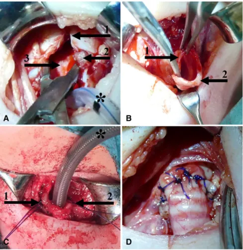
Follow-up period included regular dynamic and rigid endoscopic examinations under general anesthesia. The parents systematically registered body weight gain, length growth, swallowing or breath- ing difficulty. The voice samples were recorded with a high Fig. 3. Slide laryngotracheoplasty (intraoperative photos, patient 3)
(A) Partial anterior laryngofissure. Arrow 1: incised thyroid cartilage; arrow 2: cricoid cartilage, arrow 3: posterior wall of the laryngotracheal complex;*endotracheal tube
(B) Mobilized trachea Arrow 1: resection of the membranous part; arrow 2: anterior arch of the tracheal ring (C) The posterior wall is reconstructed Arrows 1 and 2: shows the clockwise ant counter-clockwise running threads
(D) The reconstructed anterior wall. [Colorfigure can be viewed in the online issue, which is available at www.laryngoscope.com.]
Fig. 4. Schematic picture of the anastomosis. Two double armed continuous locked sutures are running clockwise and counter- clockwise. [Colorfigure can be viewed in the online issue, which is available at www.laryngoscope.com.]
sensitivity (40–6 kHz) condenser head microphone (ATM75; Audio Technica, Machida, Tokyo, Japan) at sampling frequency of 96 kHz (24 bit US analyzed by Praat 5.3.2.9., Amsterdam, Netherlands).
The following acoustic parameters were recorded in this study:
mean pitch (F0), jitter, shimmer, and harmonics-to-noise ratio (HNR). Follow-up intervals were 80, 73, 43, 24, 21, 4, and 5 months for the patients, respectively.
RESULTS
There were no major peri- and postoperative complica- tions. The postoperative timeline of events is presented in Table II. Extubation was considered safe on the 7th, 5th,
10th, 4th, 3rd, 7th, and 5th postoperative days, for each of the seven patients, respectively. In the case of patients 3 and 6, postoperative reintubation was necessary because of their RSV infection. All patients were able to tolerate pro- gressive oral diet within 2 to 3 days post-extubation except one child, who was fed through nasogastric tube from the fifth week after birth (patient 7). The weight-for-age and length-for-age percentiles are depicted in Figure 5. The acoustic parameters, QOL scores are shown in Table III.
Compared to the preoperative values, the QOL scores improved significantly. Based on the parents’observations, the breathing was normal in all cases. Gurgling and cooing were similar to their siblings according to the parents.
In cases of two infants (patients 1 and 3) minor grade reste- nosis was detected. No patient required revision surgery.
In case of patient 4, weak voice was detected because of a 2-mm blunting at the anterior commissure.
DISCUSSION
Congenital SGS is typically cartilaginous and often associated with abnormal forms of the cricoid (small hypo- plastic cartilage) or may be associated with a dense laryn- geal web. Minimally invasive endoscopic surgery has high potential for restenosis and hence open airway reclaiming operations are preferred in such pathologies.16,17The choice between LTR and PCTR predominantly depends on the severity and length of the subglottic stenosis. LTR with an anterior graft alone is used as a single-stage operation for TABLE II.
Events of the Postoperative Period.
Name/sex
Extubation (days)
Complications and further treatment
Follow-up period (mo)
1/m 7 mild restenosis—laser 80
2/m 5 – 73
3/f 10 Pneumonia that required
reintubation (4 days), mild restenosis—laser
43
4/f 4 – 24
5/f 3 – 21
6/m 7 Pneumonia, reintubation
(2 days)
4
7/f 5 – 5
Fig. 5. Weight-for-age and length-for-age percentile. [Colorfigure can be viewed in the online issue, which is available at www.laryngoscope.com.]
the management of grade II stenosis. Grade III stenosis is likely to require both anterior and posterior grafts and can be performed in one or two stages. However, over the last decade, PCTR has emerged as a superior alternative to LTR for the treatment of grade III and IV SGSs.7,18–20The tech- nique of PCTR involves resection of the anterior cricoid arch, thinning andflattening of the posterior cricoid plate and per- forming an anastomosis between the thyroid cartilage and the trachea. The extended variant of CTR is advocated for severe SGS with additional posterior or trans-glottic steno- ses, and additionally requires a full laryngofissure, posterior cricoid split and expansion using a rib cartilage graft and a temporary laryngeal stent.12
Due to potential comorbidities, associated anatomical malformations, and complex requirements of the treatment, tracheostomy is still needed in cases of a severe grade congeni- tal SGS.1,2It severely affects the life quality of the patient and the parents as well. The complexity of the neonatal anatomy and anesthesia (small lung reserve capacity, high oxygen requirements, risk of hypo- and hyperthermia, associated comorbidities) make the surgical options even more limited in this age group. The optimal surgical intervention should be both quick, safe, and provide an immediate and adequate air- way, acceptable voice quality and good swallowing function.
Because of the above-mentioned difficulties, a definitive surgical solution is usually delayed for years, although an early tracheostomy is often unavoidable.21–24The most com- mon surgical solutions (CTR/ECTR, PCTR, LTR) are rec- ommended in general from the age of 1 year.4,11,14,15,21–27
The youngest PCTR patient described was a 1-month-old infant without any comorbidity.22However, even though the overall decannulation rate after CTR is over 80% - the rate of reoperation is between 4-41%, depending on the grade of the stenosis and the comorbidities. The rate of reoperation is between 22-45% after LTR.4,11,13–15,18,19,22,25,28
All of the presented cases included high grade, cartilagi- nous, concentric stenoses. Accordingly, no endoscopic inter- ventions, but external approach was preferred. In case of slide laryngotracheoplasty the subglottic airway is aug- mented with a good vascularized normal-size local trachea flap. This way, no donor site reaction or graft absorption occurs, and the technique provides a wide airway at the level of the subglottis and the proximal trachea, because of the par- tial resection of the posterior tracheal wall and theflexibility
of the intercartilaginous ligaments of the trachea. Using the slide tracheoplasty principle, we were able to make the tra- chea slide on a narrow cricoid and thus create a wider airway.
The cartilages in these young children areflexible and are well stabilized with a double-armed continuous knotted suture. In all our patients, the reconstructed airway has remained age-appropriate, static, and undistorted.
During the surgery, the anterior commissure remains untouched, which has an obvious positive impact on the post- operative voice quality. In the case of a severe grade subglottic stenosis, if the manipulation of the anterior commissure is unavoidable, a second stage laryngeal framework surgery (anterior commissure retrodisplacement) might be necessary to ensure a socially acceptable voice quality. However, decent voice analysis is obviously cumbersome for infants, the objec- tive voice parameters supported the patients’satisfied opinion on voice quality.
The good functional results are facilitated by the preser- vation of the entire cricoid cartilage, the recurrent nerves and the cricothyroid muscles. Since the integrity and the innervation of the posterior commissure (respiratory glottis) remains intact, the possibility of aspiration is significantly lower. Congenital SGS is not caused by scar tissue, but rather by an incomplete recanalization—hence the respira- tory mucosa is intact. Needless to say that in airway surgery if the intact respiratory mucosa is preserved, the chances of the restenosis are lower.3,5,21None of our patients had any postoperative feeding difficulties. Asymptomatic, grade I restenosis was observed in two cases, which could be effec- tively treated by single laser resection. During the further follow-up no significant restenosis was found, and the growth of the larynx seemed to be unimpaired. The surgical method presented here has the advantage of reduced risk of anasto- motic dehiscence and recurrent nerve damage that could be encountered in the conventional CTR technique.
CONCLUSION
According to our preliminary results, the single- stage slide laryngotracheoplasty might be a favorable solution for subglottic stenosis, even in early childhood.
In one step, the airway can be maintained without stenting and tracheostomy. Voice production and optimal swallowing function are well preserved. Follow-up long- TABLE III.
Functional Outcomes of the Surgery.
Patient/sex
Jitter Shimmer HNR Mean Pitch
QoL
(%) (%) (dB) (Hz)
1/m 0.8 9.9 16.2 278 7
2/m 3.3 15.9 8.6 161 7
3/f 1.2 16.1 14.3 151 6
4/f No data obtained 11
5/f 0.4 4.8 19.1 280 7
6/m 1.1 8.7 19.4 434 10
7/f 0.4 2.9 20.3 471 10
Physiological values <1.04 <3.81 >20 – Min = 6; max = 25
HNR = harmonic-to-noise ratio; QOL = quality of life score.
term outcomes and additional patients need to be studied to further validate this procedure.
BIBLIOGRAPHY
1. Holinger PH, Johnson KC, Schhiller F. Congenital anomalies of larynx.Ann Otol Rhinol Laryngol1954;63:581–606.
2. Tucker GF, Ossoff RH, Newman AN, Holinger LD. Histopathology of con- genital subglottic stenosis.Laryngoscope1979;89:866–877.
3. Hanlon K, Boesch RP, Jacobs I. Subglottic stenosis.Current problems in pediatric and adolescent health care.2018;48:129–135.
4. George M, Ikonomidis C, Jaquet Y, Monnier P. Partial cricotracheal resection for congenital subglottic stenosis in children: the effect of con- comitant anomalies.Int J Pediatr Otorhinolaryngol2009;73:981–985.
5. Landsman JS, Werkhaven EA, Motoyama K.Smith’s Anesthesia for Infants and Children. St. Louis: Mosby; 2011:786–820.
6. Niall JD, Aliza P, Cohen MA, Rutter MJ. Subglottic stenosis.Semin Pediatr Surg2016;25:138–143.
7. Monnier P.Pediatric Airway Surgery Management of Laryngotracheal Ste- nosis in Infants and Children. New York: Springer; 2011.
8. Pandian V, Garg V, Antar R, Best S. Discharge education and caregiver cop- ing of pediatric patients with a tracheostomy: systematic review.ORL Head Neck Nurs2016;34:17–18, 20–27.
9. Nakarada-Kordic I, Patterson N, Wrapson J, Rea SD. A systematic review of patient and caregiver experiences with a tracheostomy.Patient2018;
11:175–191.
10. Flynn AP, Carter B, Bray L, Donne AJ. Parents’experiences and views of caring for a child with a tracheostomy: a literature review.Int J Pediatr Otorhinolaryngol2013;77:1630–1634.
11. Yamamoto K, Monnier P, Holtz F, Jaquet Y. Laryngotracheal reconstruc- tion for pediatric glotto-subglottic stenosis.Int J Pediatr Otorhinolaryngol 2014;78:1476–1479.
12. Sandu K, Monnier P. Partial cricotracheal resection with tracheal intussus- ception and cricoarytenoid joint mobilization: early experience in a new technical variant.Laryngoscope2011;121:2150–2154.
13. Hartley EJ, Rutter MJ, Cotton RT. Cricotracheal resection as a primary pro- cedure for laryngotracheal stenosis in children.Int J Pediatr Otorhinolaryngol 2000;54:133–136.
14. Smith DF, de Alarcon A, Jefferson ND, et al. Short- versus long-term ste- nting in children with subglottic stenosis undergoing laryngotracheal reconstruction.Otolaryngol Head Neck Surg2018;158:375–380.
15. Jaquet Y, Lang F, Pilloud R, Savary M, Monnier P. Partial cricotrachealre- section for pediatric subglottic stenosis: long-term outcome in 57 patients.
J Thorac Cardiovasc Surg2005;130:726–732.
16. Quesnel MA, Gi Soo Lee, Nuss RC, Volk MS, Jones DT, Rahbar R. Mini- mally invasive endoscopic management of subglottic stenosis in chil- dren: success and failure. Int J Pediatr Otorhinolaryngol 2011;75:
652–656.
17. Maresh A, Preciado DA, O’Connell AP, Zalzal GH. a comparative analysis of open surgery vs endoscopic balloon dilation for pediatric subglottic steno- sis.JAMA Otolaryngol Head Neck Surg2014;140:901–905.
18. White DR, Cotton RT, Bean JA, et al. Pediatric cricotracheal resection: sur- gical outcomes and risk factor analysis.Arch Otolaryngol Head Neck Surg 2005;131:896–899.
19. McQueen CT, Shapiro NL, Leighton S, et al. Singlestage laryngotracheal reconstruction: the Great Ormond Street experience and guidelines for patient selection.Arch Otolaryngol Head Neck Surg1999;125:320–322.
20. Ochi JW, Evans JN, Bailey CM, Ann I. Laryngotracheoplasty and laryngotracheal reconstruction. Pediatric airway reconstruction at Great Ormond Street: a ten-year review.Ann Otol Rhinol Laryngol1992;101:
465–468.
21. Sittel C. Laryngotracheale Stenosen im kindesalter. Laryngo-Rhino-Otol 2012;91:478–485.
22. George M, Ikonomidis C, Jaquet Y, Monnier P. Partial cricotracheal resection in children: potential pitfalls and avoidance of complications.
Otolaryngol Head Neck Surg2009;141:225–231.
23. Schmidt RJ, Shah G, Sobin L, Reilly JS. Laryngotracheal reconstruction in infants and children: are single-stage anterior and posterior grafts a reli- able intervention at all pediatric hospitals?Int J Pediatr Otorhinolaryngol 2011;75:1585–1588.
24. Penchyna JG, Ortíz HE, Teyssier MG, Rivas RI, Preciado D, Alvarez- Neri H. Extended cricotracheal resection with posterior costochondral grafting for complex pediatric subglottic stenosis.Int J Pediatr Otorhinolaryngol2016;
88:213–216.
25. Yamamoto K, Jaquet Y, Ikonomidis C, Monnier P. Partial cricotracheal re- section for paediatric subglottic stenosis: update of the Lausanne experi- ence with 129 cases.Eur J Cardiothorac Surg2015;47:876–882.
26. Hartnick CJ, Hartley BEJ, Willging P, et al. Surgery for pediatric subglottic stenosis: disease-specific outcomes.Ann Otol Rhinol Laryngol2001;110:
1109–1113.
27. Sittel C. Pathologies of the larynx and trachea in childhood.GMS Curr Top Otorhinolaryngol Head Neck Surg2014;13:1011–1865.
28. Sandu K, Monnier P. Cricotracheal resection.Otolaryngol Clin North Am 2008;41:981–998.

![Fig. 5. Weight-for-age and length-for-age percentile. [Color fi gure can be viewed in the online issue, which is available at www.laryngoscope.com.]](https://thumb-eu.123doks.com/thumbv2/9dokorg/971169.58022/5.875.59.422.117.331/weight-length-percentile-color-viewed-online-available-laryngoscope.webp)
