0
THE ROLE OF ELECTROCARDIOGRAPHY AND EXERCISE TESTING
IN THE ASSESSMENT OF CARDIOVASCULAR RISK IN ATHLETES AND NON-ATHLETES
Doctoral Dissertation
Nóra Sydó, MD
Doctoral School of Basic and Translational Medicine Semmelweis University
Supervisors: Béla Merkely MD, DSc; Dávid Becker MD, PhD Consultant: Thomas G. Allison PhD, MPH
Official reviewers: Éva Martos MD, PhD, med. habil Miklós Zsuzsanna MD, PhD
Head of the Final Examination Committee:
Zoltán Benyó MD, DSc
Members of the Final Examination Committee:
József Pucsok, MD, DSc, András Zsáry MD, PhD Budapest
2018
1
Table of contents
Table of contents 1
Abbreviations 3
1. Introduction 5
1.1. Cardiovascular Adaptation to Exercise 7
1.1.1. Classificaton of Sports 8
1.1.2. Acute Adaptation to Exercise 9
1.1.3. Chronic Adaptation to Exercise 11 1.1.4. Autonomic Adaptation to Exercise 14
1.2. Sudden Cardiac Death in Athletes 15
1.3. ECG Screening in Athletes 19
1.3.1 Resting 12-lead ECG 21
1.3.2. Holter ECG 25
1.3.3. Exercise testing 30
1.4. Semmelweis Bridge Project 32
2. Objectives of our Studies 33
3. Methods 34
3.1. Participants 34
3.1.1. Study Population of Athletic ECG Projects 34 3.1.2. Study Population of Exercise ECG Projects 34
3.2. Measurements 36
3.2.1. Athletes 36
3.2.2. Non-Athletes 37
3.3. Stastistical Analyses 38
4. Results 40
4.1. Electrocardiographic Findings 40
4.1.1. Study Population 40
4.1.2. Training-related Physiological ECG Findings 40 4.1.3. Training-unrelated Pathological ECG Findings 42
4.2. Holter ECG results 45
4.2.1 Analysis by Study Group 45
4.2.2. Analysis by Sport 47
2
4.2.3. Sample individual HRV analyses 49
4.2.4. Comparison of Lower Cut-off Values 50
4.3. Exercise Test Results 51
4.3.1. Study population 51
4.3.2. General exercise test results 54
4.3.3. Peak Heart Rate by Age in Men versus Women in the Preliminary Clinical Cohort 56 4.3.4. Analysis of Factors Affecting Peak Heart Rate 56 4.3.5. Exercise Test Parameters by Age in Men versus
Women in the Pure Clinical Cohort 58 4.3.6. Exercise Testing and Mortality Risk 59
5. Discussion 63
5.1. The Role of ECG in Athletic Screening 64
5.1.1. Physiological ECG Changes 63
5.1.2. Pathological ECG Abnormalities 64
5.2. Autonomic Adaptation in Athletes 68
5.2.1. Autonomic Adaptation by Sport Intensity 69 5.2.2. Autonomic Adaptation by Sport Modality 69
5.2.3. Clinical Perspectives 70
5.2.4. Limitations 70
5.3. Exercise Heart Rate Responses in Non-athletes 71
5.3.1. Peak Heart Rate Prediction 71
5.3.2. Clinical Importance 72
5.3.3. Limitations 73
5.4. Risk prediction on Exercise Test 75
5.4.1. Effect of Cardiorespiratory Fitness on Mortality 75
5.4.2. Clinical Importance 76
5.4.3. Limitations 76
6. Conclusions 78
Summary 80
Összefoglaló – Summary in Hungarian 81
References 82
Publications related to dissertation 100 Publications not related to dissertation 101
Acknowledgement 108
3
Abbreviations
ACC American College of Cardiology AHA American Heart Association
ARVC arrhythmogenic right ventricular cardiomyopathy AV atrioventricularis
BMI body mass index
BP blood pressure
BPM beats per minute CI confidence interval
CRBBB complete right bundle branch block CRF cardiorespiratory fitness
CV cardiovascular
CVD cardiovascular disease DCM dilated cardiomyopathy
ECG electrocardiogram (also electrocardiography, electrocardiographic) ESC European Society of Cardiology
FAC functional aerobic capacity HCM hypertrophic cardiomyopathy
HF high frequency
HR heart rate
HRV heart rate variability
IRBBB incomplete right bundle branch block LBBB left bundle branch block
LF low frequency
LV left ventricle
LVH left ventricular hypertrophy
4
LVEDD left ventricular end-diastolic diameter LVEDV left ventricular end-diastolic volume MCV maximal voluntary contraction
nsVT non-sustained ventricular tachycardia
pNN50 the percentage of differences between successive RR intervals over 24 hours that are greater than 50 milliseconds
QTc corrected QT interval
RMSSD the root mean square successive difference RPE rating of perceived exertion
RV right ventricle
RVH right ventricular hypertrophy SD standard deviation
SDNN the standard deviation of normal to normal ratios
SDNN Index the standard deviation of all the 5-minute NN interval means VES ventricular extrasystole
VLF very low frequency
WPW Wolf- Parkinson White syndrome
5
1. Introduction
Electrocardiography (ECG) is an essential tool for diagnosis and management of patients with various cardiovascular (CV) diseases. With the development and increased availability of new imaging techniques, however, ECG has lost some of its diagnostic importance. (1) In athletic screening, the usefulness of ECG is also controversial. In athletes, ECG changes are common, and in most cases these reflect benign structural and electrical remodeling of the heart due to physiological adaptation to exercise, a condition which is often referred to as “athlete’s heart”. (2-4) However, the differential diagnosis between normal physiological changes and pathological abnormalities can be challenging. (5) For regular athletic screening and follow-up, widely available and inexpensive diagnostic methods are needed. In Hungary, ECG is still a cornerstone of routine athletic screening. (6,7) Its significance is based on simplicity, easy availability, short time to perform and interpret, and high informative value in special cases, as well as on the fact that with appropriate evaluation, appearance of pathological ECG signs can forecast the emergence of structural heart diseases years in advance. (8) However, the cornerstone is just part of any foundation, so additional test modalities are often necessary.
With long-term, 24-hour ECG recordings we may not only catch transient arrhythmias and ischemia, but we can also get useful information about autonomic function and predict CV risk. In case of intact autonomic regulation, simultaneous and opposite changes of sympathetic and parasympathetic activity manifest in beat-to-beat fluctuations of heart rate (HR). The extent of these fluctuations shows short-term and long-term changes due to several individual and environmental effects such as physical activity or mental stress and are reduced permanently when autonomic control mechanisms are impaired. Modern Holter monitoring systems are suitable to identify beat-to-beat perturbations of the normal RR intervals, called heart rate variability (HRV) analysis. (9) Whereas in CV screening of young athletes we are interested primarily in genetic/congenital abnormalities that manifest themselves in the ECG, in adult patients the evaluation is generally performed to discover acquired conditions that have developed over time as a result of poorly managed risk factors and unhealthy lifestyles. While the
6
ranges of symptoms in young athletes versus adult patients with CV risk factors overlap somewhat, syncope and palpitations are of greater relevance in young athletes, whereas chest pain and dyspnea on exertion are more common and of relatively greater concern in the adult population.
On an exercise test in an adult patient, interpretation of the ST segments provides useful for its diagnostic information, but more important prognostic value can be gained from variables such as functional aerobic capacity (FAC), peak exercise HR and HR recovery.
The prognostic predictive value of these parameters has been studied in a wide range of CV populations and has proved to be an independent powerful predictor of mortality. (10- 12) Both FAC and HR responses are affected by autonomic function with FAC and peak exercise HR driven by sympathetic tone, while HR recovery is principally related to parasympathetic tone.
Cigarette smoking has profound effects on health. Besides CVD, lung cancer and chronic obstructive pulmonary disease are disabling and life-shortening consequences of cigarette smoking. (13,14) Cigarette smoking reduces FAC and impairs the HR response to exercise. These are important early markers of CV risk. (15,16) Smoking cessation, on the other hand, frequently leads to weight gain and weight-associated comorbidites including hypertension and diabetes. (17) The interplay between smoking status and the predictive exercise factors including FAC and HR recovery are of great potential importance in assessing risk and establishing the benefits of smoking cessation.
The purpose of our studies was to emphasize the importance of ECG in athletic screening, to show the additional diagnostic potential of long-term ECG recording in athletes, to conversely present the predictive value of non-ECG factors – FAC and HR responses – on the exercise test in an adult referral population and to define the role of FAC and HR responses in predicting risk according to smoking status.
7
1.1. Cardiovascular Adaptation to Exercise
Physical activity is movement by contraction of specific skeletal muscles of the body, which requires more energy expenditure than resting status. (18,19) This definition includes daily sport activity, physical work, regular training, competitive sports and as well as rehabilitation movement program. All forms of exercise have an effect on the body, specific structural and functional adaptation can be observed according to the type and intensity of sport activity. (2,18) As a result of training, depending on its amount and frequency, acute and chronic sport adaptation can be observed. (2,20) Exercise affects most body systems, including profound effects on the CV system.
As differentiating physiological and pathological sport adaptation is still challenging, we need athletic normal values from studies on large sample size. Because we do not really have such normal values, one possible differential diagnostic method is deconditioning.
Cardiac adaptation to exercise may appear after six weeks of intensive training, but as a result of deconditioning these changes disappear. Athletic adaptation is reversible while signs of stuctural heart diseases are irreversible, therefore it may help in the differential diagnosis of borderline cases. (21,22)
8
1.1.1. Classification of Sports
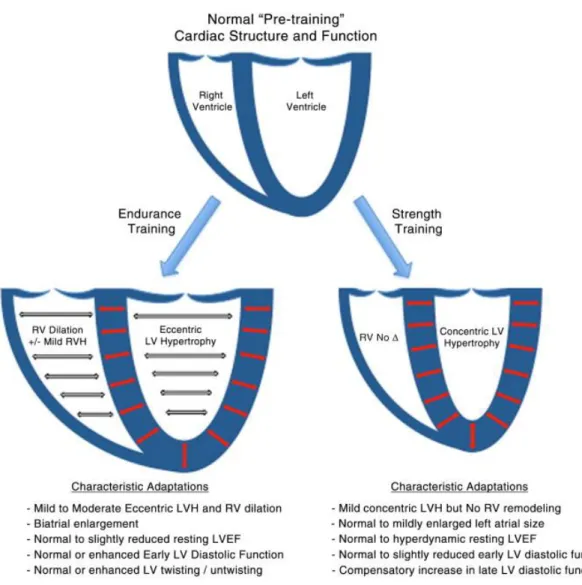
Physical activity can be divided into dynamic and static categories according to metabolic characteristics. The dynamic component can be described by predicting maximal oxygen uptake, while the static by maximal contraction volume. According to the ”Morganroth hypothesis”, endurance (dynamic, isotonic, aerobic) sports are for example swimming and long-distance running, while weight-lifting ranks among strength sports (static, izometric, anaerobic). (6,23,24) Dynamic sports cause volume overload, while static ones produce pressure overload on the CV system. The specific cardiac chamber morphology changes are shown in Figure 1.
Figure 1. Summary of sport type specific athletic ventricular remodeling. (25)
LV = left ventricular, LVH = left ventricular hypertrophy, RV = right ventricular, RVH
= right ventricular hypertrophy, LVEF = left ventricular ejection fraction.
9
In compliance with the 36. Bethesda Conference recommendation, we can classify sports by the proportion of dynamic and static components Figure 2. (2,18,26-28)
Figure 2. Classification of sports based on peak static and dynamic components achieved during competition. The increasing dynamic component is defined in terms of the estimated percentage of maximal oxygen uptake (VO2max) achieved and results in an increasing cardiac output. The increasing static component is related to the estimated percentage of maximal voluntary contraction reached and results in an increasing blood pressure load. The lowest total cardiovascular demands (cardiac output and blood pressure) are shown in the palest color, with increasing dynamic load depicted by increasing blue intensity and increasing static load by increasing red intensity. Note the graded transition between categories, which should be individualized on the basis of player position and style of play. *Danger of bodily collision. †Increased risk if syncope occurs. (27,28)
Cycling or rowing are good examples of combining static and dynamic activity, but in most sports endurance and isometric elements are both represented in the training program. (18) Moreover, different position players may have quite different CV loads.
Nevertheless, there are some weaknesses of this classification, namely that there is no guideline about psychic stress during competition and about different environmental circumstances. (28)
10
1.1.2. Acute Adaptation to Exercise
To provide enhanced energy of muscle contraction during physical activity, breathing intensity increases with more oxygen uptake in the lungs. As pulmonary blood flow increases with enhanced breathing, other parts of the vascular system show adaptation to exercise. (20,29,30) Acute CV adaptation includes chronotrop mechanisms – increased HR – and inotrop mechanisms – enhanced contractility power. (29,31) HR increase during exercise is regulated by the autonomic nervous system characterized by rapid and sustained parasympathetic withdrawal coupled with sympathetic activation. Peak HR is affected by several factors like age and sex and may actually slightly decrease with aerobic training. Stroke volume increases significantly both at rest and during exercise with prolonged aerobic training. Cardiac output – the product of stroke volume and HR – may increase 5- to 6-fold during a maximal exercise effort. Increased cardiac output and redistribution of circulating blood provides increased perfusion of muscles and heart with stable cerebral blood flow (Figure 3). (29,31) Blood flow in the gut and skin decrease during heavy exercise, though skin blood flow will increase during prolonged exercise in hot environments, while blood flow in other organ systems remains relatively constant.
Figure 3. Distribution of cardiac output at rest and during heavy exercise. (32)
11
1.1.3. Chronic Adaptation to Exercise
As a result of long, repeated bouts of physical exercise, structural and functional adaptation can be observed in the CV system. This includes prominent cardiac remodeling called athlete’s heart (Figure 4). (2,30,33) The type and magnitude of changes depends on the type and amount of exercise and also on sex and genetic factors.
Figure 4. Morphology of left ventricular hypertrophy in athletes. (33)
Cardiac hypertrophy is the most pronounced feature of athlete’s heart, which affects the entire heart but mostly left ventricular (LV) wall thickening can be observed. Besides muscle mass increase, capillary density increases too. (2,29,33,34) While wall thickening as a consequence of static exercise is due to pressure overload, dynamic exercise results in volume overload, which manifests itself in increased diameters. Henschen’s definition of athlete’s heart: “an enlargement of the heart which can perform more work than a normal heart.” (2,35) It is very important to distinguish the physiological left ventricular hypertrophy (LVH) of athlete’s heart from the pathological LVH of hypertrophic cardiomyopathy (HCM); this can be accomplished with appropriate diagnostic methods.
(34,36) The difference between physiological athlete’s heart and pathological LVH is the structure and amount of pathological cells and tissue, whose consequences worsen first the diastolic and then the systolic function in pathological LVH. (30)
In recent years, cardiac remodeling has been studied with several imaging techniques.
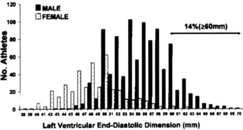
Echocardography studies showed that ventricular diameters are related to sports type and intensity. (4,37) The most pronounced increases can be observed in rowing, cross country
12
skiing, cycling and swimming. However, adaptative changes may be different, as almost all athletes have some evidence of athlete’s heart including LV dilatation and increased wall thickness. (21) Marked (> 60 mm) LV end-diastolic diameter dilatation may be present in only 15% of athletes, while > 13 mm wall thickness is only present in 2% of the athletes (Figure 5/A and 5/B). These two parameters are generally lower in women compared to men. (2,34)
Figure 5/A. Left ventricular end-diastolic dimensions in male and female athletes by echocardiography. (2)
Figure 5/B. Left ventricular wall thickness in male and female athletes by echocardiography. (2)
Increased LV diameter can be observed after one week of endurance training, while the increase of myocardial mass is slower, correlating with training years and maximal oxygen uptake. Enhanced blood volume increases volume overload of the heart and
13
contributes to the rapid increase of heart dimensions. Athletes have higher LV end- diastolic volume with increased cardiac stroke volume, so they can provide the resting cardiac output with lower HRs. (2,3,30)
It is important to mention here that these structural adaptations to chronic exercise also affect the resting ECG, making it potentially more challenging to distinguish healthy athletes from those with cardiac diseases.
14
1.1.4. Autonomic Adaptation to Exercise
Cardiovascular autonomic function also shows acute and chronic adaptation to physical exercise through the regulation of HR, blood pressure (BP) and circulatory redistribution mediated by sympathetic and parasympathetic nervous systems.
At the beginning exercise, HR increases rapidly, mainly due to the withdrawal of vagal activity. As the workload increases, HR increases due to further vagal withdrawal and concomitant sympathetic activation. (38-41)
The complexity of exercise induced long-term CV adaptation is well reflected by lower HR at rest and during submaximal exercise with unchanged or even slightly lower peak HR remains. (2,3,18) As a consequence of structural adaptation, in athletes a given increase in cardiac output requires less increase in HR due to the maintenance of a larger stroke volume. On the other hand, HR may be reduced during submaximal exercise due to a lower intrinsic HR, as a result of sympathetic activity decrease with less circulating catecholamines and a greater parasympathetic influence. (42) At the same time, regular physical activity also reduces the sympathetic effect on the sinoatrial node. (39,40) Besides lower HRs, chronic autonomic adaptation may also manifest in conduction abnormalities such as transient second degree AV block and increased ventricular ectopy that might mimic underlying cardiac disease.
Underdeveloped cardiac autonomic adaptation may be a warning sign of underlying cardiac abnormality as sympathovagal imbalance and impaired baroreceptor function may preceed ventricular arrhythmias and myocardial ischemia and have been associated with an increased risk of mortality. (43-45)
15
1.2. Sudden Cardiac Death in Athletes
In recent years, sudden cardiac death (SCD) has become a center of focus in the media, as several prominent US collegiate and professional athletes literally “died on the screen”.
Although, the number of athletic SCDs is relatively small, these deaths represent an important and emotionally charged public health issue.
The incidence of SCD is greater in athletes compared to non-athletes who perform recreational sports. (46-50) Competitive sports activity enhances the risk of SCD by 2.5- fold in adolescents and young adults. (47,51-53) The difference in incidence may be explained by the fact that the intensity and duration of physical activity is likely much higher in competitive versus recreational sports, but use of performance-enhancing substances may also play a role in some of the deaths. We also cannot rule out an ascertainment bias in that competitive sports are more closely scrutinized and deaths witnessed and well-documented, whereas deaths in recreational activity may more likely be undocumented and unreported. According to a prospective population-based study from Italy, the incidence of sudden death is 2.3 (2.62 in males and 1.07 in females) per 100 000 athletes per year from all causes, and 2.1 per 100,000 athletes per year from CVDs. (51) In the United States the prevalence of SCD is in the range of 1:100,000 to 1:300,000 high school–age athletes, while among older athletes the frequency of SCD due principally to coronary artery disease (CAD) may exceed that of younger athletes (1:15,000 joggers and 1:50,000 marathon runners). (52,54-56) The difference in incidences between Italy and the United States includes that studies from the United States probably underestimated the prevalence of sports-related SCD because they relied on reports from individual schools and institutions, or on media accounts. Another reason could be the different underlying pathological substrates which might reflect differences also in ethnic and genetic factors (specifically arrhythmogenic right ventricular cardiomyopathy - ARVC) along with higher mean age and higher level of participation of Italian competitive athletes compared with U.S. high school and college participants.
(52,57,58)
SCD in athletes shows a clear gender difference with male predominance up to 10:1.
(50,52,59,60) This may be explained by the higher participation rate of male compared to female athletes in competitive sports, by the higher intensity with more intense pressure
16
of training load and level of athletic achievement of males, longer competitive seasons, as well as the lower prevalence of cardiac abnormalities capable of causing SCD in females. (61) Greater use of performance-enhancing drugs and supplements among male athletes may also play a role. More recently, male gender was reported to be, in itself, a risk factor for sports-related SCD. (48,49,52,57,58)
In Hungary, there are no published statistical data about the incidence of SCD during sports, but the estimated frequency of sports-related death is not likely higher than the Italian experience. (18,62)
SCD occurs more frequently in certain sports. In the United States, basketball – primarily men’s basketball (63) – has the greatest incidence, whereas in Europe soccer predominates. (52)
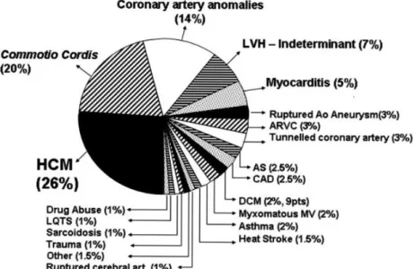
SCD is usually the result of an interaction between an acute trigger and underlying heart disease (substrate). (64,65) Emotional stress, environmental factors, myocardial ischemia, sympathetic-vagal imbalance, and hemodynamic changes may be acute triggers for life-threatening ventricular tachyarrhythmias and SCD during sports. (66) In young athletes (<35 years of age), the most common cause of SCD is underlying congenital cardiac disease, such as cardiomyopathies and congenital coronary anomalies (Figure 6).
(67)
Figure 6. Causes of sudden cardiac death in young athletes. (2,67)
HCM = hypertrophic cardiomyopathy, LVH = left ventricular hypertrophy, ARVC = arrhythmogenic right ventricular cardiomyopathy, AS = aortic stenosis, CAD = coronary artery disease, DCM = dilated cardiomyopathy, LQTS = long QT syndrome.
17
In the presence of heart disease, intensive and systematic athletic training itself may promote progression or worsening of the arrhythmogenic substrate (either structurally or electrically) therefore increasing the risk of SCD. (66) For example, in patients with HCM, recurrent episodes of exercise-induced myocardial ischemia during intensive training may result cell death and myocardial replacement fibrosis, which in turn enhances ventricular electrical instability. (68) In patients with ARVC, regular and intense physical activity may provoke RV volume overload and cavity enlargement, which in turn may accelerate fibrofatty atrophy. (69)
Geographical differences have been reported in the causes of SCD between Europe and the US in young athletes. In the US the most common cause is HCM, while in Europe ARVC. These differences are more likely to be related to the differences in pre- participation screening methods in the identification of athletes at a higher risk, rather than reflecting a truly different etiology. In the US, African-American males have higher rates of SCD during sports than white males.
Other common causes are genetic diseases as ion channelopathies (long QT syndrome, Brugada syndrome, short QT syndrome and cathecholaminerg polymorphic ventricular tachycardia). These deaths occur most commonly in team sports such as basketball and football, which have the highest levels of participation. (52) Blunt chest trauma also may cause ventricular fibrillation in a structurally normal heart; this is known as commotio cordis. (70,71)
In older athletes (35 years and older), atherosclerotic CAD is the most common cause (85%) of SCD.(56) They are individual athletes – such as long distance runners – rather than members of organized team sports. (52) Besides known risk factors of ischemic heart disease, in athletes myocardial ischemia may be induced by increased wall stress from increased HR and BP, exercise induced coronary artery spasm, decreased coronary perfusion as a result of shortened diastolic filling time and increased myocardial oxygen demand. These mechanisms may induce acute coronary artery plaque disruption, including plaque rupture or erosion, with acute thrombotic occlusion. (72) An increase in thrombogenicity also could contribute to coronary thrombosis after plaque rupture or erosion. (73) Myocardial ischemia causes electrically inhomogeneous areas, scars which can be disposed to develop malignant ventricular arrhythmias and fibrillation. All of the
18
above-mentioned abnormalities may induce malignant ventricular arrhythmias, mainly (65-80%) ventricular fibrillation, which is the definite cause of SCD.
Diagnosing underlying cardiac diseases could potentially prevent some cases of SCD during sports. Athletes could be treated for example with surgery for anomalous coronary arteries in younger athletes or percutaneous coronary intervention or bypass surgery in older athletes. Other athletes with less treatable conditions like HCM could be disqualified or rechanneled into less strenuous sports. Recently published guidelines from ACC/AHA also allow continued participation for some athletes with arrhythmogenic substrate after the institution of appropriate therapies including implantable cardiac defibrillators.(74,75) Therefore, all athletes should undergo some sort of pre-participation CV screening. However, the elements of the screening have not been standardized across countries. Moreover, evaluation of athletes raises diagnostic difficulties, particularly differentiating between physiological adaptation to exercise and pathological conditions, so some normal athletes may inadvertently be disqualified while some abnormalities might be classified as “normal variant”. Economics of screening is a major issue, considering the high cost of some cardiac tests versus the very low rates of SCD (1-2 per 100,000 athletes per year) during sport. In US, screening is conducted according to an algorithm whereby history and physical exam determine who gets an ECG or further cardiac tests, whereas general screening with ECG ± echocardiography may be employed in Europe or in some American professional sports (Women’s NBA, for example).(76) Other questions to be addressed include frequency of screening and at what age screening begins. Is one evaluation to look for cardiac abnormalities when the athlete first begins high level training and competition sufficient, or does evaluation need to be repeated annually? There may also be medical-legal issues in the case of athletes who experience SCD after being screened or with athletes who are disqualified from sports but independently produce contrary evidence that they do not have cardiac abnormalities.
19
1.3. ECG Screening in Athletes
The role of pre-participation screening is to identify or raise the suspicion of those underlying CVDs, abnormalities and CV risk factors which are responsible for athletic field deaths so as to disqualify – or in some cases provide ameliorative treatment to – athletes at risk. We expect that applying this strategy may prevent some cases of SCD.
(77-80) In Hungary, similarly to other developed European countries, pre-participation screening is mandatory once a year for athletes over 18 years of age and every six months under 18 years of age. (7,81) The basic examinations include family and personal history, physical examination with BP measurement, and resting 12-lead ECG. Personal history is performed on a detailed questionnaire which contains several questions about the health status of the athlete, such as symptoms during exercise, CV risk factors, and medications.
In addition to personal history, there is a separate section for family history designed to identify possible inherited CVDs and assess the presence of SCD in the family. (7,81) In Hungary, resting 12-lead ECG is an essential part of pre-participation screening.
Nevertheless, imaging diagnostic techniques such as echocardiography or cardiac MR, are also performed in case of symptoms or positive findings on basic exams as shown on Figure 7. Athletes diagnosed with clinically relevant CV abnormalities are managed according to available guidelines for assessing athletic risk.
20
Figure 7. Flow diagram illustrating pre-participation screening protocol for young competitive athletes. (80)
EMB = endomyocardial biopsy, EPS = electrophysiological study, MRI = magnetic resonance imaging.
21
1.3.1. Resting 12-lead ECG
ECG changes in athletes are common and in most cases reflect benign structural and electrical remodeling of the heart due to physiological adaptation to physical training, as a part of “athlete's heart”. (2,3,5,82) The majority of disorders associated with an increased risk of SCD can be suggested or identified by resting ECG abnormalities.
Moreover, ECG changes may preceed years in advance of the abnormalities caused by structural heart disease.(8) Resting ECG is an essential part of pre-participation screening in Hungary, but the US and Europe have treated the need for universal ECG differently.
In Europe, long-term Italian (specifically, Veneto region) experience has demonstrated that pre-participation screening with medical history and physical examination has limited sensitivity to detect athletes at high risk, but the implementation of ECG to the screening protocol increases its sensitivity and is an effective way of identifying athletes with potentially lethal CVDs and ultimately saves lives. (83-87) This screening algorithm, which has been used for pre-participation screening in Italy for over a 25-year period, has proved adequate sensitivity and specificity for detecting athletes affected by potentially dangerous cardiomyopathy or arrhythmia, and has led to a decreased mortality of young competitive athletes by ∼90%, mostly by preventing SCD from ARVC which is common in this region but has a low prevalence elsewhere. Figure 8. (80,88-90)
Figure 8. Annual incidence rates of sudden cardiac death in screened competitive athletes and unscreened nonathletes aged 12 to 35 years in the Veneto Region of Italy (1979- 2004). (89)
22
However, in the US concerns have been raised and the usefulness of ECG was questionable because of cost-effectiveness and low specificity with high level of false positive results. (78,91-93) Americans may be more sensitive to the issue of inappropriate disqualifications and generation of expense downstream evaluations and the anxiety they produce in otherwise healthy athletes with isolated ECG abnormalities. In recent years some American groups have accepted the use of universal ECG screening and have stated that ECG added to focused CV history and physical examination may be cost-effective and improve the overall sensitivity of pre-participation screening. (94,95)
Digital ECG recording has been spreaded recently, therefore serial comparison of previous ECG recordings may help to identify and highlight changes in order to make clinical decisions and it may decrease the high false positive results.
The original recommendations for ECG interpretation in athletes were published in 2010 by the European Society of Cardiology (ESC). (96) Athletic ECG changes are common and usually reflect structural and electrical remodeling of the heart. (97) However, ECG abnormalities may be an expression of an underlying heart disease. (98) It is important to correctly distinguish between physiological ECG changes and potentially pathological ECG abnormalities. Over the last decade, ECG interpretation standards have evolved quickly. (99) In 2013, an international group of sports cardiology experts updated contemporary standards for ECG interpretation in athletes, called ”Seattle Criteria”.
(100) The ”Seattle Criteria” significantly improved the specificity of ECG screening for detecting CVD associated with SCD, by accounting for specific benign repolarization anomalies associated with black ethnicity and designating less conservative limits for an abnormal QT interval. (101,102) In 2014, the ”Refined Criteria” have been associated with further reduction in false positives - isolated atrial enlargement, axis deviation and right ventricular hypertrophy (RVH) - without compromising sensitivity. (101) While comparing the three guidelines, the application of ”Refined Criteria” results in a significant increase in specificity when compared to the “ESC” and the “Seattle Criteria”, while retaining the same sensitivity. This reduces the amount of abnormal ECGs from 12.7% - 22.3% with the ESC criteria to 1.9% - 6.6% with the “Refined Criteria”.
Moreover, new ECG interpretation guidelines are associated with a cost reduction of up to 21% without compromising sensitivity to detect serious cardiac disease. (103)
23
In 2017, an international consensus statement was published about ECG interpretation in athletes, which provides expert opinion-based recommendations of specific ECG abnormalities and secondary evaluation for conditions associated with SCD. (104) Figure 9 lists the international standards for ECG interpretation. (104) The consensus document distinguishes normal, abnormal and borderline ECG findings. Athletes having two or more borderline ECG findings require further evaluation.
Figure 9. International consensus standards for electrocardiographic (ECG) interpretation in athletes. (104)
AV = atrioventricular, LBBB = left bundle branch block, LVH = left ventricular hypertrophy, RBBB = right bundle branch block, RVH = right ventricular hypertrophy;
PVC = premature ventricular contraction, SCD = sudden cardiac death.
24
On a characteristic athletic ECG (Figure 10) resting sinus bradycardia, first degree AV block and early repolarization are common ECG signs resulted by autonomic adaptation of the heart (increased vagal tone and decreased sympathetic activity). Besides these evidences of electrical sport adaptation, structural adaptation can also be observed as signs of LVH. These physiological changes have to be differentiated from pathological abnormalities such as pathological T-wave inversion, q waves, conduction abnormalities and arrhythmias, which may be a sign of structural heart disease. (105-108)
Figure 10. Resting ECG recording of a Hungarian Olympic champion. Sinus bradycardia, heart rate: 51/min, marked sinus arrhythmia, central axis, left ventricular hypertrophy (Cornell criteria), V2-V3: 1 mm J-point elevation.
25
1.3.2. Holter ECG
Besides evaluation of morphologic ECG changes on the resting ECG, periodic alterations of ECG rate, rhythm, and morphology could provide information about underlying conduction diseases and transient ischemic changes of the heart. For this we need long- term ECG recording and detailed analyses of indirect signs such as HRV. Another advantage of Holter ECG is that we can monitor ECG features during different parts and activities of day and night. Most widespread functions of Holter ECG are evaluation of HR profile during resting and training, detecting brady-and tachyarrhythmias and recognizing transient ST-T changes. Arrhythmias can be described thoroughly with onset, lengths, frequency, relation to exercise and symptoms. Besides these basic, widely known functions, modern Holter systems have special functions such as QT analysis, T-wave alternant analysis and evaluation of autonomic function with HRV.
We have some earlier unpublished data about Holter ECG changes in athletes. On a Holter screening of healthy, asymptomatic athletes, we revealed ECG changes in more than 60%
of the cases, which could not be detected by a standard resting ECG. Certainly, Holter changes we detected were mostly non pathological. Intermittent first degree AV-block, transient junctional rhythm and Wenckebach phenomenon with non-significant pausa are common signs of a consequence of increased vagal tone. However, Holter screening revealed pathological, sport-unrelated changes too, such as atrial and ventricular arrhythmias, transient high degree AV block and intermittent repolarization changes.
Athletes with these abnormalities underwent a detailed cardiac examination, but no structural heart disease was verified. We recommended regular follow-up with Holter ECG control. In two asymptomatic athletes, Holter ECG identified accessory pathway conduction. Electrophysiology exam and catheter ablation was performed.
26 Heart Rate Variability (HRV) Analysis
With the availability of new, digital modern 24-hour, multichannel ECG recorders, HRV has the potential to provide additional valuable insight into physiological and pathological conditions and to enhance risk stratification. (9) SCD may be triggered by changes of autonomic regulation. In case of intact autonomic nervous function, simultaneous and opposite changes of sympathetic and parasympathetic activity manifest in oscillation in the interval between consecutive heartbeats. The extent of these fluctuations shows short- term and long-term changes due to several individual and environmental effects such as physical activity or mental stress and is reduced permanently when autonomic control mechanisms are impaired. Modern Holter monitoring systems are suitable for detecting beat-to-beat fluctuations of RR intervals (Figure 11), called HRV analysis. (9)
Figure 11. Beat-to-beat perturbations of RR intervals. (9)
The physiological background of HRV can be defined as the autonomic nervous system with sympathetic and parasympathetic regulation maintaining CV homeostasis by responding to beat-to-beat perturbations sensed by baroreceptors and chemoreceptors.
(109) Arterial and venous BP are altered continuously as a result of the cyclic variation in intrathoracic pressure associated with respiratory movements, and also because of the fluctuations in peripheral vascular resistance resulting from regional blood flow autoregulation. (110)
The variations in HR may be evaluated with several methods. The RR variations can be evaluated by time domain and frequency domain methods. In a continuous ECG recording, each QRS complex is detected, normal-to-normal (NN) intervals (that is, all intervals between adjacent QRS complexes resulting from sinus node depolarizations) are determined. Simple time domain variables that can be calculated for example, the mean NN interval, the mean HR, the difference between the longest and shortest NN interval.
Short time HRV parameters (RMSSD and pNN50) reflect parasympathetic activity, while SDNN and SDNN Index show all frequency changes of the examined time Table 1.
(9,110)
27
Table 1. Time domain parameters of heart rate variability and their definitions. (9) Time- domain
parameter
Definition
SDNN (ms) the standard deviation of normal to normal ratios
SDNN Index (ms) the standard deviation of all the 5-minute NN interval means RMSSD (ms) the root mean square successive difference
pNN50 (%) the percentage of differences between successive RR intervals over 24 hours that are greater than 50 ms
While analyzing the HRV frequency domain, the spectral analysis of HR signals displays their power spectrum density and marks in a plot the relative contribution (amplitude) of each frequency, each plot including a minimum of three peaks. The fast periodicities in the range 0.15-0.4 Hz (HF) are the result of the influence of the respiratory phase on the vagal tone. The low frequency periodicities (LF) situated in the range of 0.04-0.15 Hz, are produced by baroreflex feedback loops, affected by both sympathetic and parasympathetic modulation of the heart, and very low frequency periodicities.
Accordingly, in the frequency range less than 0.04 Hz, the periodicities have been variously ascribed to modulation by chemoreception, thermoregulation and the influence of vasomotor activity. The total spectrum (TP 0.01-1.0 Hz) reflects the total variability, which includes VLF besides LF and HF. The area that can be found under the power spectral curve in a particular frequency band is considered to be a measure of HRV at that frequency. According to the Task Force report, the ECG signals analyzed have to meet several technical requirements in order to obtain reliable information. For grounded results, the ectopic beats, arrhythmic events, missing data and noise effects should be properly filtered and omitted. While carrying out short-term investigations, frequency domain methods must be preferred, additionally frequency and time domain methods are well correlated to each other. SDNN correspond to TP, SDNN Index is correlated well with VLF and LF while pNN50 and RMSSD is proportional to HF. (111)
Intensive research of HRV - as a marker of cardiac autonomic function - has been carried out in the past few decades. (40,41,53) HRV has been utilized to characterize adverse CV adaptation in certain disease entities such as ischemic heart disease and heart failure.
(112,113) The alteration of autonomic influences cannot just present a marker for psychophysiological state but can also be a trigger mechanism of life-threatening
28
arrhythmias in specific situations. (114,115) The clinical importance of HRV became accepted in the late 1980s, after it was confirmed that HRV is a strong and independent predictor of mortality after an acute myocardial infarction. (116-118) Presumably in connection with its arrhythmogenic effects, decreased HRV has been shown to be a strong and independent predictor of mortality in other CVDs as well. (119)
Several trials examined the influences of physical activity on HRV. The enhancing effect of training on HRV was observed in healthy participants as well as in CAD, heart failure, obesity, type-2 diabetes mellitus and following myocardial infarction. (120,121) In healthy participants, baseline autonomic status has been shown to be an important determinant of cardiac autonomic response to exercise. (122)
HRV analysis has also been used for the testing and follow-up of athletes. It is well known that long-term training increases HRV. (123) Training for sports competition promotes a long-term increase in parasympathetic tone with concomitant inhibition of sympathetic tone in the resting state. (124) Athletes generally manifest resting bradycardia - often with first or transient second degree AV blocks and nodal rhythms. (125) With exercise, however, HR increases appropriately, and the conduction abnormalities disappear. As a characteristic of athletic training, HRV analysis may be useful for following CV fitness development and determining optimal training intensity. (126-128) Short-term HR monitoring and calculation of frequency-domain parameters of HRV has been suggested to be a useful method for training load quantification (129), the testing of psychophysiological adaptation to different phases of training (130) and recovery monitoring of injured athletes. (131) On the other hand, the decrease of HRV is used as a marker of overtraining and exhaustion. (114,119) Following high eccentric strength training, the decrease of specific high frequency domain HRV parameters beside the increase of low frequency HRV values was supposed to indicate autonomic imbalance towards sympathetic modulation predominance in healthy older men. (132) Moreover, those athletes who were trained taking into account the changes of their HRV parameters showed better athletic achievements compared to the ones with fixed training plans that did not include adjustment for HRV. (115)
However, the methodology of measuring and calculating HRV has not been standardized due to differing recording time and the variability of measured parameters. Short-term
29
HRV measurements of 1 to 10 minutes can be affected by several transitional effects even in maximally controlled testing conditions. For this, long-term HRV analysis during all- day activity of up to 24 hours using Holter monitoring could be a more stable and reproduceable measure of the actual autonomic state of athletes, besides several other advantages of Holter ECG screening. Moreover, while short-term HRV measurements are often limited to frequency-domain analysis, long-term HRV measurements offer the opportunity of accurate evaluation of time-domain HRV parameters as well. However, there are at present very limited data on long-term HRV measures and time-domain HRV parameters of athletes, although time domain measures are considered to be more consistent than frequency domain parameters to describe chronic autonomic adaptations in athletes. (133)
30
1.3.3. Exercise Testing
Exercise testing is one of the most common non-invasive techniques to diagnose CAD, determine its prognosis and follow patients longitudinally. Applying exercise testing, we can assess FAC, exercise related symptoms and hemodynamic changes (HR and BP). In athletes, we can detect ECG abnormalities not present at rest, including exercise induced arrhythmias, conduction disorders and myocardial ischemia. We can also identify masked hypertension. Nevertheless, exercise testing is not the part of routine pre-participation screening of athletes. It requires special facilities, adds expense to the screening, and brings up interpretative conundrums similar to the resting ECG and various cardiac imaging tests. In non-athletes, particularly older individuals with CAD risk factors and, especially, symptoms that suggest CAD, exercise testing is widely employed.
Exercise HR responses have been studied for many years, without full agreement about differences in HR by age in men versus women. Peak exercise HR has been estimated as 220 minus age for both men and women since an initial report in the 1970s by Fox et al,(134-138) though more recent studies question the accuracy of this formula, particularly in women. (139) The number of women undergoing exercise testing has increased in recent years, so laboratories have more experience testing women. Also, sports activities for women have increased, so women are more accustomed to high levels of exertion. Although these important facts are known, women have been underrepresented in most CV studies, including those involving exercise testing.
HR responses to exercise have been studied in different patient populations and have proved to identify higher risk patients. (11,12,116,128,135,140-142) Smoking is one of the major risk factors for CV disease (CVD) and all-cause mortality. As the leading preventable cause of death, smoking accounts for more than 480,000 deaths every year in the United States.(143) Therefore, smoking cessation is one of the most important steps in CVD prevention. However, quitting smoking may cause weight gain and increase the risk of weight-associated comorbidities such as diabetes and hypertension, thus potentially attenuating the benefits of quitting. (144-146) Moreover, obesity itself is also a strong risk factor for incident CVD. Exercise is another most important tool of both primary and secondary prevention. It can help in weight loss and has several well-known protective effects. Lee et al. proposed that exercise may have a protective effect in men
31
regarding cancer mortality by smoking status, after his study there was only one large study which investigated the associations between exercise and smoking habits. (147) The researchers found that exercise reduces mortality by around 30% in smokers and past smokers. (148)
32
1.4. Semmelweis Bridge Project
Semmelweis Bridge Project (TÁMOP 4.2.2-08/1/KMR) was established to perform translational research in the area of CVDs through international scientific cooperations.
It provides possibilities to athletic screening with the highest technological equipment to assess CV risk. At Semmelweis University Heart and Vascular Center we aimed to perform detailed basic and clinical research of athletic adaptation with differentiating physiological changes and pathological abnormalities. In the frame of this extended CV screening, all athletes underwent personal and family history screening, physical examination, ECG, laboratory testing, 24-hour Holter monitoring, echocardiography and cardiac MR.
In order to investigate CV risk in athletes, non-athletes and patients, we initiated a cooperation with the Mayo Clinic Cardiovascular Health Clinic, which has the largest exercise test laboratory in the world. The Mayo Integrated Stress Center Database (MISC) contains exercise testing data about almost 140.000 patients with more than 200.000 exercise tests covering approximately 32 years, which has thus provided a solid resource basis of the different composite populations for our own research.
33
2. Objectives of our Studies
1. Our first aim was to study electrocardiographic adaptation to sports-specific training.
We conducted a detailed prospective observational study of athletes in which we performed ECG analysis
We documented the prevalence of physiological, common ECG changes, potentially pathological, training-unrelated ECG changes and pathological abnormalities in a composite sample of healthy, asymptomatic Hungarian athletes. (149)
2. Our hypothesis was that training adaptation of the ECG depends upon autonomic nervous system adaptation, which can be determined by analysis of HRV.
Consequently, we measured HRV in our prospective cohort of young asymptomatic elite and masters athletes and young non-athletic controls during long-term Holter ECG monitoring.
Our second aim was therefore to investigate training-related, sport- specific differences in HRV.
Moreover, we targeted to determine normal distribution curves and lower cut-off values for the studied HRV parameters to provide the lower limit of normal athletic values in the elite athletes. (150)
3. Subsequently, we also analyzed a large cohort of adult, non-athletic patients without CVD, who underwent clinical exercise testing.
Our aim was to determine factors including age, sex, cardiorespiratory fitness (CRF) and comorbidities that affect exercise HR.
After the exclusion of HR modifying factors we aimed to determine exercise HR responses in men and women according to age (151)
4. Finally, using our large exercise testing database with a fully ascertained total, CV, and cancer mortality during long-term follow-up
We proposed to study the effect of smoking on weight-associated comorbidities and mortality in patients without baseline CVDs.
We equally sought to determine whether better CRF on a stress test reduces mortality risk in past and current smokers.
34
3. Methods
3.1. Participants
3.1.1. Study Population of Athletic ECG Projects
Healthy asymptomatic elite, non-elite and master athletes and controls were studied. Our athlete group was stratified according to training intensity and frequency. Athletes were categorized into two groups according to their level of activity assessed in a detailed questionnaire (registering the year they started the competitive sport activity, the number of training sessions per day and per week, lengths and types of training, phase of training and best results). The studied elite athletes were members of national teams, and they trained more than 10 hours per week. Masters were defined as athletes over 30 years of age who had been former national team members, were still participating in master championships and their amount of training was between 6 and 9 hours per week. Healthy volunteers not participating in competitive sports composed the control group recruited from employees of Semmelweis University, mainly medical students and residents. Their physical activity was less than 3–4 hours per week according to the questionnaire mentioned above. The upper limit of training time during Holter monitoring was maximum 2 hours. Individuals were excluded if they had any history of heart disease, diabetes, or still existing systemic disease.
3.1.2. Study Population of Exercise ECG Projects
Patients who underwent exercise treadmill testing between September 21, 1993, and December 20, 2010, were identified retrospectively using the Mayo Integrated Stress Center (MISC) database in Rochester, Minnesota, USA. This computerized database contains prospectively collected demographic and clinical information about patients.
For our exercise HR project we included patients aged 40 to 89 years who had performed non-imaging treadmill tests according to the Bruce protocol. Exclusion criteria to define our preliminary cohort were: (1) documented history of known CVD, including ischemic heart diseases, heart failure, cardiac surgery, structural or valvular heart diseases, major arrhythmias, defibrillator or pacemaker, congenital heart diseases, cerebrovascular
35
diseases, and peripheral vascular diseases; (2) use of any HR attenuating or rhythm- modifying agents, including beta blockers, calcium channel blockers, sotalol, and amiodarone, at the time of the exercise test; (3) patients younger than 40 years because reasons for exercise testing in younger patients were different and the number of younger patients was relatively small; (4) the test was not symptom limited but stopped because of ST changes, major arrhythmias, or abnormal BP response; and (5) for patients who underwent multiple exercise tests during the study period, only the initial exercise test was included.
For our smoking project the study population was basically the same, but we included patients older than 30 years and we did not exclude patients using HR attenuating or rhythm-modifying drugs.
Demographic and relevant clinical characteristics extracted from the database included hypertension (defined by previous diagnosis or receiving antihypertension medication), diabetes mellitus (defined by previous diagnosis), obesity (defined as body mass index of 30 kg/m2), and current smoking. Smoking status was defined according to the Centers for Disease Control (CDC) definitions. (152) Never smokers have not smoked 100 cigarettes in their lifetime and do not smoke now. A smoker was considered past smoker if smoked at least 100 cigarettes but does not smoke any now. Current smokers have smoked at least 100 cigarettes in their lifetime and currently smoke. We also identified patients who were grossly unfit or unable to exercise adequately as having a FAC of < 80%. Patients were divided into 3 groups by smoking status, and then these groups were sub-divided into 3 groups according to CRF based on FAC on the exercise test: poor CRF < 80%, reduced CRF 80 – 99%, normal CRF ≥ 100%.
Ethical approval for the Hungarian research projects was obtained from the Central Ethics Committee of Hungary (13697- 1/2011-EKU[443/PI/11.]) and all participants gave informed consent. Exercise ECG investigations were a retrospective database studies approved by the Institutional Review Board of Mayo Clinic, Rochester, Minnesota.
Subjects who did not consent to have their data used in research under Minnesota Statue (§144.335) were excluded. (153)
36
3.2. Measurements
3.2.1. Athletes
Resting 12- lead ECG
Standard resting 12-lead ECGs were recorded (BTL – 08MT). Detailed ECG analysis was performed using the 2010 recommendations for ECG interpretation of athletes by the European Society of Cardiology. (96)
Holter ECG
Three-channel Holter ECG screening was applied (ScottCare Cardioview DxSuite, USA) using modified V5, V1 and III leads. (Figure 12)
Figure 12. Electrode position of three-channel Holter ECG recording.
Automatic HRV analysis was carried out using software HolterCare ver. 10.2.2, 32 bit Windows 7 Enterprise, Service Pack 1 in the system DELL Optiplex 755 [Intel(R) Core(TM) 2 Duo CPU E 8400 @ 3.00 GHz 2.99 GHz, 2.00 GB]. After the automatic analysis, all recordings were examined by a physician to verify premature beats and artifacts. During the test the participants were asked to do their daily routine as usual, including training, food intake and sleeping.
37 Holter ECG Variables
Short- and long-term time-domain parameters were studied excluding premature and artifacts beat as follows: (1) The standard deviation of all normal-to-normal intervals (SDNN), which is a global index of HRV reflecting all circadian changes during the whole recording period. It characterizes the overall variability of HR, which means it can be influenced both by parasympathetic and sympathetic stimulation. (2) The mean of the 5-min standard deviation of the normal-to-normal intervals (SDNN Index), which measures the variability of 5-min cycles. It integrates the fast and intermediate components of HRV. (3) The square root of the mean squared differences between adjacent NN intervals (RMSSD) and (4) the percentage of successive normal-to-normal interval differences that are greater than 50 ms (pNN50) primarily reflects the parasympathetically mediated short-term alterations of autonomic tone (9).
3.2.2. Non-Athletes
Exercise Treadmill Test Protocol
Symptom-limited treadmill exercise testing was performed using the standard Bruce protocol according to the American College of Cardiology / American Heart Association guidelines. (154,155) Where multiple qualifying tests were available for a given patient, the first test chronologically was chosen to maximize follow-up. Symptoms, BP, HR, rating of perceived exertion (RPE), and workload were electronically entered into the database during the final minute of each stage of exercise, peak exercise, 1 and 3 minutes of active recovery, and 6 minutes after peak exercise in passive recovery. Tests were supervised by a certified exercise specialist or a cardiac nurse and interpreted by a cardiologist, who was immediately available for emergencies.
Exercise Treadmill Test Variables
Exercise data used in analyses included percentage of predicted FAC calculated from published equations from our laboratory with adjustment for age and sex. (137) Patients with an FAC of 80% were considered either grossly unfit or unable to exercise adequately to achieve peak HR. A positive exercise ECG was defined by standard criteria. An abnormal exercise ECG was defined as any test with 1.0 mm ST deviation irrespective of whether the resting ST segments were normal. Resting HR obtained in the standing
38
position and peak HR were identified and used to calculate the HR reserve (difference between peak and resting HR). The HR recovery was defined as peak HR minus HR at 1 minute of active recovery (1.7 miles per hour/0% grade) and was considered abnormal if it was less than 12 beats/min. (10)
Outcomes
Outcomes were taken from Mayo Clinic patient records and the Minnesota Death Index, which was reviewed in March, 2016. A death was considered to be CV-related if a CV condition was included among the first three listed causes in the Minnesota Death Index.
CV mortality data was classified using International Classification of Diseases (ICD) 9 (391, 391.9, 394-398, 402, 404, 410-414, 415-417, 420-429, 430-438, 440-448, 451-454, 456-459) and ICD 10 (I101, I05-I09, I11, I13, I20-I25, I26-I28, I30-I52, I60-69, I70-I79, I80-89) codes. Cancer death was also defined by ICD 9 (173, 235-239, 273, 569, 602, 622-624) and ICD 10 (C00-C26, C30-C41, C43-C58, C60-C80, C7A, C7B, C81-C96) codes.
3.3. Statistical analyses
Athletes (Research Aims 1-2)
Statistical analyses were performed using SPSS 15.0 program package.
Age was compared using two-sample t-test, while gender and ECG characteristics were compared using Fisher’s exact test. Differences between groups were evaluated using one-way analysis of variance (ANOVA) (grouping variable: either sport intensity or kind of sport activity; dependent variable: age or recording time), while gender distributions were compared using Fisher’s exact test (rows: pairs of either sport intensity or kind of sport activity, columns: gender). Differences between HRV parameters of the studied groups were assessed after normalizing square-root transformation by analysis of covariance (ANCOVA) with age as a covariate (grouping variable: either sport intensity or kind of sport activity, dependent variable one of HRV parameters). Pairwise comparisons of age-corrected means were performed using Tukey–Kramer post hoc test.
For significant differences shown in text effect sizes (e.s.) are also presented. Data are expressed as mean ± standard deviation (SD) or, categorical data are presented as
39
percentages; in case of square-root transformed data, back-transformed (squared) mean and confidence interval (CI). Normal distribution curves were fitted by square root transformation, and lower cut-off values were determined at 5 %.
Non-Athletes (Research Aims 3-4)
For the exercise testing data, statistical analyses were carried out using SAS Studio Version 5. Statistical analyses proceeded in 2 parts: (1) create a preliminary cohort of patients without CVD or drugs affecting HR and identify the comorbidities that influence peak HR and (2) determine the relationship between exercise HR and sex in a pure cohort with those comorbidities eliminated. Baseline characteristics and exercise test variables in the preliminary cohort were compared by sex using 2-sided t tests for continuous variables and the chi-square test of continuity for categorical variables. Analysis of variance using the general linear model was used to identify factors affecting peak HR with adjustment for sex and age. Factors included in the model were diabetes, hypertension, hyperlipidemia, obesity, and unfit status (FAC< 50%).
Patient characteristics, outcomes, and exercise data were also analyzed by smoking status.
Differences among continuous variables were assessed by analysis of variance under the general linear model (SAS PROC GLM) with multiple comparisons handled by Tukey’s method, while logistic regression (SAS PROC LOGISTIC) was used to determine differences in discrete variables according to smoking status. For the primary analysis of smoking-related comorbidities diabetes, hypertension, and obesity, logistic regression models were adjusted for age and sex. Differences among smoking groups in total, CV, and cancer death were tested using Cox proportional hazards regression (SAS PROC PHREG) with adjustment for age, sex, and the 3 smoking-related comorbidities. Finally, we also analyzed mortality by smoking status and level of CRF. Never smokers with normal CRF formed the referent group, then the differences between hazard ratios were tested within the groups with the reference of normal CRF never, past and current smokers.
P < .05 was considered significant for all analyses.
40
4. Results
4.1. Electrocardiographic Findings
4.1.1. Study Population
Standard ECG screening was applied in 227 Caucasian athletes (male: 180, age: 27.2 ± 8.7). Elite athletes (n = 155) including several members of the Hungarian Olympic team, masters (n = 16), non-elite athletes (n = 56) and 89 controls (male: 57, age: 28.1 ± 6.8) were examined. No difference was found in age between athletes and controls, but the rate of female gender was higher in controls. Athletes performed various sports, mainly kayaking and canoeing, water polo and rowing. They were in diverse phases of training, with 51.5% in training period, 13.2% in speed up period and 10% competing.
4.1.2. Training-related Physiological ECG Findings
According to our results, almost all athlete had at least one training-related ECG sign.
Sinus bradycardia, early repolarization and isolated voltage criteria of LVH were common and found more often in athletes compared to the control group (Table 2, Figure 13). Incomplete right bundle branch block and 1st degree AV block were often seen as well, but no differences were found between groups. Marked sinus arrhythmia and transient junctional rhythm also appeared in some athletes and controls (Figure 14).
Table 2. Incidence of physiological, training related ECG changes.
ECG findings Athletes Controls
N % N %
Sinus bradycardia 116 51.1** 12 13.5
Early repolarization 146 64.3** 32 35.9
Left ventricular hypertrophy× 52 22.9** 7 7.8 Incomplete right bundle branch block 70 30.8 28 31.5
1st degree AV block 25 11.0 6 6.7
Junctional rhythm 2 0.9 1 1.1
(× isolated voltage criteria) ** P < .005 versus controls