THEODORE L.
JAHN
Department of Zoology, University of California, Los Angeles, California
The movement of protoplasm usually can be detected by the move- ment of granules and/or crystals that are bound or enmeshed in the protein framework of the cell. This is especially true of Amoeba proteus and other amebae, in which the cytoplasm abounds in granules and crystals. These objects are very prominent under dark-field illumination and the larger ones are easily photographed by a variety of methods, using either cinematographic or single-frame photographs of either brief or long exposures which produce streak pictures of the tracks of the granules. During the past 2 years granular movement has been photo- graphed in my laboratory by Dr. Robert A. Rinaldi and Mr. James R.
Fonseca, and movement of carmine particles attached to the pellicle has been studied by Mrs. Jane Jorgensen Russell.
I plan to discuss some of these results in relation to the contraction- hydraulic and frontal-contraction theories of movement of Amoeba.
My conclusions, as already expressed (Jahn and Rinaldi, 1963; Rinaldi and Jahn, 1962), are that our records of granule paths support the con- traction-hydraulic theory and cannot be explained on the basis of frontal-contraction. I also plan to present some old data of other investi- gators, which essentially invalidate the frontal-contraction theory, even
though some of these data have been interpreted previously (Allen, 1961a, b) in favor of this theory.
Contraction-Hydraulic versus Frontal-Contraction Mechanisms Since the term "fountain streaming" is a misnomer and therefore is likely to be confusing, and since the contraction-hydraulic theory is sometimes erroneously called the "sol-gel" theory, it seems wise to point out the essential similarities and differences of these theories. In both theories it is assumed that both the sol and the gel are composed of a spongy contractile protein network in which the various granular inclu- sions are enmeshed. This gives both the sol and the gel the well-recog- nized characteristics of non-Newtonian flow. Also it is assumed in both theories that the difference between sol and gel is the denseness of the
l Aided by National Institutes of Health, grant # 6 4 6 2 .
279
protein framework, i.e., in the number of protein fibers per unit volume of the framework. Many of the enmeshed granules are free to undergo Brownian movement in both the sol and the gel (Mast, 1926), but the range of movement of any given granule in gel is more restricted as would be expected on the basis of a greater number of fibers (or of alveolar walls) per unit of volume.
In brief, the contraction-hydraulic and the frontal-contraction the- ories, as now expressed, differ primarily on two basic points: (1) The site of the contractile force, i.e., front versus rear; and (2) The effect of the force on the plasmasol, i.e., pull versus push.
Relative Motion of Parts of the Ameba
MOVEMENT OF THE PELLICLE
Movements of the pellicle seem to be complex, especially in poly- podial amebae, and to a certain extent undetermined. There are two principal schools of thought, namely, that (1) new pellicle is formed at the anterior end and old pellicle is incorporated into the gel at the posterior end, and (2) the pellicle moves forward at the same speed as the net movement of the protoplasm so that particles attached to the pellicle at a given distance from the tip remain in the same location relative to the ameba as it moves forward. The first idea is strongly supported by the observations of Goldacre (1952a, b, 1961) and the second by the work of Griffin and Allen (1960), who studied the movements of particles attached to the surface of the ameba. Two years ago we made serial photographs of carmine particles near the advancing tip of A.
proteus and Chaos carolinensis which could support the contention of Griffin and Allen (1960) to the extent that new plasmalemma did not seem to be forming at or near the advancing tip. However, our results also indicate that new plasmalemma may be formed slightly behind the advancing tip, i.e., more than one diameter back from the tip.
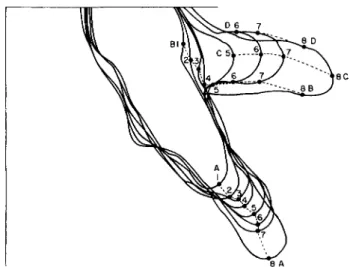
For example, in Fig. 1, granule A, attached to the particle, moved through positions 1 to 8 in successive photographs taken at 10-sec intervals. One might assume that, if new membrane were formed at the tip, this granule would have moved to one side of the pseudopodium.
The statement can be made for the successive positions of granules C, D, and the later positions of B, since all of them seem to have remained in more or less the same position in relation to the tip of the pseudopod.
Furthermore, granules B, C, and D are almost in the same relationship to each other in position 8 as in positions 6 and 7. However, it is obvious that granules A and Β are much farther apart in position 8 than they were in position 1. Therefore, new membrane must have been added
someplace between granules A and B as they moved from position 1 to position 8. We have a considerable amount of similar data which show the same thing.
From these data it is not possible to say exactly where this new membrane was formed, and, unfortunately, we have not been able to continue these observations. However, it is obvious that the whole pellicle cannot move forward without the addition of new areas. This is contradictory to the suggestion of Griffin and Allen (1960). New pellicle must be formed, and, in the organism shown in Fig. 1, this presumably occurred within a short distance, possibly one or two diam- eters behind the advancing tip.
FIG. 1. Successive positions of carmine particles on pseudopodia of A. proteus.
Goldacre (1961) has offered an alternative electrical interpretation for experiments involving the use of carmine granules, basing his con- clusions on the use of oil droplets. In his published oil-droplet experi- ments the drop at the anterior tip tended to remain at the tip, just as the carmine granules in our own experiments. However, regardless of the relative merits of carmine and oil, the results of Goldacre (1961) with the use of several techniques (oil droplets, glass filaments, wrinkle contours) convincingly demonstrate that new plasmalemma must be formed someplace in the advancing pseudopodium. In a polypodial ameba, formation of new plasmalemma seems necessary even if a mod- erately unreasonable amount of plasmalemma deformation is assumed.
The same must be true of Mayorella, because it seems inconceivable that the whole membrane would move forward in relation to the almost vertical, determinate pseudopodia, which are formed in the second
quarter of the advancing fan-shaped cell, and in relation to the cell certainly move posteriorly through the third and fourth quarters to be retracted and absorbed at the posterior end. If the idea of Griffin and Allen (1960) were applied to the whole membrane of Mayorella we would have to visualize a movement of particles of membrane anteriorly over
R
A Β FIG. 2. ( A ) Diagram of relative movement of protoplasm in an ameba attached
near middle of body. Wavy line denotes region of solation. (B) Same for ameba attached at rear: X , Nonmoving protoplasm; R, region of attachment; S, shear zone.
the peak of each of the determinate pseudopodia as the pseudopodia move backward. This would require more stretching and contraction of the surface of the membrane than seems possible with the assumption of anything like a surface area in which the various sections are fixed in position in relation to each other.
One possibility in polypodial Amoeba and Chaos, and also in May- orella is that (1) new membrane is formed whenever and wherever the
existing membrane is placed under sufficient tension, and (2) old mem- brane is absorbed whenever the tension is sufficiently decreased, especially in the posterior end. This possibility does not seem to be contradictory to the data of Goldacre (1952a, b, 1961, 1962).
MOVEMENT OF THE G E L
The direction of protoplasmic flow that might be expected in a monopodial ameba on the basis of the contraction-hydraulic theory is shown in Figs. 2A and B. In Fig. 2A it is assumed that the ameba is attached someplace in its mid-region and in Fig. 2B it is assumed to be attached at the rear end.
A polypodial A. proteus or C. carolinensis really moves in three di- mensions so that locomotion is more of a "walk" or a roll, as described by Wilber (1946). Mr. Fonseca and I have taken cinematographs from the side of C. carolinensis and have confirmed most of Wilber's obser- vations. Whenever an ameba (Amoeba or Chaos) is observed moving monopodially the location of the attachment usually is not determined, and furthermore it often cannot be determined by simple observation.
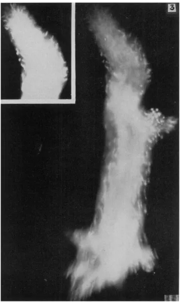
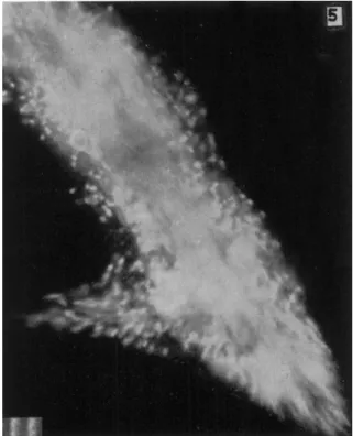
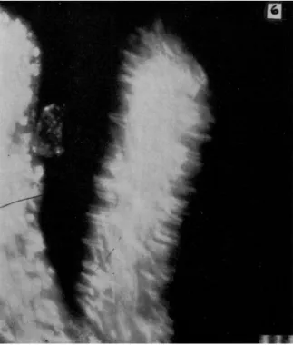
In Fig. 2A the gel is shown as moving forward in the anterior and posterior regions but not in the mid-region. This can be seen in streak (8-sec, dark-field) photographs. An example is shown in Fig. 3 in which there is a short mid-lateral pseudopodium in and near which the granules are not moving in the main direction of locomotion. However, in the posterior and anterior regions of this ameba, the granules of the gel are moving forward. Forward movement also is noticeable in the insert of Figs. 3 and 4. These streak pictures alone do not tell us the direction of movement, but the forward flow can easily be seen in some of the cinematographs of A. proteus and also of Mayorella in which the speed of motion is increased by a factor of 8. The relative immobility of the granules of the mid-gel region also can be seen in Fig. 5 in which the granules of the major posterior end (and also of a small withdrawing pseudopodium) are moving rapidly.
Figure 6 shows one type of movement in a retracting pseudopod of a polypodial ameba as the pseudopod undergoes a rapid decrease in diameter, presumably by elastic as well as active contraction. An inter- pretation of lateral movement adequate to explain the lines at right angles to the major axis is precluded by the fact that the center of the lower part (flowing sol) shows no evidence and the upper end shows relatively little evidence of lateral movement. The numerous short lines at right angles to the major axis are interprétable as evidence of radial contraction of the gel tube caused by a sudden drop of pressure of the sol such as that resulting from formation of a new lateral pseu-
dopodium. This type of movement can occur easily at the beginning of a pseudopodial retraction and is rapidly converted into the more charac- teristic type shown in Figs. 3 and 5.
If the sol were actually pulling the closed posterior end of the gel tube forward, as postulated by the frontal-contraction eversion hypoth-
FIG. 3. Streak photograph of moving A. proteus. Note that movement is more rapid at anterior and posterior ends than in the mid-body region. Exposure: 7 sec.
Scale: 10 μ. (After Rinaldi and Jahn, 1963.)
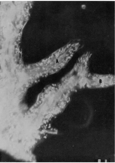
FIG. 4. Advancing pseudopodia of A. proteus. In A the granules of the new gel are advancing and also moving peripherally. Exposure: 8 sec. Scale: 10 μ. (After Rinaldi and Jahn, 1963.)
esis, we would have to postulate attachment only at the extreme posterior end in Fig. 6, but attachment over a much longer area in order to explain the data of Figs. 3 and 5. For reasons to be discussed later, it is highly improbable that any adequate attachment exists.
It is possible, under certain conditions, for the movement of proto- plasm to be as shown in Fig. 2B. This type of flow looks exactly like a
FIG. 5. Central and tail regions of A. proteus. Note that gel movement is rapid in the posterior region but almost zero in the mid-body region. (Rinaldi and Jahn, 1963.)
fountain, and we have an excellent moving picture showing this type of flow in C. carolinensis. However, organisms with this type of flow definitely are not locomoting, and are obviously anchored at some point.
In Fig. 2B the anchor point is shown at the posterior, but it is possible that blockage at the anterior end, or friction along the sides, especially in a tight-fitting glass tube, could produce the same effect. It is inter- esting that this fountain-type flow can easily be explained on the basis of the contraction-hydraulic theory, with the driving force being exerted at the base of the fountain—a position where it occurs in real fountains.
This has been observed by several investigators and is described for A. proteus by Mast (1926, p. 400) as follows:
'Occasionally, however, monopodal specimens are found in which all but the tip of the posterior end is free and in which this end is so attached that locomotion is prevented. In such specimens the movements of the plasmasol and the plasmagel are precisely the same as they are in moving specimens; but in reference to points in space, the plasmagel
FIG. 6. Pseudopod which has just begun to withdraw. Showing inward radial movement of granules. Exposure: 5 sec. Scale: 10 μ. (After Rinaldi and Jahn, 1963.)
actively moves backward instead of being at rest. This gives the appear- ance of typical fountain streaming, so much discussed in the literature on Amoeba/'
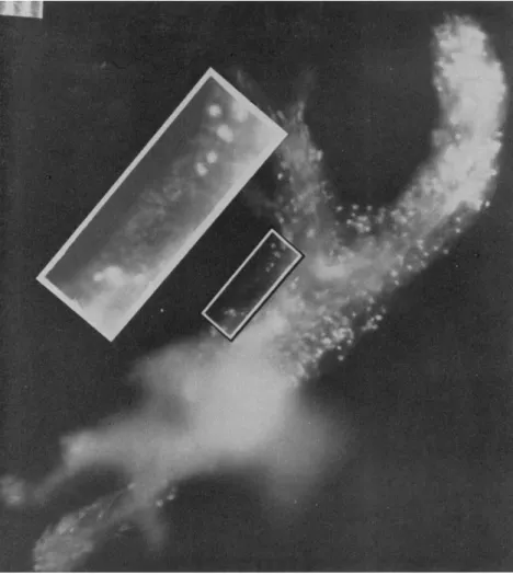
MOVEMENT OF VERY SMALL GRANULES THROUGH THE G E L AND THE PERIPHERAL HYALOPLASM
In Fig. 7 a section is outlined in which there are small granules that move forward along the edge of a gel region, between the plasmagel and plasmalemma, in the hyaline ectoplasm. Forward flow is not re- stricted to granules of this very small size, as frequently a few isolated
FIG. 7. Small granules in the gel region of A. proteus. Exposure: 8 sec. Scale:
10 μ. (After Rinaldi and Jahn, 1963.)
(Fig. 7) one can observe a number of the small granules. They move forward and may leave faint streaks. They move much slower than granules of the central region and in general faster than the larger granules of the peripheral region.
granules 3-4 μ in diameter can be seen flowing in the plasmagel. T h e smallest granules can be observed only by means of dark-field micros- copy. They are difficult to observe, but they can be seen clearly by careful focusing, especially with a cardioid condenser. In streak pictures
The important thing about the movement of these small granules is that they move in the direction of the main stream of flow of the plas- masol. Furthermore, they move forward, not only between the plasmagel and the plasmalemma, but also along zigzag paths through the interstices of the plasmagel. Such small moving granules can be seen to move past and around many of the larger fixed granules of the gel, as if they were bumping into invisible fibers of the spongy structure of the gel. This type of movement of granules in A. proteus has not been described pre- viously, but Abé (1961) has described somewhat similar movement of granules in A. striata.
MOVEMENT OF THE SOL
The fact that the sol moves forward is, of course, easily observed and can be seen in streak photographs (e.g., Fig. 4). When the sol approaches the tip of the pseudopodium it spreads laterally as shown in Fig. 3, insert, and sometimes there is a backward movement near the periphery. These movements also can be demonstrated by means of shadowgraphs made from cinematographs.
A backward movement might occur on the basis of either the hy- draulic-contraction or the frontal-contraction theories, but on the basis of the frontal-contraction theory a backward movement is an essential part of the locomotor mechanism. I have not found any statement to this effect in Dr. Allen's publications, but a backward movement of the contracting sol is definitely shown in a figure (Allen, 1961b, Fig. 26) demonstrating the postulated contraction of the gelating sol.
In order for such a frontal-contraction eversion system to work, the contraction of fibers must occur during the eversion. Each fiber is assumed to turn 180 degrees and to be shortened and fastened in position in the gel at the end of the turn. This means that the shortening must continue until the fiber is finally fastened into position. Since the everting material has a definite thickness (the radius of the pseudopod) the material from the center of the sol is everted to the periphery and that from the periphery to the inner portion of the gel tube. The ma- terial that travels from the center of the sol to the periphery of the gel must move in a greater arc than the material from the periphery of the gel. Consequently this central material must be pulled backward at the periphery in order to complete the arc.
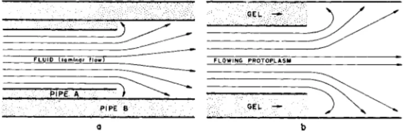
However, it also is possible to explain the backward flow on the basis of hydrodynamics, and furthermore to explain why it does not always occur. Figure 8 is modified from a summary of fluid mechanics by Kenyon (1960, p. 183) in a section concerned specifically with the pattern
of flow due to a sudden expansion by fluid flowing in pipes. As illus- trated by the diagram in Fig. 8a, there is a striking and noticeable back- ward flow upon an expansion of a laminar flowing fluid at the orifice where the fluid begins its expansion from the diameter of pipe A to pipe B.
Figure 8b depicts protoplasm flowing as a fluid. When the proto- plasm leaves the plasmagel tube the cross-sectional area of flowing increases because it is no longer restrained at the tip by the tube. Con- sequently, this would result in a backward flow at this region similar to that of any laminar flow through an orifice. T h e orifice in the case of an ameba is the simultaneously formed (plasmagel) tube. In addition, the plasmagel tube through which the stream is flowing forward in an
α b FIG. 8. Behavior of a fluid upon expansion from a smaller to a larger tube,
illustrating the turbulence (backward flow) produced at the orifice, (a) Flow of fluid in a pipe. (After Kenyon, 1960.) (b) Flow of protoplasm. (After Rinaldi and Jahn, 1963.)
ameba can itself move slightly forward by stretching under hydraulic pressure (indicated by the arrows) and also is being extended by gelation at the end of the tube.
If the profile of new gel being formed (Fig. 8b) is sloping forward and outward toward the plasmalemma rather than perpendicular as shown, there will be no necessity for a backward movement. Some of the shad- owgraphs of Rinaldi and Jahn (1963) show forward movement only.
Radial movement, of course, also occurs but cannot be shown in this type of shadowgraph used.
Allen (1962) states that the "endoplasm must shorten as it thickens just before or during its turning at the front of the cell to form the ectoplasmic tube." If we assume tentatively that a frontal-contraction eversion system were to exist, the contraction would have to continue during the eversion, regardless of whether it had started previously.
Therefore, we can disregard the possibility that it shortens "just before . . . its turning" as inconsequential.
GENERAL DIRECTION OF MOVEMENT OF GRANULES IN ANTERIOR THIRD OF AN ADVANCING PSEUDOPODIUM
The results described previously demonstrate that there is a flow of small granules forward, not only in the central plasmasol, but also in the hyaline ectoplasm on the outside of the plasmagel tube, and furthermore, through the interstices of the spongy structure of the gel.
In brief, there is a forward movement of tiny granules throughout the entire cross section in the anterior third of an advancing pseudopodium but it is most obvious in newly formed, lateral pseudopodia when the total length of pseudopodium is only 1-2 times its diameter. This for- ward flow is exactly the type that can be expected on the basis of the Mast (1926) theory; it is merely a flow of syneretic material from the con- tracting region. It is not predictable from the frontal-eversion assump- tion (Allen, 1961a), which requires that there be a posteriorly directed flow of syneretic material. In a later paper (Allen et al., 1962, p. 116) it stated that the hyaline cap is formed from syneretic fluid from the eversion zone and that: "It seems likely that hyaline cap fluid is recircu- lated toward the tail in a channel beneath the plasmalemma." This channel would be the lateral hyaline ectoplasm. These same authors further aver: "The plasmalemma slides over the ectoplasmic tube, pro- viding a channel through which the hyaline cap fluid may (in fact must) circulate toward the tail, where it somehow finds its way through the weakened ectoplasmic tube to rejoin the ectoplasmic stream." A later figure (Allen, 1962, pp. 116-117) shows arrows indicating a reverse flow in the hyaline ectoplasmic layer, but there has never been presented any evidence that such a reverse flow exists. Our observations show that the small granules of the hyaline ectoplasm actually move forward rather than backward. Therefore no reverse flow of syneretic fluid is present, and no amount of supposition can produce it.
The general forward flow of material which occurs throughout the entire cross section of the ameba (plasmasol, plasmagel, hyaline ecto- plasm) can be predicted from contraction-hydraulic postulates. It cannot be predicted nor accounted for by the present version of the frontal eversion idea which requires forward flow in the plasmasol only, no forward flow in the gel, and a reverse flow of the hyaline ectoplasm along a postulated but unproved route. For these reasons it is concluded that the posterior contraction-hydraulic pressure explanation agrees best with the facts.
Analysis of Other Data Pertaining to Mechanism of Movement
TRANSMISSION OF TENSION TO THE POSTERIOR END
According to the frontal-contraction eversion concept, the contracting plasmasol at the anterior end exerts a tension which is assumed to pull axial plasmasol forward. In order for a force so positioned to pull the posterior end of the ameba forward it must be transmitted to the pos- terior plasmagel. However, evidence has been given (Allen, 1960) that the area between the posterior end of the axial plasmagel and the pos- terior plasmagel (i.e., the solation or so-called "recruitment zone") is one of very low consistency. T o pull the tail the tension must, therefore, be transmitted across this area of solation, which, together with the shear zone, has the lowest consistency (i.e., viscosity) of the entire ameba (Allen, 1960). Since no bridge is therefore provided between the pos- terior gel and sol, then either the gel must move forward by contraction, or else be sucked forward by axial plasmasol. If the posterior gel con- tracts, then a frontal contraction and eversion pulling from the anterior end is superfluous. If the posterior gel is drawn forward by suction, then the same suction should pull the posterior portion of the shear zone backward, and there is no evidence available that this occurs.
Therefore until evidence is presented that the area in question is actu- ally bridged by fibrous structures of sufficient strength to maintain a frontal eversion while causing and accompanied by a posterior inversion, the mere conjecture of their presence (Allen, 1961a) against evidence to the contrary (Allen, 1960) renders the scheme unacceptable.
Furthermore, if the back end of the ameba is being pulled forward by suction, cutting a small hole in the plasmagel in this region should result in a flow of external medium into the hole rather than a flow of protoplasm into the medium as ordinarily seen by micrurgists, and which has been demonstrated very elegantly by Goldacre (1961).
Another interesting consideration is that the unlimited posterior narrowing and elongation of the tail, described by Goldacre (1958), would be impossible if the tail were being pulled forward by the sol instead of actually contracting as described by Goldacre.
FORMATION OF LATERAL RIDGES
It is generally recognized (Leidy, 1879; Mast, 1926; Schaeffer, 1920) that the lateral ridges on the pseudopodia of A. proteus are formed by the flow of granular plasmasol through large interstices of the gel to the outside of the plasmagel tube and thence forward between the plasmalemma and the gel tube, forming a ridge, which later consists of gel with a core of flowing plasmasol, and which may beome a secondary
pseudopodium. T h e fact that during formation these ridges progress forward, with little or no circumferential spreading, is a strong indi- cation that the driving force must be pressure from the posterior end.
This is the same pressure that drives the small granules forward through- out the entire cross section of the ameba.
ORIGIN OF SYNERETIC FLUID
It has been calculated on the basis of measurements or refractive index (Allen and Roslansky, 1958) that the protein content of a moving ameba is about 3.95% at the posterior end, 3.33% at the anterior part of the sol, and about 1.00% in the hyaloplasm.
It is generally assumed that during the folding of an elongated protein molecule some of the intermolecular bonds become intramolecular, and consequently there are fewer exposed groups to which water and other small molecules could be attached. Therefore, the amount of water and other smaller molecules attached to the protein is reduced. This idea was applied to Amoeba by Goldacre and Lorch (1950) and Goldacre (1952b). Conversely, it is assumed that water would become attached to the protein during the unfolding process.
In the contraction-hydraulic theory it is assumed that part of the framework folds and becomes detached, thereby going into solution, at the time and also at the site of contraction, in the mid-posterior region.
It is also assumed on the basis of the bond conversion mentioned pre- viously that water is squeezed out of the molecular framework during contraction. If the contraction is at the posterior end and is a gradual process involving slow, molecule by molecule, dissolution of part of the protein framework, some water would be released from partly folded molecules which would still be attached to the framework. This would provide surplus water which would be pushed forward ahead of the protein molecules which would be released only after they are completely folded and detached from the protein framework. Water carried by this forward flow would accumulate at the front end of the ameba until it is used during gelation and in the hardening process of the gel which follows. T h e amount of water absorbed during gelation would include the excess water released during the solation process and forced forward as part of the flowing sol, and that which filters forward through the gel, and also that which flows forward in the peripheral hyaloplasm. In this way a gradient of water would be maintained throughout the length of an ameba.
In the fountain-streaming or frontal-contraction eversion theory the framework is assumed to become more compact by folding of either or both molecules and fibers at the anterior "fountain" zone, thereby
squeezing water of syneresis into the hyaline cap. It is also assumed that this syneretic fluid flows backward in the lateral hyaline layer between the gel and the plasmalemma.
If one assumes that contraction is at the anterior end and that the water is released during contraction, the surplus water at the anterior end is easily explained. However, the next question is how it is trans- ported to the posterior end so that it can be available during solation.
Since the actual movement of granules in the peripheral hyaline zone is
s 9
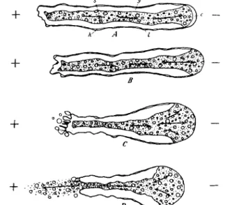
FIG. 9. T h e effect of galvanic current on a monopodial ameba moving toward the cathode. —, Cathode; -f, anode; arrows, direction of streaming. A, very weak current; B, C, D, progressively stronger current. Note that in a current of moderate density the hyaline cap disappears and the plasmasol extends to the plasmalemma, and that in stronger current the cathodal end expands and the anodal end contracts and finally breaks after which the granules in the plasmasol flow toward the anode, indicating that they are negatively charged. (After Mast, 1931c.)
the reverse of that postulated in the frontal-contraction eversion hypoth- esis; it cannot move backward as proposed by Allen (1961, 1962). Further- more, even if it were transported posteriorly in the peripheral hyaline zone it would have to cross the wall of the gel tube in order to reach the solation zone, where according to the fountain-zone theory the folded molecules of the gel presumably become unfolded. The only other possi- bility would be a reverse flow through the gel itself, and this also has been disproved.
For these reasons, it seems as if the data of Allen and Roslansky (1958) as far as these data pertain to the origin and the cycling of syneretic
fluid, are best explained on the basis of the contraction-hydraulic theory as originally explained by Allen and Roslansky (1958) rather than on the basis of the frontal-contraction eversion hypothesis as suggested by Allen (1961).
EFFECT OF ELECTRIC CURRENT
The effect of direct electric current on locomotion of ameba has been studied by Mast (1931c) and Hahnert (1932). These investigators have
FIG. 10. T h e effect of alternating current. A, before circuit was closed; B-F, successive stages after closure; arrows, direction of streaming; double-ended arrows, direction of the current. Note that the ameba orients perpendicularly to the direction of the current, that the plasmagel in the pseudopods at this time is very thin or absent, that the plasmagel contracts violently at the surface directed toward the poles, that blisters are formed on these surfaces, and that the ameba eventually breaks here and then disintegrates. (After Mast, 1931c.)
demonstrated that in an organism moving toward the cathode the effect of current is to inhibit gelation at the cathodal (i.e., anterior) end.
However, this inhibition of gelation does not stop the ameba (Fig. 9) which continues to move forward in spite of the fact that the gel tube is not extended, and the anterior advancing portion is all sol. Since there is no new gel tube to confine the sol it spreads out to form a large low- viscosity structure which may have more than twice the diameter of the ameba. On the basis of the fountain-zone contraction theory this would be impossible; nevertheless, it occurs. A possible alternative suggestion
(Heilbrunn and Daugherty, 1939) that cathodal movement is caused by a positive charge on the particles is disproven by Mast's (1931c) dem- onstration that the charge on the granules is negative.
Mast (1931c) also demonstrated that an alternating current causes a rounding of the ameba and then a contraction of the gel. This con- traction causes the formation of long pseudopodia at right angles to the electric field (Fig. 10). These pseudopodia contain no gel and, therefore, could not possibly be extended by means of a frontal-contraction ever- sion mechanism.
EFFECT OF ILLUMINATION
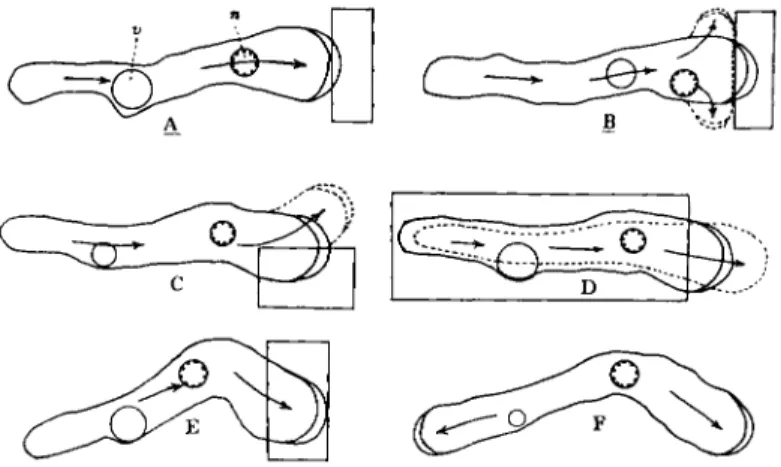
It is well known that the effect of illumination of the advancing tip of a pseudopodium, i.e., of the fountain-zone region, causes immediate
FIG. 11. Response of Amoeba to localized illumination. Only the rectangular areas were brightly illuminated. Α-D, same specimen; F , same specimen as E , a few moments later. (After Mast, 1932.)
gelation of the anterior end (Mast, 1932). However, this gelation does not inhibit forward flow of the main stream of sol which continues for- ward, unimpeded, and branches out laterally, as shown in Fig. 11, through weak places in the end of the gel tube. If the sol were being pulled forward, in accordance with the fountain-zone contraction theory, a gelation of the anterior end should block the forward movement, at least temporarily, or until new contracting forward areas could be formed. However, this does not occur.
Furthermore, if the whole body of an ameba locomoting in very dim light is brightly illuminated, the sol of the anterior tip is gelated, exactly as described previously (Mast, 1931b). However, when the anterior end gelâtes, the closure of the gel tube is complete; there are no weak places, as mentioned previously, and the newly formed closed end of the tube
begins to contract (Fig. 12). This causes a reverse flow and this reversed flow progresses backward exactly like the upstream waves of reversal so characteristic of Physarum, described by Kamiya (1959; and earlier) and Stewart and Stewart (1959) and by other investigators. It seems to me as if this single observation of Mast completely invalidates the frontal- contraction eversion theory, and it is especially intriguing because the only experimental manipulation necessary is opening of the condenser diaphragm of the microscope. We often have observed such reversals
FIG. 12. Processes observed in a pseudopod in the response to rapid increase in illumination. Arrows, direction of flow in the plasmasol; x, regions at rest. Note that the response begins by cessation in streaming at the tip of the pseudopod imme- diately back of the hyaline cap and continues by cessation farther back accompanied by streaming in the reverse direction at the tip. (After Mast, 1931b.)
when we switch by means of a mirror from tungsten filament lamps used for visual observations to carbon arcs for cinephotomicrography. A com- parable diagram showing flow from both ends toward the middle was published by Mast in another paper (Mast, 1931a) with the comment that it was "repeatedly observed."
Still another figure (Mast, 1931b, Fig. 2) shows essentially the same phenomenon except that when the two streams meet they cause a bal- looning of the body because of the increased hydraulic pressure in that region (Fig. 13). Mast (1931b, p. 141) described this as follows:
"A few moments after the increase in illumination streaming stopped, first immediately back of the hyaline cap at the tip of both pseudopods, then successively farther back, but before streaming stopped at the pos- terior end it had begun again at the anterior end. Here, however, it now continued in the opposite direction. Soon after this it also began again at the posterior end, but in the original direction. There was thus at this time backward streaming in the anterior portion and forward streaming in the posterior portion of the amoeba (Fig. 2,b). This resulted in the formation of a marked enlargement near the middle.
A few moments later there was a sudden break in the plasmagel far- ther forward (Fig. 2,b,x) and the plasmasol immediately rushed to-
FIG. 13. Processes observed in the response of an ameba to rapid increase in illumination. Arrows, direction of flow in the plasmasol; x, regions at rest. Note that the response consisted of a complex series of phenomena and that in this series there was at no time complete cessation of flow in the organism. (After Mast, 1931b.)
ward this break, resulting in the formation of another enlargement which eventually contained nearly all the substance in the amoeba (Fig. 2,d). From this, new pseudopods developed, after which the amoeba soon returned to its original form (Fig. 2,f)."
It also seems quite significant that illumination of all of an ameba except the fountain-zone area and the advancing tip increases the rate of streaming and causes a decrease in diameter of the organism, as well as increases in the thickness of the gel tube and in the gel-sol ratio (Mast, 1932). These facts are easily explainable on the basis that light converts the layer of sol next to the gel into gel, as explained by Mast (1932). However, if we assume the contractile force is in the unillumi- nated anterior end, it is difficult to see how illumination of the posterior 90% or more could increase the rate of streaming, especially since the diameter of the flowing stream is decreased.
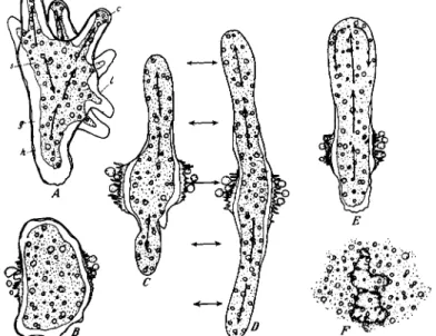
FIG. 14. Changes of form and activity in A. proteus during and after exposure to a pressure of 6000 psi at 25 ° C (A) Normal form at atmospheric pressure. ( B ) Same specimen at 6000 psi, maintained for 5 min. Note the rounded form of the main cytoplasmic mass and the pinched-off part of the large pseudopodium (above). (C) Fif- teen minutes later pressure still maintained. Specimen has rotated about 90°. Note complete rounding of the main mass and of the pinched-off part of the cytoplasm which now lies to the left. ( D ) Fifteen seconds after pressure was reduced to the atmospheric level. Note the marked contraction of the granular cytoplasm (plasmagel) and the development of a broad hyaline zone between the granular cytoplasm and the cell membrane. (E) Ninety seconds after decompression. Note first signs of ameboid activity. (F) Seven minutes after decompression. Ameboid activity now quite vigorous.
(From Landau et al., 1954.)
Some of the above experiments of Mast (1931a, b) have been misin- terpreted by Allen (1961a) who states, "There are many aspects of the ameba's responsiveness to stimuli which lend excellent support to the
fountain zone contraction theory. First, both mechanical stimuli and light are effective only if applied locally to the fountain zone." Mast's observations, corroborated by our own, demonstrate that light also is very effective when applied to the whole organism. Furthermore, these observations do not "lend excellent support to the fountain zone con- traction theory" but are quite contradictory to the fountain-zone theory.
EFFECT OF HIGH HYDROSTATIC PRESSURE
One type of evidence which supports the contraction-hydraulic theory is the definite correlation between contraction and solation. This is easily observable in the posterior end of an ameba if one believes in the theory, but to nonbelievers the observation may seem equivocal. An ex- planation on a molecular basis was given by Goldacre and Lorch (1950).
I H B O N OS
».
, T H I OL
> B O N OS ( ? )|
, P E P T I DE I B O N 0 S ( ?)
GEL STATE
3 - D I M E N S I O N AL P R O T E IN N E T W O RK
P R O P ER I O N IC E N V I R O N M E NT
- Δ V Ρ H Y D R O L Y S IS ( ?)
CONTRACTED GEL
M E C H A N I C AL W O RK
N E T W O RK A ND A G G R E G A TE ( ?)
B R E A K D O WN
SOL STATE
P R O T E IN M O L E C U LE M O L E C U L AR A G G R E G A TE
( M Y O S INL I Κ Ε ?)
FIG. 15. A schematic representation of the sol-gel phenomenon. (After Landau, 1959.)
This explanation is still completely valid in principle, although I would tend to apply it to some rather than to all the framework of the gel, leaving a loose spongy network in the sol as assumed by Mast (1926).
Furthermore, the correlation of solation and contraction is strongly sup- ported by studies on the effect of high hydrostatic pressures on ameba protoplasm (Landau et al., 1954), especially during conditions under which the ameba is not moving. If an ameba is subjected to pressures of 6000 psi (Fig. 14) all the protoplasm becomes a sol of low viscosity and the ameba becomes spherical. If the pressure is suddenly released the sol is converted into gel and the gel contracts violently with one massive contraction, leaving a layer of hyaline material around the periphery.
Within seconds after this contraction occurs the granules move into the hyaline zone, indicating that the granular region that was gel is now at least partly solated. Shortly afterward it is obvious that the ameba has normal sol-gel ratios. On the basis of this and other evidence the
correlation between contraction and solation was incorporated into the diagram of sol-gel relationships (Fig. 15) devised by Landau (1959). The current uncertainties that concern this diagram seem to involve the role of adenosine triphosphate (ATP) and the mechanics of contraction, as pointed out by Landau (1959), rather than the relationship between con- traction and solation, which seem to have a very solid foundation.
If we were to assume that the anterior contraction theory is true, we would have to discard this diagram (Fig. 15) almost completely and invent a new one in which gelation and contraction would be correlated with each other, as would solation and molecular extension. The as- sumption that these proposed correlations are true would then necessi- tate a re-evaluation of the theory of high-pressure effects on protoplasm.
This is a task which I gladly leave for consideration by the experts on high pressure who, of course, have the alternative of retaining their pres- ent interpretations which strongly support the contraction-hydraulic theory.
Summary
It has been demonstrated by studies of the movement of carmine granules attached to the membrane of A. proteus and C. carolinensis that new membrane is not formed at the advancing tip of a pseudopod but that it may be formed a few diameters behind the tip.
It is demonstrated that, in the cross-sectional area of the anterior third of large advancing pseudopodia and especially in newly formed, lateral pseudopodia, all the protoplasm is moving forward, including the sol, gel, peripheral hyaloplasm, pellicle, and syneretic fluid through the gel.
A reanalysis is presented of data of other investigators concerning (1) the low viscosity of the posterior solation zone, (2) the formation of lateral ridges, (3) the origin and cycling of the syneretic fluid, (4) the effect of direct and of alternating electric current, (5) the effect of illu- mination, and (6) the effect of changes in hydrostatic pressure.
It is pointed out that all of these data can be explained on the basis of the contraction-hydraulic theory of posterior contraction of the gel tube and that most of the data discussed are contradictory to the foun- tain-zone or frontal-contraction eversion theory. It is concluded, there- fore, that the fountain-zone contraction theory is invalid.
REFERENCES Abé, T . H. (1961). Cytologia (Tokyo) 26, 378.
Allen, R. D. (1960). / . Biophys. Biochem. Cytol. 8 , 379.
Allen, R. D. (1961a). Exptl. Cell Res. Suppl. 8 , 17.
Allen, R. D. (1961b). In "The Cell" (J. Brächet and A. E. Mirsky, eds.), Vol. II, pp. 135-216. Academic Press, New York.
Allen, R. D. (1962). Sei. Am. 2 0 6 , 112.
Allen, R. D., and Roslansky, J . D. (1958). / . Biophys. Biochem. Cytol. 4 , 517.
Allen, R. D., Cooledge, R. R., and Hall, P. J . (1962). / . Cell. Biol. 1 2 , 185.
Goldacre, R. J . (1952a). Symp. Soc. Exptl. Biol. 6 , 128.
Goldacre, R. J . (1952b). Intern. Rev. Cytol. 5 2 , 135.
Goldacre, R. J . (1958). Intern. Congr. Cybernetics, 1st, Namur, 1956, p. 715. Gauthier- Villars, Paris.
Goldacre, R. J . (1961). Exptl. Cell Res. Suppl. 8 , 1.
Goldacre, R. J . (1962). In "Biological Structure and Function" (T. W. Goodman and O. Lindberg, eds.), Vol. I I , pp. 633-643. Academic Press, New York.
Goldacre, R. J . , and Lorch, I . J . (1950). Nature 1 6 6 , 497.
Hahnert, W. F. (1932). Physiol. Zool. 5 , 491.
Heilbrunn, L. V., and Daugherty, K. (1939). Physiol. Zool. 1 2 , 1.
Jahn, T. L., and Rinaldi, R. (1963). Ann. Meeting Biophys. Soc, Ith, New York, 1963, Abst. MDL
Kamiya, N. (1959). Protoplasmatologia, 8 , 1.
Kenyon, R. A. (1960). "Principles of Fluid Mechanics." Ronald Press, New7 York.
Landau, J . V. (1959). Ann. Ν. Y. Acad. Sei. 7 8 , 487.
Landau, J . V., Zimmerman, A. M., and Marsland, D. A. (1954). /. Cellular Comp.
Physiol. 4 4 , 211.
Leidy, J . (1879). U. S. Geol. Surv. Territories 1 2 , 1.
Mast, S. O. (1926). J. Morphol. Physiol. 4 1 , 347.
Mast, S. O. (1931a). Protoplasma 1 4 , 321.
Mast, S. O. (1931b). Z. Vergleich. Physiol. 1 5 , 139.
Mast, S. O. (1931c). Z. Vergleich. Physiol. 1 5 , 309.
Mast, S. O. (1932). Physiol. Zool. 5 , 1.
Rinaldi, R. Α., and Jahn, T . L. (1962). / . Protozool. 9 , (Suppl.), 16.
Rinaldi, R. Α., and Jahn, T . L. (1963). / . Protozool. 1 0 , 344.
Schaeffer, A. A. (1920). "Amoeboid Movement." Princeton Univ. Press, Princeton, New Jersey.
Stewart, P. Α., and Stewart, Β. T. (1959). Exptl. Cell Res. 1 7 , 44.
Wilber, C. S. (1946). Trans. Am. Microscop. Soc. 6 5 ( 4 ) , 318.