Microarchitectural study of the augmented bone following ridge preservation with a porcine xenograft and
a collagen membrane: prospective clinical, histological and micro-CT analysis
Ph.D. Thesis
Kivovics Márton
Semmelweis University PhD School of Clinical Medicine
Supervisor: György Szabó, MD, DSc.
Ferenc Dőri, DMD, Ph.D.
Opponents: Katalin Nagy, DMD, Ph.D.
Krisztina Márton, DMD, Ph.D.
Chairman of the Examination Committee: Árpád Fazekas DMD, DSc.
Members of the Examination Committee: Ádám Kovács, MD, Ph.D.
Zsuzsanna Tóth, DMD, Ph.D.
Budapest 2017
1 1. Introduction
The alveolar ridge is a tooth-dependent structure in the jaw. Its development is induced by tooth eruption, and loss of teeth results in resorption and atrophy of the alveolar ridge. Severe atrophy of the ridges prevents favourable prosthetic positioning of endosseus implants, which may lead to esthetic and loading compromise; therefore, ridge augmentation might be necessary for implant placement.
Preservation of the alveolus following tooth extraction predictably maintains the bone volume. Several techniques and biomaterials for alveolus preservation have been described in the literature. Studies show that the use of bone substitute materials with or without bioresorbable or non-resorbable barrier membranes significantly reduces the horizontal and vertical bone loss following extraction. There are reports of successful application of several bone graft materials in ridge preservation. However, used as fillers in the alveolus all these materials interfere with normal extraction socket healing.
2. Aim of the study
The aim of this prospective study is to clinically examine and then study the integration of a porcine xenografts in alveolar ridge preservation by histological and microCT analysis. Our aim is to determine whether socket grafting interferes with natural bone healing.
1. Does the bone substitute material induce any undesirable inflammatory response or foreign body reaction?
2
2. Does the bone substitute material used in socket preservation integrate in the socket with newly formed bone or connective tissue formation?
3. Is the bone formation homogenous in the grafted socket?
4. Is the microarchitecture of the particles of the bone augmentation material different from that of the newly formed bone in the preserved socket?
5. Is the microarchitecture of the grafted socket different from that of the native bone?
6. Is the microarchitecture of the newly formed bone in the grafted socket different from that of the native bone?
3. Materials and Methods
3.1. Patients
Patients who were periodontally healthy, more than 18 years of age, able to sign an informed consent form, and needed implant- supported restoration were included in this study. The exclusion criteria were as follows: history of systemic diseases that would contraindicate oral surgical treatment, unwillingness to return for follow-up examinations, smoking, and pregnancy. The procedures included in the study were thoroughly explained to the patients, and they signed informed consent forms. The study was approved by the Regional and Institutional Committee of Science and Research Ethics and the Hungarian Office of Health Authorisation and Administrative Procedures (identification number of the study: 39993/2013/OTIG), and conducted in accordance with the Helsinki declaration.
The patients were categorized into three study groups. Group 1 (test group) consisted of 9 patients that required ridge
3
preservation to maintain sufficient bone volume for implant placement. Sockets in the test group showed a three-wall bone morphology. Following tooth removal immediate GBR treatment was carried out to preserve the sockets of Group 1.
Group 2 (positive control group) consisted of 8 patients that did not require ridge preservation to maintain the bone volume for implant placement. These patients showed an intact and thick buccal bone after tooth removal, and their alveoli presented a four-wall bone morphology. Sockets of Group 2 were left to heal the natural way.
Group 3 (negative control group) consisted of 9 patients. These patients, just like patients of the test group showed a three-wall bone morphology. In the sockets of Group 3 a flap was raised and mobilised to achieve primary closure.
3.2. Surgical procedures
All patients rinsed with 0.2% chlorhexidine solution for 1 minute before the surgery. Removal of teeth was carried out in an atraumatic manner in local anaesthesia. The sockets were thoroughly debrided to remove all the soft tissues. Socket morphology was examined and sites were allocated to study groups.
In Group 1 (test group) and Group 3 (negative control group) an intrasulcular incision with or without crestal incision and two releaving incisions were made to raise a full-thickness flap. In Group 1 the corticocancellous porcine bone (Gen-Os;
Osteobiol, Tecnoss Dental, Torino, Italy) graft was packed in the socket and a porcine collagen membrane (Evolution;
Osteobiol, Tecnoss Dental, Torino, Italy) was used as the occlusive barrier. In Group 1 and Group 3 the buccal flap was mobilized to allow tension-free primary closure. The margins were stabilized with single interrupted sutures.
4
In Group 2, sockets were left to heal without the use of socket preservation techniques.
Antibiotics (1 g amoxicillin-clavulanate twice a day for 5 days, or in case of side effects or known allergy to penicillin, 300 mg clindamycin 4 times a day for 4 days), anti-inflammatory drugs (50 mg diclofenac 3 times a day for 3 days), and chlorhexidine mouthwash (twice a day for 21 days) were prescribed to patients assigned to all groups. During the healing period, patients either received temporary prostheses that were in no contact with the surgical area or did not wear temporary prostheses at all.
3.3. Reentry Procedure
After 6-month healing period, clinical examination and the surgical re-entry procedure were performed under local anaesthesia. A bone core biopsy was taken with a trephine with an external diameter of 3.5 mm and an internal diameter of 2.5 mm (330 205 486 001 025 Hager & Meisinger GmbH, Neuss, Germany) and implants of at least 4.0 mm of diameter were placed into the grafted alveolus. Final prosthetic restorations were cemented after 3 months. Patients were recalled for follow-up every 6 months.
The bone core biopsy samples were removed, pushed out from the trephine, and placed in sample holders filled with 4%
formaldehyde solution in 0.1M phosphate buffer saline (PBS), pH 7.3, stored at 4°C.
3.4. Histologic Evaluation
Bone core biopsy material was fixed in 10% buffered formaldehyde solution. Following dehydration, the biopsy material was embedded in paraffin and 6-µm sections were
5
prepared with a Diatome. The sections were stained with routine haematoxylin-eosin stain. Slides were examined with light microscopy in magnification 40-400x.
3.5. Microcomputed tomography analysis
The bone core biopsy samples were scanned using a μCT scanner (Skyscan 1172 X-Ray microtomograph, Kontich, Belgium). The X-ray tube potential (peak) was 70 kV and the X-ray intensity was 124 µA. Bone core biopsy samples were scanned in a 4% buffered formaldehyde solution. Scanning was carried out in 5.90-µm resolution. A 0.5-mm aluminium filter was used to remove image noise. The ring artefact correction was 10, and the beam hardening correction was 61%. Average scan duration was 25 minutes. After scanning, reconstruction of 3D microstructural image data was carried out using the
“Skyscan NRecon” software. The reconstruction duration per slice was 1.59 seconds.
From the tomographs of each specimen, the region of interest (ROI) was set so that it would contain the newly formed bone as well as the particles of the bone graft material. In order to differentiate newly formed bone from particles of the bone graft and connective tissue from the newly formed bone, segmentation of the images was carried out by thresholding.
The thresholds for connective tissue, bone, and bone graft material were determined by analysing the appearance of the particles of the bone graft material in visual images and the intensities (grey level [GL]) of the different structures by the means of a histogram.
3.6. Statistical Analysis
Micromorphometric data was collected by evaluation of the augmented bone of the test group. The augmented bone was
6
segmented to newly formed bone and particles of the xenograft.
Additional micromorphometric data were recorded by separately analysing these components of the augmented bone.
Another set of data were collected by the evaluation of the native bone of the control groups.
The microarchitectural parameters of
a) the newly formed bone and particles of the bone substitute material within the augmented area
b) augmented bone and native bone of the control groups c) and newly formed bone within the augmented area and
native bone of the control groups
were compared.
The results were analysed statistically in consultation with an experienced statistician using the “Statistica 12” data analysis software program (StatSoft, Hungary). The Levene test of homogeneity of variances was carried out to determine whether to use the parametric or non-parametric statistics. Analysis of variance was used as the method for parametric and Mann- Whitney U test was used as the method for non-parametric statistics when comparing two sets of data. Analysis of variance (Oneway ANOVA) and Bonferoni test, was used as the method for parametric and Kruskal-Wallis test was used as the method for non-parametric statistics when comparing three sets of data.
Values of P < 0.05 were considered significant.
4. Results
A total of 26 patients were included in the study.
In Group 1, 12 bone core biopsy samples were taken from the preserved sockets of 9 patients (5 male and 4 female patients)
7
and 12 implants were placed in the augmented sites. At the follow-up visits, clinical examination and radiological evaluations were performed. At the 2-year follow up, 1 of 12 implants failed to osseointegrate in the augmented area.
In Group 2 (positive control group), 12 bone core biopsy samples of native bone were obtained from 8 patients (5 male, 3 female) and 12 implants were placed. At the 2-year follow up, all implants survived.
In Group 3 (negative control group), 12 bone core biopsy samples of native bone were obtained from 8 patients (5 male, 3 female) and 12 implants were placed.
At the 2-year follow up, all implants survived.
4.1. Histological Results
At the time of re-entry care was taken in order to preserve the anatomical orientation of bone core biopsies. Histological analysis of the bone core biopsy samples obtained from the augmented sites of Group 1 concluded that in 8 out of the 12 cases, particles of the bone substitute material were surrounded by newly formed trabecular bone. In these cases the apical and central parts of the biopsy samples comprised of particles of the xenograft surrounded by mostly woven and also some lamellar bone with marrow spaces. However, in the coronal part bone graft particles were surrounded by connective tissue with fibroblasts collagen fibres and blood vessels with no inflammatory reaction present. However, in the remaining 4 cases, the particles of the porcine xenograft were encapsulated by granulation tissue, with no sign of inflammation or foreign body reaction.
Histological analysis of the bone core biopsy samples obtained from the non-augmented sites in Group 2 and Group 3 presented healthy bone formation in the extraction sockets.
8
4.2. Results of the microCT Analysis
4.2.1. Qualitative Analysis of the microCT Images The microCT images for 8 out of the 12 bone core biopsy samples obtained from the augmented sites in Group 1 showed newly formed bone trabecules in contact with or surrounding the particles of the porcine xenograft in the apical and central parts of the specimens. In the coronal parts of the biopsy samples particles of the xenograft were surrounded with non- mineralized tissue. In the remaining 4 samples, the microCT images showed particles of the bone graft surrounded by non- mineralized tissue.
MicroCT analysis of the 12 bone core biopsy samples obtained from the non-augmented sites in Group 2 and Group 3 presented newly formed, trabecular bone, which consisted of mineralized trabecules and non-mineralized tissue (marrow spaces). Thus, the findings of the microCT analysis were consistent those of the histological analysis.
4.2.2. Bone Microarchitecture Analysis
In the μCT images from Group 1 (test group), the particles and the newly formed bone were segmented by thresholding. The morphometric parameters of the bone graft particles and the newly formed bone were compared. The values of the morphometric parameters that showed significant difference between the two sets of data and the results of the statistical analysis are presented in Table 1.
9 Table 1.
P values are presented with a cross if the statistical analysis used was the Mann-Whitney U test. Elsewhere, the statistical analysis used was the analysis of variance. N shows the number of bone core biopsy samples harvested.
n
Tb.N (M) 1/mm
Tb.N (SD) 1/mm
Tb.N (p- value)
Tb.Pf (M) 1/mm
Tb.Pf (SD) 1/μm
Tb.Pf (p- value)
SMI (M)
SMI (SD)
SMI (p- value) Newly
formed
bone and 8 1,01 0,872 -15,7 20,3 0,181 1,51
Particles of the xenograft in the bone core biopsy samples of
Group 1 8 0,328 0,349 0,0499+ 21,6 23,6 0,00447 2,08 1,02 0,0103
n
Po.N (cl) (M)
Po.N (cl) (SD)
Po.N(cl) (p-value)
Po.V(cl) (M) mm3
Po.V(cl) (SM) mm3
Po.V(cl) (p-value)
Po.S (cl) (M) mm2
Po.S(cl ) (SD) mm2
Po.S(cl) (p-value)
Newly formed
bone and 8 1260 1680 0,246 0,422 30,6 34,8
Particles of the xenograft in the bone core biopsy samples of
Group 1 8 32,6 39,1 0,0312+ 0,000949 0,00105 0,00195+ 27,0 0,309 0,00741+
The morphometric parameters of the augmented areas in Group 1 and the native bone in Group 2 and Group 3 were compared.
The values of the morphometric parameters that showed significant difference between the three sets of data and the results of the statistical analysis are presented in Table 2.
10
Table 2. P values are presented with a cross if the statistical analysis used was the Kruskal-Wallis test. Elsewhere, the statistical analysis used was the analysis of variance (Oneway ANOVA) and the Bonferoni test.
Bone Volume Fraction
Intersection Surface
Bone Surface Density
Trabecular Number
Trabecular Bone Patter Factor
BV/TV i.S BS/TV Tb.N Tb.Pf
% mm2 mm2/mm3 1/mm 1/mm
Bone core biopsy samles
of Mean (M) 9,0764 1,8081 2,7850 0,6075 1,9900
Group 1
Standard deviation
(SD) 9,1315 1,6845 2,1669 0,5054 12,7735
Bone core biopsy samles
of Mean (M) 4,3424 0,9490 1,1175 0,2408 -3,1800
Group 2
Standard deviation
(SD) 1,8082 1,3834 0,3973 0,1082 10,9826
Bone core biopsy samles
of Mean (M) 18,3355 4,3979 5,2358 1,0967 -12,8117
Group 3
Standard deviation
(SD) 7,6189 1,3856 2,3504 0,5309 10,8500
P-value of significance between Group 1 and 2
no statistically significant difference between the groups
no statistically significant difference between the groups
no statistically significant difference between the groups
no statistically significant difference between the groups
no statistically significant difference between the groups P-value of
significance between Group 1 and
3 0,014 0,002 0,018 0,046 0,021+
P-value of significance between Group 2 and
3 0,000 0,000 0,000 0,000 0,038+
11
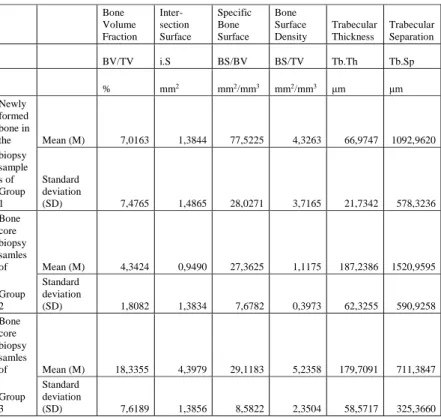
The morphometric parameters of the newly formed bone that surrounded the bone graft particles in Group 1 and the parameters of the native bone of Group 2 and Group 3 were compared. The values that showed significant difference between the three sets of data and the results of the statistical analysis are presented in Table 3.
Table 3. P values are presented with a cross if the statistical analysis used was the Kruskal-Wallis test. Elsewhere, the statistical analysis used was the analysis of variance (Oneway ANOVA) and the Bonferoni test.
Bone Volume Fraction
Inter- section Surface
Specific Bone Surface
Bone Surface Density
Trabecular Thickness
Trabecular Separation
BV/TV i.S BS/BV BS/TV Tb.Th Tb.Sp
% mm2 mm2/mm3 mm2/mm3 μm μm
Newly formed bone in
the Mean (M) 7,0163 1,3844 77,5225 4,3263 66,9747 1092,9620 biopsy
sample s of Group 1
Standard deviation
(SD) 7,4765 1,4865 28,0271 3,7165 21,7342 578,3236
Bone core biopsy samles
of Mean (M) 4,3424 0,9490 27,3625 1,1175 187,2386 1520,9595 Group
2
Standard deviation
(SD) 1,8082 1,3834 7,6782 0,3973 62,3255 590,9258
Bone core biopsy samles
of Mean (M) 18,3355 4,3979 29,1183 5,2358 179,7091 711,3847 Group
3
Standard deviation
(SD) 7,6189 1,3856 8,5822 2,3504 58,5717 325,3660
12
Bone Volume Fraction
Inter- section Surface
Specific Bone Surface
Bone Surface Density
Trabecular Thickness
Trabecular Separation
P-value of significanc e between Group 1 and 2
no statisticall y significant difference between the groups
no statisticall y significant difference between the groups
+ 0,000+
no statisticall y significant difference between the
groups+ 0,0000 no statisticall y significant difference between the groups
P-value of significanc e between Group 1
and 3 0,007+ 0,002+ 0,000+ no statisticall y significant difference between the groups
+ 0,000
no statisticall y significant difference between the groups
P-value of significanc e between Group 2
and 3 0,000+ 0,000+ no statisticall y significant difference between the groups
+ 0,000+
no statisticall y significant difference between
the groups 0,001
Trabecular Number
Closed Porosity
Open Porosity
Total porosity
Euler- Number
Connec- tivity
Tb.N Po(cl) Po(op) Po(tot) EuN Conn.
1/mm % % %
Newly formed bone in
the Mean (M) 1,0088 3,6160 92,7568 92,9837
- 9073,0000
11240,375 0 biopsy
sample s of Group 1
Standard Deviation
(SD) 0,8720 4,6563 7,7007 7,4765 9415,9846 9341,8397 Bone
core biopsy samles
of Mean (M) 0,2408 0,0866 95,6531 95,6576
-
1831,3333 2529,8333 Group
2
Standard Deviation
(SD) 0,1082 0,0938 1,8120 1,8082 2504,6223 3369,2606
13
Trabecular Number
Closed Porosity
Open Porosity
Total porosity
Euler- Number
Connec- tivity Bone
core biopsy samles
of Mean (M) 1,0967 0,0926 81,6460 81,6645
-
4347,9167 5789,0833 Group
3
Standard Deviation
(SD) 0,5309 0,0716 7,6286 7,6189 3935,4133 4371,3358
P-value of significanc e between Group 1 and 2
no statisticall y significant difference between the groups
+ 0,000+
no statisticall y significant difference between the groups +
no statisticall y significant difference between the groups
+ 0,020 0,005+
P-value of significanc e between Group 1 and 3
no statisticall y significant difference between the groups
+ 0,000+ 0,009+ 0,007+
no statisticall y significant difference between the groups
no statistically significant difference between the groups +
P-value of significanc e between Group 2
and 3 0,000+
no statisticall y significant difference between the groups
+ 0,000+ 0,000+
no statisticall y significant difference between
the groups 0,021+
5. Discussion
Histological analysis of the bone core biopsy samples obtained from the augmented sites of the test group concluded that in 8 out of the 12 cases, particles of the bone substitute material were surrounded by newly formed trabecular bone. However, in the remaining 4 cases, the particles of the porcine xenograft were encapsulated by granulation tissue, with no sign of inflammation or foreign body reaction.
14
Hindered healing of the augmented bone might be due to the unfavourable morphology of the grafted socket. Loss of the buccal wall creates a three-wall bone defect. The presence of more bony walls allows more predictable healing of the socket.
Regeneration in a thick four-wall extraction socket most often results in a ridge with abundant height and width for implant placement without the use of biomaterials or regeneration techniques. If a three-wall bone morphology is observed after tooth removal, bone augmentation techniques are to be utilised to obtain sufficient bone volume for implant placement. When the buccal bone plate is missing, short, or thinner than 1 mm, the conditions favour repair rather than regeneration. In these cases, bone loss due to resorption is inevitable without the use of ridge preservation techniques.
Histological findings in the test group at re-entry suggest that in the coronal part of the grafted socket particles of the xenograft failed to integrate in bone tissue despite using a collagen membrane as cell occlusive barrier. Instead after a 6 month healing period particles of the graft were encapsulated by connective tissue with no sign of inflammation or foreign body reaction. The coronal part of the socket is the furthest from the bony walls. During healing the bony walls of the alveoli are the source of osteoprogenitor cells which might explain the absence of bone surrounding the graft particles in the coronal part of the sockets. Our findings of the apico-coronal inhomogeneity of tissues surrounding the particles of the bone substitution materials in socket preservation are consistent with the findings of previous studies.
Various morphometric parameters (Trabecular Number Tb.N, Trabecular Bone Pattern Factor Tb.Pf, Structure Modell Index SMI, Number PoN[cl], Volume PoV[cl], and Surface PoS[cl]
of Closed Pores) showed statistically significant differences between the newly formed bone and the particles of the bone graft material within the augmented sites. Statistically
15
significant differences in these parameters can be explained by the appearance of the two structures. The structure of the bone graft particles consists of separate sphere like objects, while structure of the newly formed bone consists of rod and plate like trabecules in complex interconnection. Neither bone, nor graft particles are characterised by closed porosity. However, in those instances where the newly formed bone completely surrounds a bone graft particle segmentation of the image results in a closed pore within the newly formed bone structure.
Various morphometric parameters (Bone Volume Fraction BV/TV, Intersection Surface i.S, Bone Surface Density BS/TV, Trabecular Number Tb.N, Trabecular Bone Pattern Factor Tb.Pf) showed statistically significant differences between the augmented bone of the test group and the native bone of the negative control group. The microarchitecture of the preserved sockets is different from the sockets of Group 3. Sockets of both groups were three wall defects and covered by the flap. The microarchitecture of the immediate GBR treated sockets resembled that of the sockets of the positive control group.
Therefore, microCT analysis confirms the osteoconductive property of the xenograft.
Various morphometric parameters (Bone Volume Fraction BV/TV, Intersection Surface i.S, Specific Bone Surface BS/BV, Bone Surface Density BS/TV, Trabecular Number Tb.N, Trabecular Thickness Tb.Th, Trabecular Separation Tb.Sp, Open Po[op], Closed Po[cl], and Total Porosity Po[tot], Connectivity Conn. and Euler Number Eu.N) showed statistically significant differences between the newly formed bone within the augmented bone of the test group and the native bone of the control groups. These results indicate that particles of the bone graft material interfere with the natural bony
16
healing. This raises the question whether these differences shall disappear with time due to remodelling.
Various morphometric parameters (Bone Volume Fraction BV/TV, Intersection Surface i.S, Specific Bone Surface BS/BV, Bone Surface Density BS/TV, Trabecular Number Tb.N, Trabecular Separation Tb.Sp, Open Po[op], and Total Po[tot]
Porosity, Connectivity Conn.) showed statistically significant differences between the positive and negative control groups.
These findings indicate that the bone wall configuration of the defects and whether a flap is elevated and mobilized to allow for closure of the socket directly affects the microarchitecture of the bone. Complete remodelling occurs during socket healing only if the socket has a four bone wall morphology after tooth removal.
6. Conclusion
6.1. Conclusions based on the histological and qualitative microCT findings
1. The porcine xenograft integrated in the augmented sites with no sign of inflammation or foreign body reaction.
2. In 2/3 of the cases particles of the xenograft integrated in the augmented sites with formation of bony trabeculae and marrow spaces. However, in 1/3 of the cases particles of the xenograft were encapsulated in connective tissue.
3. In the apical and central parts of the socket the bone graft material integrated with newly formed bone. However, in
17
the coronal part of the socket particles of the xonograft were encapsulated in connective tissue.
6.2.Conclusions based on Bone Microarchitecture Analysis
4. Various morphometric parameters (Tb.N, Tb.Pf, SMI, PoN[cl], PoV[cl], PoS[cl]) showed statistically significant differences between the newly formed bone and the particles of the bone graft material within the augmented sites. Therefore, the microarchitecture of the two structures are different, which can be explained by the different appearance of the newly formed bone and bone graft particles. This conclusion is important to be able to explain further findings
5. Various morphometric parameters (BV/TV, i.S, BS/TV, Tb.N, Tb.Pf) showed statistically significant differences between the augmented bone of the test group and the native bone of the negative control group. Therefore, microCT analysis confirms the osteoconductive property of the xenograft.
6.a. Various morphometric parameters (BV/TV, i.S, BS/BV, BS/TV, Tb.N, Tb.Th, Tb.Sp, Po[op], Po[cl], Po[tot], Conn., Eu.N) showed statistically significant differences between the newly formed bone within the augmented bone of the test group and the native bone of the control groups. These results indicate that particles of the bone graft material interfere with the natural bony healing.
6.b. Various morphometric parameters (BV/TV, i.S, BS/BV, BS/TV, Tb.N, Tb.Sp, Po[op], Po[tot], Conn.) showed
18
statistically significant differences between the positive and negative control groups. These findings indicate that the bone wall configuration of the defects and whether a flap is elevated and mobilized to allow for closure of the socket directly affects the microarchitecture of the bone.
6.3. New results
1. In 2/3 of the cases particles of the xenograft integrated in the augmented sites with formation of bony trabeculae and marrow spaces. However, in 1/3 of the cases particles of the xenograft were encapsulated in connective tissue. In some cases, if alveolar ridge preservation of three wall sockets is carried out by immediate GBR treatment with the combination of the porcine xenograft and collagen membrane the bone graft material integrates with connective tissue encapsulation.
2. If socket preservation of three wall sockets is carried out with the combination of the porcine xenograft and collagen membrane the particles of the xenograft will integrate with newly formed bone in the apical and central parts of the socket.
In the coronal part of the socket connective tissue encapsulation of the particles can be observed.
3. The microarchitecture of the grafted three bone wall sockets closed with a flap is different from that of the three bone wall sockets treated without GBR and closed with a flap and similar to that of the four bone wall sockets left to heal without flap elevation and closure. Therefore, microCT analysis confirms the osteoconductive property of the xenograft.
19
4. The microarchitecture of the newly formed bone within the preseved three bone wall sockets is different from that of the three bone wall sockets treated without GBR and that of the four bone wall sockets left to heal without flap elevation and closure.
These results indicate that particles of the bone graft material interfere with the natural bony healing.
5. The microarchitecture of the three bone wall sockets treated without GBR and closed with a flap is different than that of the four bone wall sockets left to heal without flap elevation and closure. These findings indicate that the bone wall configuration of the defects and whether a flap is elevated and mobilized to allow for closure of the socket directly affects the microarchitecture of the bone.
7. Publications
7.1. Thesis-related publications
1. Kivovics M. (2013) „Osteobiol” és a Magyarországon alkalmazott csontpótló anyagok (irodalmi összefoglalás). Fogorv Szemle, 106:33- 38.
2. Nemeth O, Kivovics M, Pinke I, Marton K, Kivovics P, Garami M.
Late effects of multiagent chemotherapy on salivary secretion in children cancer survivors. J Am Coll Nutr, 2014. 33:(3):186-9.
3. Kivovics M, Szabó B T, Németh O, Tari N, Dőri F, Nagy P, Dobó- Nagy C, Szabó G. (2017) Microarchitectural study of the augmented bone following ridge preservation with a porcine xenograft and a collagen membrane: preliminary report of a prospective clinical, histological, and micro-computed tomography analysis. Int J Oral Maxillofac Surg, 46(2):250-260.
20 7.2. Other publications
1. Kivovics M, Mihályi Sz, Suba Zs, Gyulai-Gaál Sz. Sövettani vizsgálatok az ambuláns szájsebészeti gyakorlatban. Fogorv Szemle, 2012. 9-12.
2. Németh O, Kivovics M, Garami MI. Late Oral Consequences of Chemotherapy in Children Cancer Survivors. J Dent Res, 2013. 92:
Paper 55.
3. Strasser-Solti F, Makra D, Kivovics M, Kivovics P. Cystaszűkítő készülékek formái és alkalmazási lehetőségei. Magyar Fogorvos, 2016. (6): 298-300.
4. Bécser J, Kiss D, Kivovics M. Parodontitis és periimplantitis diagnosztizálása az aktivált mátrix-metalloproteináz-8 (aMMP-8) kimutatására alkalmazott teszt segítségével, és ennek népegészségügyi jelentősége. Magyar Fogorvos, 2016. 25:(3) 142- 144.