Novel methods for evaluation of osseointegration, bone regeneration and oral flap management
PhD thesis
Dr. Sándor Farkasdi
Doctoral School of Clinical Medicine Semmelweis University
Supervisor: Gábor Varga, D.Sc
Official reviewers:
Kinga Laczkóné Turzó, Ph.D József Tímár, MD, D.Sc
Head of the Final Examination Committee:
Tibor Fábián, DMD, Ph.D
Members of the Final Examination Committee:
Senior Zoltán Rakonczay, D.Sc Árpád Joób-Fancsaly, DMD, Ph.D
Budapest, 2019
1
TABLE OF CONTENTS
LIST OF ABBREVIATIONS 4
1. INTRODUCTION 5
1.1 Intraosseous implants for tooth replacement and their regulation 6
1.2 Bone healing and regeneration 8
1.3 Osseointegration 12
1.4 Dental implantology 15
1.5 Preclinical studies to provide a basis for human bone regeneration and
osseointegration 23
1.6 Surgical wound closure in humans 27
2. OBJECTIVES 29
3. MATERIALS AND METHODS 30
3.1 The refinement of original in vivo rat tail implant model for quantitative and
qualitative monitoring of osseointegration 30
3.1.1 Development of an implant design that is suitable for the investigation of the effect of surface modifications to osseointegration 30 3.1.2 Development of complex biomechanical evaluation by the combination of resonance frequency analysis and pull-out techniques 34
3.1.2.1 Validation of the individually developed connection between SmartPeg
and customized implant using RFA 34
3.1.2.2 Evaluation of implant-hook connection during pull-out tests 36 3.1.2.3 Evaluation of three implant geometries with RFA 37 3.1.3 Combination of biomechanical evaluations with structural tests for reliable and complex monitoring of the osseointegration process using
“Direct OSSI” model 38
3.1.3.1 Experimental animals for “Direct OSSI” model 38
3.1.3.2 Mini-implant design 39
3.1.3.3 Surgical procedure in “Direct OSSI” experimental model 40
3.1.3.4 Postsurgical treatment 41
2
3.1.3.5 Sample harvesting and evaluations for “Direct OSSI” experimental
model 42
3.2 Development of a preclinical model for quantitative, qualitative
monitoring of the regeneration of multiple bone defects and the integration of simultaneously-placed several implants perpendicular to the rat tail 47
3.2.1 Ex vivo developments for rat caudal vertebrae bone drilling to create
transversal defects 47
3.2.2 Experimental animals for “BD OSSI” and “Gap OSSI” models 48
3.2.2.1 Experimental setup for “BD OSSI” model 49
3.2.2.2 Experimental setup for “Gap OSSI” model 50
3.2.3 Surgical interventions 51
3.2.3.1 Surgical procedure of the “BD OSSI” 51
3.2.3.2 Surgical procedure of the “Gap OSSI” 51
3.2.4 Postsurgical treatment 52
3.2.5 Sample harvesting and evaluations for “BD OSSI” and “Gap OSSI” 53 3.2.5.1. Radiological visualization with micro-CT 53
3.2.5.2 Histomorphological visualization 53
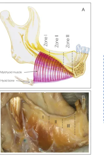
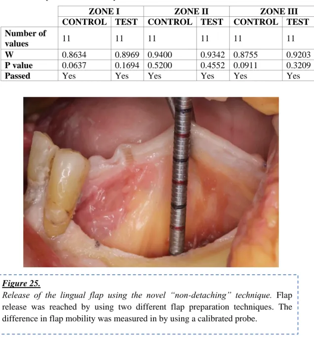
3.3 Evaluation of the effectiveness of two oral different flap designs for the improvement of lingual flap release, applying fresh human cadaver heads 54
3.3.1 Sample and randomization 54
3.3.2. Flap management technique 54
3.3.3 Outcome measurements 56
3.4 Statistical analysis of in vitro and in vivo evaluations 57 3.4.1 Statistical analysis for the refinement of original in vivo rat tail implant
model 57
3.4.1.1 Statistical analysis of in vitro RFA stability results measured in PUF blocks 57 3.4.1.2 Statistical analysis of biomechanical and structural evaluation of
osseointegration in different endpoints using “Direct OSSI model” 58 3.4.2 Analysis of self-regeneration of the bone in “BD OSSI” and
osseointegration of customized implants in “Gap OSSI” model 58
3
3.4.3 Statistical analyses for the evaluation of the differences in flap mobility
after two flap preparation techniques 58
4. RESULTS 59
4.1 Quantitative and qualitative monitoring of osseointegration using “Direct OSSI” model to refine the original in vivo rat tail implant model 59
4.1.1 Validation measurements of an implant design suitable for the
investigation of the effect of surface modifications to osseointegration in rat
tail 59
4.1.2 Development of complex biomechanical evaluation by the combination of resonance frequency analysis and pull-out techniques 60 4.1.3 Complex monitoring of osseointegration with biomechanical and structural tests; assessment of improved surgical conditions and
postsurgical care 62
4.1.3.1 Biomechanical evaluation of implant osseointegration 62 4.1.3.2 Structural evaluation of implant osseointegration 63 4.2 Development of “BD OSSI” and “Gap OSSI” experimental models 67 4.3 Ex vivo evaluation of oral-surgical flaps mobility following “non-
detaching” and “detaching” techniques for the mylohyoid muscle 68
5. DISCUSSION 71
5.1 Quantitative and qualitative monitoring of osseointegration using the
“Direct OSSI” rat tail implant model 71
5.2 Importance of the newly developed “BD OSSI” and “Gap OSSI”
experimental models 77
5.3 Importance of differences between “non-detaching” and “detaching”
techniques for the mylohyoid muscle 80
6.CONCLUSIONS 82
7. SUMMARY 84
8.ÖSSZEFOGLALÓ 85
9. BIBLIOGRAPHY 86
10. LIST OF OWN PUBLICATIONS 118
11. ACKNOWLEDGEMENTS 120
4
LIST OF ABBREVIATIONS
ALP – alkaline phosphatase ANOVA – analysis of variance
ASTM – American Society for Testing and Materials
BD – bony defect
BD OSSI – rat tail based experimental model for bone regeneration evaluation BIC– bone to implant contact
BV/TV – bone volume/tissue volume CBCT – cone-beam computed tomography
CSD – critical size bone defect cPTi – commercially pure titanium D – density
DALY – disability-adjusted life year Direct OSSI - rat tail based experimental model for evaluation direct ossteogenesis
Gap OSSI - rat tail based experimental model for evaluation distant osteogenesis around implants
GBR – guided bone regeneration GDP – gross domestic product HU – Hounsfield units
IF – Immunofluorescence i.m. – intramuscular i.p. – intraperitoneal IT – insertion torque
i.S/TS – intersection surface/tissue surface
ISQ – implant stability quotient
Micro–CT – microcomputer tomography
mPa – millipascal N – Newton n – number
Ncm – Newton centimeter
OSSI – osseointegration experimental model
p – probability value
PTS - Pammer torque system PUF – polyurethane blocks
RFA – resonance frequency analysis ROI – region of interest
RP – retromolar pad
RPM – revolutions per minute Runx2 – Runt-related transcription factor 2
Ti – titanium TV – tissue volume SD – standard deviation
SEM – standard error of mean V – version
WHO – World Health Organisation
5
1. INTRODUCTION
The function of lost tissues can be restored by three fundamentally different methods:
reparation, replacement, regeneration. This PhD thesis addresses the open questions of tooth replacement with intraosseous implants and bone regeneration at the preclinical level.
Nowadays, in practical healthcare, the regeneration of simple tissues by methods such as transplantation is more and more feasible. But the engineering of complex organs consisting of multiple tissues is still restricted in most medical professions, including dentistry and oral and maxillofacial surgery.
In oral and maxillofacial medicine and dentistry, tooth disorders occur the most frequently. Tooth loss is the most frequent disease which affects a considerable rate of the population of the world (Kassebaum et al., 2017). The disorder which the most frequently affects the erupted part of the tooth is dental caries, which is primarily an infectious disease. Caries is a mechanical and chemical breakdown of teeth due to acids and bacteria (Silk, 2014). Its complications can include the inflammation process of the tooth and the periodontal tissue, tooth loss, or even abscess formation (Laudenbach et al., 2014). At the end of the twentieth century, the incidence and prevalence of caries showed a decreasing tendency in countries with high gross domestic product (GDP). According to World Health Organisation (WHO) data, this tendency was a consequence of governmental regulations regarding caries in these countries but the incidence is still very high (Kassebaum et al., 2017). The biggest side effect of tooth extraction/tooth loss is that it has a significant impact on functionality, aesthetic, and social status of people affecting the quality of their lives (Batista et al., 2014; Gerritsen et al., 2010). According to recently published data, tooth loss is responsible for 7.6 million disability-adjusted life years (DALY) (Kassebaum et al., 2017).
The incidence and prevalence of periodontal diseases are also very high.
Accordingly, half of the US population over the age of 30 has periodontitis based on data published by the “Center for Disease Control and Prevention” (Eke et al., 2015).
Neglected oral hygiene, inadequate nutrition, smoking and stressful lifestyle are important triggers for the development of periodontal disease (Albandar, 2014; Kinane et al., 2017; Kwon et al., 2014; Larsen et al., 2017). During the development of periodontal
6
disease, inflammation causes massive, irreversible tissue destruction in the alveolar bone, in the periodontal ligament and in the cement tissue and even in the gum (Kinane et al., 2017). The pathological cascades can lead to tooth loss in untreated cases. Despite the improvements in the quality of dental healthcare, policies to expand public health insurance coverage, increasing accessibility to dental clinics and advanced treatments, the rate of tooth extractions are continuously increasing due to periodontal disease (Lee et al., 2017). Periodontitis itself causes serious aesthetic and functional problems, and may also be associated with systemic diseases such as diabetes (Demmer et al., 2010), cardiovascular disorders (through the stimulation of the development of atherosclerosis) (Lockhart et al., 2012) (Humphrey et al., 2008; Iniesta et al., 2012), with adverse pregnancy (Chambrone et al., 2011) and immunosuppressive conditions (stress, HIV infection) (Kinane et al., 2001). Beside the socio-economic impact, numerous pieces of evidence demonstrate that chronic periodontitis can be a risk factor for cancer as well (Fan et al., 2016; Michaud et al., 2013; Michaud et al., 2007; Michaud et al., 2016;
Michaud et al., 2008; Ogrendik, 2017).
As a result of both dental caries and periodontal inflammation, both soft and hard tissue loss will most probably appear. One of the greatest challenges of modern dental and craniofacial research is to restore the function of lost tissues.
1.1 Intraosseous implants for tooth replacement and their regulation
There is a strong need for the replacement of missing teeth. Unfortunately, it is not possible with regeneration yet. Not even the regeneration of separate parts/tissues of the tooth (such as the enamel or dentine) is available for standard clinical practice. The intraosseous implant is the gold standard for the replacement of the extracted tooth. Its application is the major example for reparative medical technology besides fillings and crowns. The clinical success of using artificial intraosseous implants for the rehabilitation of the human body is based on osseointegration (see part 1.3).
Intraosseous implants, which are used in dental practice, are called dental implants.
A dental implant is the root part of the artificial tooth, which is in the bone. The superstructure, which is directly connected to the top of a dental implant, has two parts.
One is the crown and the other is an abutment which connects the crown to the dental
7
implant. These structures can be replaced. But the dental implant of the artificial tooth should remain in the person’s body during the whole of his life (Hof et al., 2014; Korfage et al., 2018).
The clinical efficiency of various dental implants is different (De Angelis et al., 2017; Jimbo et al., 2015; Marcello-Machado et al., 2018; Moraschini et al., 2015). This can be due to the different patient background (patient’s general and local status, habits, additional treatments) (Baig et al., 2007; Candel-Marti et al., 2011; Gelazius et al., 2018;
Javed et al., 2010), surgical protocol (Chrcanovic et al., 2015; Pjetursson et al., 2008; Tan et al., 2008) and loading protocol (Al-Sawai et al., 2016). Also, a significant impact on osseointegration is made by different surface treatments of the dental implants (Calvo- Guirado et al., 2015; Le Guehennec et al., 2007). In term of success and survival rates of implants the surface modifications plays one of the mayor role (Jimbo et al., 2015).
The majority of intraosseous implants are classified as medical devices. In clinical practice, the rules for new medical devices, instruments and methods have been becoming more and more rigorous during the last 100 years. Mostly, new regulations limit the access to new devices on the market based on new findings in clinical experience.
In the European Union, the Medical Devices Directive 93/42/EEC regulates the market for medical devices (EuropeanParliament, 1993). This directives continuously change according to technical innovations and research developments. However, it is still relatively easy to introduce a new medical device into the market. For instance, implant manufacturers still have the possibility to launch new implant products on the market easily by avoiding important preclinical and clinical evaluations. The manufacturer of a dental implant can apply for a declaration of conformity, CE marking and, after successful registration, put the device on the market (based on Directive 93/42/EEC Annex III, module B) (EuropeanParliament, 1993). The actual regulation for the assessment of a medical device does not fully take into account the biological reaction in response to the transplantation of the newly developed product. This is because the current regulation is safety-centered and basically does not put too much emphasis on demonstrating efficiency. Therefore, there is a strong need for a standard evaluation of an upcoming medical device in experimental circumstances.
From the spring of 2020, a more rigorous guideline will come into effect regarding the regulation of new medical devices on the EU market. At present, this new law is in a
8
transitional period. According to Directive 93/42/EEC, the regulations of the medical device industry in Europe, which have been relatively unchanged since the 1990s, will be modified considerably. Incidents at the beginning of the 21st century, including the breast implant crisis (Heneghan, 2012) and the hip replacement problems (Cohen, 2012;
Howard, 2016), have provoked urgent reforms in the regulation of medical devices (French-Mowat et al., 2012; Heneghan, 2012). Accordingly, the post-marketing activities and midterm onsite controls of manufacturing should be significantly improved. Based on the above-mentioned problems, the European Commission have come up with proposals for the regulation of medical devices (EU MDR) (EuropeanParliament, 2017).
The recent problems with breast implants (Poly Implant Prothese (Greco, 2015)) and hip implants (metal on metal hip implants) reflected only a small part of the whole “iceberg”
of necessary recalls from the medical market (Heneghan, 2012; Heneghan et al., 2012).
The purpose of the new regulations is to create better evidence-based control than before. It is worth noting that a really big difference between the current and next regulations will be that the manufacturer will not only have to prove the safety of his/her own medical device but also its efficacy. This will bring the regulation of medical devices closer to drug regulations. This target can be achieved if scientifically proved checklists of preclinical evaluations have been introduced. Scientific authorisation can be done for certain medical devices only if the particular device has been validated with well- described and standardly reproducible experimental models, experimental work to ensure repeatability. To prove the efficacy and compare it with similar medical devices, standardised screening experimental models will be needed. Moreover, often there is no clinical evidence of which commercially available implant and which surface modification of implant is better. For that, preclinical, commercially independent evaluations will be needed. This is also important for a financially more predictable social insurance support in case of treatments for possible complications.
1.2 Bone healing and regeneration
One of the thoroughly investigated tissues for regeneration is the bone (both in general medicine and dentistry). Firstly, this is because bone regeneration is a physiological process that leads to bone union (Tsiridis et al., 2007). It can be part of the repair process
9
after a trauma (such as a fracture or bone grafting) or the process of skeletal development, as well as continuous bone remodelling throughout human life. Secondly, bone regeneration is the second most frequent tissue transplantation in clinical practice after blood transfusion (Campana et al., 2014). Thirdly, the bone has become the most frequent hosting tissue for reconstructive intraosseous implants. Natural bone regeneration (after fracture) and man-made bone regeneration (after bone-grafting procedures and intraosseous implant placement) are among the few processes in the human body which can heal without the formation of a fibrous scar tissue (Marsell et al., 2011). But errors may happen during this process that can generate a delay in healing, and even develop pseudo-joints or non-unions (Marsell et al., 2010).
The process of normal bone-healing after a trauma, fracture, intraosseous implant placement and grafting procedure usually tends to repeat the process of bone formation (Caplan, 1987; Marzona et al., 2009; Shah et al., 2019; Shapiro, 2008; Vortkamp et al., 1998). Bone-healing in the damaged site involves the cooperation of inflammatory cells, stem cells, osteoblasts, chondrocytes, osteoclasts, and endothelial cells with surrounding pericytes, cytokines and growth factors (Frost, 1989a, 1989b; Marsell et al., 2011). As a result of the reunion of bone after fracture (Marsell et al., 2011) and even after bone regeneration, osteon formation takes place (Fernandez et al., 2014; Makihara et al., 2018).
The fundamental unit of bone is the osteon (Haversian systems). Bone remodelling goes parallel to the streamline of the osteon system. When the bone equilibrium is breached, bone-healing starts immediately. Irrespective of the cause of the breach, the immune response starts with angiogenesis and recruitment of progenitor cells. One of the key elements for successful bone-healing is continuous blood supply (Carano et al., 2003).
Regarding the mechanism, bone-healing can follow two pathways (Marsell et al., 2011; Ono et al., 2017). The most common among fractures is indirect bone-healing or secondary bone-healing (Marsell et al., 2011). Indirect or secondary healing happens, when there is a gap and an insufficient stability of the fractured edges (Marsell et al., 2011). During indirect healing mostly endochondral ossification takes place (Marsell et al., 2011). There are three phases of secondary bone-healing (Sfeir C, 2005). The first one is the inflammatory phase, which ends up in granulation tissue formation (Schell et al., 2017; Sfeir C, 2005). The second phase is the reparative phase when first fibrocartilage callus and then bony callus develops (Marsell et al., 2011; Sfeir C, 2005).
10
During this process the hyaline cartilage and the woven bone is replaced by lamellar bone (Marsell et al., 2011; Sfeir C, 2005). The third step is bone remodelling and addition of compact bone to the mechanical stressors. Finally, the fractured site is totally remodelled by the balanced work of osteoclasts and osteoblasts into a new shape, which almost repeat the original shape and strength (Marsell et al., 2011; Sfeir C, 2005).
The second type of bone-healing after bone integrity impair is referred to as direct bone-healing (Marsell et al., 2011; Ono et al., 2017). Direct healing happens when there is no gap between the fractured edges at all (Marsell et al., 2011) or when the rigid fixation of the fractured fragments is done (Marsell et al., 2011) or in case of alveolar bone-healing after tooth extraction (Vieira et al., 2015), implant placement and bone-grafting (Colnot et al., 2007; Shah et al., 2019). During the direct healing of the bone intramembranous ossification takes place (Marsell et al., 2011). Bone fragments are joined by continuous ossification avoiding woven bone or cartilaginous bone formation, and the remodelling of the newly-formed bone happens to a tiny extent (Shapiro, 2008).
Blood clots play an important role in the initial phase of bone-healing (Schell et al., 2017). Further on, sufficient blood supply and a gradual increase in mechanical stability are needed for an uncompromised remodelling phase (Einhorn et al., 2015;
Marsell et al., 2011; Runyan et al., 2017). Accordingly, the angiogenesis goes parallel with all phases of fracture healing. At the end of the bone healing, the Haversian system is re-established by the creation of remodelling units called «cutting cones» (McKibbin, 1978).
The difference between fracture-healing and bone-healing after tooth extraction is that in case of fracture-healing (closed fracture) the endochondral ossification happens in sterile conditions (Vieira et al., 2015). In case of alveolar healing intramembranous ossification happens with constant microbial challenge from the oral cavity (Vieira et al., 2015). The same microbial challenge influences the osseointegration of dental implants after implantation. There are differences between intramembranous ossification and new bone formation after tooth extraction and implantation (Shah et al., 2019). They are due to the presence of titanium, a foreign body between the “fractured” edges of the bone. By implant insertion the fractured gap is considerably reduced (Shah et al., 2019).
Accordingly, the area needed for regeneration decreases. The titanium surface modifies local cell response in the site of the fracture (Schwarz et al., 2007; Shanbhag et al., 2015).
11
The titanium and titanium alloys are unique, because when they are exposed to air or water, a spontaneous formation of native titanium oxide (TiO2) layer (5-10 nm) occurs (Shah et al., 2019; Wang et al., 2016). This TiO2 layer prevents the release of metallic ions from the implant surface (Okazaki et al., 2005) and also prevents adverse reactions of the body (Tengvall et al., 1992; Wang et al., 2016). Accordingly, the TiO2 layer is needed for successful osseointegration and becomes a biologically inert metal (Wang et al., 2016). We know that changing the properties of titanium oxide surface leads to biocompatibility having an effect on the titanium intraosseous implant (Wang et al., 2016).
As a result of osteotomy, haemostasis and blood clot formation happen in the presence of bone debris (Shanbhag et al., 2015). After osteotomy, local hypoxia starts due to the disruption of the local vasculature (Potier et al., 2007; Shanbhag et al., 2015). After surgical placement of the implant oxidization continues and the hypoxic environment can be enhanced. The hypoxic environment induces angiogenesis. Osteogenesis starts simultaneously with angiogenesis (Mamalis et al., 2013). The complete understanding of osseointegration remain unclear (Shah et al., 2019). For a clear analysis of the effect of the different surface modifications, standardised experimental models are needed, which allow the evaluation of pure biological integration without any additional influence of the geometrical design (threads, holes, self-tapping).
The mechanical strain and the distance between the two fractured edges of the bone are the two crucial factors, which affect the quality of healing after the initial trauma (Claes et al., 1998; Ghiasi et al., 2017). In case of an intraosseous implant, the mechanical strain can lead to micromotion. If micromotion is more than 100-200 m, fibro- osseointegration occurs, which means the failure for the placed implant (Brunski, 1999;
Szmukler-Moncler et al., 1998). The increase in the size of the bone defect directly correlates with a delay in the healing process (Claes et al., 1998). The minimum size of bone defect, which is large enough not to be able to heal spontaneously, is called “critical size bone defect” (CSD) (Agarwal et al., 2015). The size of CSDs is different depending on the location and live specimen (Harris et al., 2013).
After severe bone traumas, complicated tooth extractions can lead to CSD formation; the method for bone reunion can be guided bone regeneration (GBR). The GBR procedure is considered to be one of the most widespread dental surgical procedures for rebuilding the width and the height of the alveolar ridges before implant placement
12
(Elgali et al., 2017; Khojasteh et al., 2017; Windisch et al., 2017). GBR is characterized by using barrier membranes with or without particulated bone grafts or/and bone substitutes (Elgali et al., 2017; Liu et al., 2014). The membrane is mainly applied for eliminating the penetration of non-osteogenic tissues into the grafted area and for space maintenance (Elgali et al., 2017; Liu et al., 2014). Particulated bone-grafting materials are used as scaffolds to facilitate bone formation. These grafts are biodegradable, have no antigen-antibody reaction, and serve as a mineral reservoir (Kumar et al., 2013;
Thrivikraman et al., 2017). Bone-grafting materials are usually classified according to their origin. They are called autografts when harvested from the same individual as the one receiving the graft; allografts when harvested from the same species (i.e. from other humans); xenografts when harvested from a species other than a human; synthetic when produced in laboratory conditions and finally the combination of grafting materials, which are listed above (Campana et al., 2014; S. Titsinides, 2018).
1.3 Osseointegration
The incorporation of titanium implants into the living bone utilises direct bone-healing;
this process is called osseointegration. Osseointegration is defined as the development of a direct structural and functional contact between the artificial implant surface and the living bone tissue (Branemark, 1983). Furthermore, the term also refers to the process of forming this direct fixation which has a high dependency on the preceding surgical procedure and preoperative circumstances (Trisi et al., 2009). The process of osseointegration is defined as bone growth or bone bonding from the broken ends of the drilled site to the artificial implant surface without any intermediate fibrous tissue formation. The osseointegration of implants has further key steps: inflammatory phase, proliferative phase and maturation phase. After the osseointegration, bone remodelling happens lifetime. The initial tissue response around the inserted implant is the inflammatory response involving the release of growth factors and cytokines (Bielemann et al., 2018; Kuzyk et al., 2011), which lasts for the first three days (Bielemann et al., 2018; Kuzyk et al., 2011). As a result of the initial tissue response, extracellular matrix and haematoma are formed (Davies, 2003). Then the process of adhesion and aggregation leads to a fibrin matrix formation (Davies, 2003; Kuzyk et al., 2011). During the
13
osseointegration process the implant surface acts as biomimetic scaffold (Davies, 2003).
This scaffold promotes migration, proliferation and differentiation of cells (Davies, 2003;
Kuzyk et al., 2011). Then angiogenesis around the implant starts in the next four days (Kuzyk et al., 2011). In the meantime, mesenchymal progenitor cell differentiation results in osteoblast formation until the end of the second week (Biguetti et al., 2018). Within this period, osteoblasts form calcified collagen fibers and early mineralisation begins at the bone-implant interface (Biguetti et al., 2018; Kuzyk et al., 2011). As a result, a woven bone is formed (Kuzyk et al., 2011). Finally, the woven bone is transformed to a lamellar bone (Biguetti et al., 2018; Davies, 2003).
Bone-bonding to the implant can be considered to be completed when there is no motion between the bone and the implant at all. After healing, that connection can be loaded (Cochran et al., 1998; Ledermann et al., 1998). Bone healing/regeneration and bone-bonding to the implant surface are somewhat different in various bone types. Bones are classified into three main types which are complementary to each other (Chugh et al., 2013) (Figure 1).
It is essential for dentists to recognize bone types during their clinical practice.
Classification of bones according to density and their typical locations in the jaws are presented in Figure 1.
14 Figure 1.
Description of bone densities based on Lekholm&Zarb, Misch classifications. Visual comparison of different bone densities: A. Description by Lekholm and Zarb based on conventional radiographs and histological components / by Misch based on descriptive morphology combined with clinician’s tactile sensation (Anitua et al., 2015; Blazsek, 2008; Misch, 1989, 1990; Zarb et al., 1985). B. Hounsfield units (HU) (Shapurian et al., 2006; Schreiber et al., 2011; Turkyilmaz et al., 2008). Different bone densities of edentulous jaws can be assigned to different Hounsfield units (HU) (Shapurian et al., 2006;
Turkyilmaz et al., 2008) C. Most frequent localisations in adults. D. Bone quality classification and visualisation according to Lekholm & Zarb (1985) based on conventional radiographs. E. Bone quality classification and visualisation by Misch (1988) based on macroscopic cortical and trabecular bone characteristics divided into four types. F. Screenshots from CBCT viewer program (Osirix, Pixmeo SARL) of horizontal cross-sections of the human mandible and maxilla showing the Hounsfield units (from the left to the right: inter-foraminal region of the mandible, lateral zone of the mandible, frontal area of the maxilla, lateral area of the maxilla, tuberosity area of the maxilla).
15
Osseointegration comprises two parallel processes: contact and distance osteogenesis, which lead to new bone formation around the titanium implants (Davies, 2003; Mavrogenis et al., 2009). Contact osteogenesis is the new bone formation on the implant surface, which is to be colonized by bone cells before bone matrix formation (Davies, 2003; Mavrogenis et al., 2009). Contact osteogenesis is also called “de novo bone formation” (Davies, 2003). Distance osteogenesis is the process of new bone formation on the surface of the old hosting bone (Davies, 2003; Mavrogenis et al., 2009).
Both types of osteogenesis result in a direct bone to implant contact (Sivolella et al., 2012). The process of osseointegration starts when the primary stability of the implant is achieved by mechanical fixation (Berglundh et al., 2003). Then bone regeneration and remodelling proceed continuously, which finally leads to a rigid and stable fixation of the implant into the surrounding bone tissue. After the initial bone-healing around titanium implants, bone remodelling is practically lifelong (Haga et al., 2009). In spite of continuous efforts, the course of osseointegration, bone remodelling and regeneration around the implants have not yet been fully understood (Pearce et al., 2007; Yin et al., 2016). To more extensively investigate this complex process, there is a need for developing reliable and reproducible preclinical and clinical methods.
On the whole, bone regeneration and implant integration depend on many factors.
The main factors are the location of the defect, its extent, the mobility of edges, the presence or absence of infection, general diseases of the patient and the types of used reconstructive materials.
1.4 Dental implantology
Dental implant placement is one of the most common procedures in oral healthcare. At present, there are only a few absolute contraindications for implantation. They include intravenous bisphosphonate treatment during cancer therapy (for instance radiotherapy protocols in the head or neck region); patients who are unable to comprehend dental treatment logically; immunosuppressed conditions (when the total white blood count falls below 1500–3000 cells/mm3); acute bleeding (a loss of 500 mL of blood); one month after repair of cardiac or vascular defects, and less than one year since myocardial infarction or a cerebrovascular accident (Gomez-de Diego et al., 2014; Hwang et al.,
16
2006). Indeed, oral bisphosphonate intake seems to be an important contraindication for treatment with dental implants (Gomez-de Diego et al., 2014). Also, relative contraindications for dental implantations are adolescence, ageing, osteoporosis, smoking, diabetes, human immunodeficiency virus, cardiovascular disease, hypothyroidism and interleukin-1 genotype (Hwang et al., 2007).
The crucial element for intraosseous implant placement is its integration into the living tissues, which is osseointegration. Since PI Branemark introduced osseointegration as a rigid fixation of an implant in bone tissue more than half-century ago (Branemark et al., 1969), numerous in vitro, in vivo preclinical and clinical studies have been carried out to investigate this process.
The proper planning of implant placement procedure is a key factor for the success of the procedure. There are different bone diagnostic approaches for planning the surgical procedure of implantation, such as palpation bone sounding, x-ray evaluation, planning the positioning of the implant digitally or conventionally (Anwandter et al., 2016; Greenberg, 2015; Katsoulis et al., 2009). Based on x-ray diagnostics, the clinician can first choose the most appropriate position of the implant surgically and prosthetically (Karami et al., 2017; Katsoulis et al., 2009). Second, the dentist can modify the time needed for osseointegration by considering the fact that the higher the primary stability, the faster the secondary stability is achieved (Anitua et al., 2015; Javed et al., 2013). But one always has to keep in mind that primary stability has to be sufficient to avoid micromotion (Javed et al., 2013) but not large enough to prevent bone resorption of the hosting bone (Wang et al., 2017). Beside knowing the parameters of the implant site, the surgeon should know the difference in the behavior of different implant geometries in various bone conditions.
The major bone condition, which directly influences implant stability is bone density (Merheb et al., 2016). Based on this, recommendations exist for which density which implant should be inserted. As it was described above, there are three main bone density classifications (Lekholm U, 1985; Misch, 1990; Schreiber et al., 2011; Shapurian et al., 2006). The classification of density is relative because in humans there are variations of densities even within one region of interest (Figure 1.F).
Clinically there is a need to choose the proper implant geometry for the planned, specific implant site. Only sufficient primary stability guarantees a high resistance of the
17
implant in response to external micro-movements. The local milieu of the hosting implant is the bone bed/implant site. The bone bed is one of the most influencing factors for primary stability (Alsaadi et al., 2007). Primary stability is the mechanical retention of the implant in the bone bed without mobility after implantation (Sennerby et al., 1998).
Primary implant stability during the implant placement is documented as peak insertion torque (IT) (Alsaadi et al., 2007; Meredith, 1998). Sufficient insertion torque prevents the appearance of micromotion (Trisi et al., 2009). Micromotion, which is more than 100- 200 m, can lead to fibro-osseointegration, which is actually a failure of the placed implant (Brunski, 1999; Szmukler-Moncler et al., 1998). The peak insertion torque is influenced by macro-, as well micro-designs (Dos Santos et al., 2011). Macro-design includes implant body shape, thread intensity (pitch) and thread geometry (Gehrke et al., 2015). Surface modifications are considered to be the micro-design variations of implants. There is a generally accepted rule that the insertion torque predicts the outcome of implantation and determine submerged healing time (Walker et al., 2011). There are factors, which influence the longitudinal success rate of implants. Endogenous factors include bone density, cortical bone thickness and osseointegration. Exogenous factors include thread pitch, thread depth, the diameter of implant neck and body size (Cheng et al., 2015).
Nowadays the number of implant factories is higher than 200 worldwide (Yakir).
Each implant factory has a couple of implant systems. As a result, there are thousands of clinically available implant systems. The majority of dental intraosseous implants share the characteristics that they have threads to reach primary mechanical retention.
Modern radiological pre-surgical diagnostic methods such as cone-beam computed tomography (CBCT), provide three-dimensional morphologic analysis of the cortical and trabecular alveolar bone of the hosting side of dental implants (Salimov et al., 2014). Based on well-described clinical protocols, a proper prognosis of primary implant stability can be estimated before surgery (Cortes et al., 2015; Dorogoy et al., 2017; Salimov et al., 2014; Wada et al., 2016). By receiving all data from x-ray analyses, the proper implant geometry, drilling protocol can be planned. The images provided by CBCT give very precise data (Anderson et al., 2014). Based on these precise images, the exact copy of the clinical reality to prepare surgery can be printed in 3D (Huang et al., 2016; Shui et al., 2017). Having a 3D model before surgery, the surgical procedure can
18
be improved in accuracy, time and predictability. Recently published data on intra- operatory bone density measurement system allow to measure the density with a special probe connected with a surgical/implant motor (Di Stefano et al., 2015; Iezzi et al., 2015).
This bone density system can be applied using the 3D printed copies of the planned surgical site. For this, the surgeon can receive all the relevant information before surgery and plan the procedure properly. The motor is capable of measuring instantaneous torque.
This torque is a function of the friction exerted by the bone walls of the implant bed. The average torque along implant bed significantly correlates with the bone density of the surrounding bone wall (Di Stefano et al., 2015; Iezzi et al., 2015).
Apart from primary stability, the success of the osseointegration process depends on a number of factors (Figure 2). It is strongly influenced by the performed medical procedure and the selected implant. The important factors here are the applied implant surface modifications (Campana et al., 2014), the type of implant placement immediately after tooth extraction or later, conventionally (Pal et al., 2011) and bone debris (Bosshardt et al., 2011). Osseointegration can be influenced by growth hormone supplementation (Abduljabbar et al., 2017), smoking (Ekfeldt et al., 2001) and the patient's medical conditions. For example, AIDS, uncontrolled diabetes mellitus, osteoporosis, corticosteroids, bisphosphonate therapy, collagen disorders and other adverse conditions can strongly influence the initial healing process of the bone (Sakka et al., 2012). The list of important factors affecting primary and secondary implant stabilities are shown in Figure 2.
19
Implant stability is an important indicator of the level of osseointegration.
Osseointegration can be assessed by invasive and non-invasive ways. The invasivity in this classification is distinguished based on the destruction of the bone to implant connection during the analysis or saving it for further analysis. Non-invasive methods include radiological analysis/diagnostics (Atsumi et al., 2007), resonance frequency analysis (RFA) (Huwiler et al., 2007), “damping characteristics” (Cranin et al., 1998) and also the perception of the surgeon (Swami et al., 2016). Radiological analysis is considered to be one of the first and oldest methods for determining implant stability (Atsumi et al., 2007). The main disadvantage of x-ray diagnostics for osseointegration analysis is the metal artifact (Ritter et al., 2014). The 3D x-ray diagnostics with high resolution (such as micro-CT) can eliminate artifacts and provide a more precise morphological picture of the surroundings of the implant (Kang et al., 2015). The most widely available and most investigated methods are RFA and “damping characteristics”
(performed with a “Periotest” device, among others) validations.
Figure 2.
Parameters influencing primary stability of implants.
Factors in this figure are categorized based on whether the clinician can influence them or not. Factors which are painted with red cannot be controlled by the expert and those with green should be controlled by the surgeon. Factors which are labeled with orange depend on the patient as well as on the surgeon.
20
RFA is an easy-to-use and standardly reproducible method. RFA is considered to determine the stability of implants and the time of their loadability. Damping characteristics are not as reliable for the assessment of implant stability (Zix et al., 2008).
Accordingly, this method is not the most suitable approach for analyzing osseointegration. Meanwhile, there are new approaches on the horizon, which are at a developing stage. These methods do not induce the implant with electromagnetic waves, but with an electrically-controlled rod that punctures the implant similarly to “Periotest”.
On the dental market this device is called “Implomates” (Biotech One Inc., San Chung, Taiwan). Another representative of the new methodologies for non-invasive testing of implant stability is based on quantitative ultrasound (Mathieu et al., 2011a, 2011b). This device generates a broadband ultrasonic pulse through the transductor (6-14 MHz) (Zanetti et al., 2018). The perception of surgeons is very subjective and not reproducible.
Thus, implant stability evaluation should not rely on this methodology.
Invasive techniques include pull-out/push-in tests (Blazsek et al., 2009; Brunski et al., 2000; Swami et al., 2016), reverse/removal torque measurement (Carvalho et al., 2010), cutting torque resistance (Friberg et al., 1999), tensional or micromotion test (Chang et al., 2010; Trisi et al., 2011) and histology/histomorphometry (Bernhardt et al., 2012; Bissinger et al., 2017; ISO/TS_22911:2016, 2016). Invasive methods are not applicable for clinical monitoring and diagnostic procedures. Therefore, the refinement of non-invasive methods is of great significance for human application (Davies, 2007;
Rodrigo et al., 2010).
The pull-out test is the most reliable biomechanical test for implant stability evaluation (Bell et al., 2014; Sivolella et al., 2015). This is because the main loading on implants arrives vertically. Accordingly, using pull-out tests with non-threaded implants, we can precisely evaluate the bone anchorage strength to the implant surface (Bell et al., 2014). In studies on the use of non-threaded implants (Nonhoff et al., 2015; Seong et al., 2013; Stubinger et al., 2016) it was shown that the pull-out test is a very reliable method for the follow-up of osseointegration. Reverse torque evaluation is the process of unscrewing the implant from the bone when we can detect the minimally needed level of torque for destroying bone implant contact (BIC) (Atsumi et al., 2007). This method evaluates osseointegration indirectly (Atsumi et al., 2007). The assessment of reverse torque can serve as control for osseointegration at the level of loading the implant. It is
21
suggested that trying to unscrew the implant with 30 Ncm can be an acceptable force for evaluating osseointegration (Simeone et al., 2016). Reverse torque assessments can be done only with threaded implants. Even getting the peak value for destroying the BIC during reverse torque is difficult to translate to any mechanical property. The cutting torque resistance analysis describes the energy needed by a surgical motor for cutting off one unit of bone during implant bed preparation (Atsumi et al., 2007). Consequently, this method allows the analysis of bone density in the hosting area of the implant. The disadvantage of this method is that it strongly depends on the operator and it cannot describe implant stability precisely (Friberg et al., 1995). With tensional tests or micromotion testing we can evaluate the lateral resistance of bone to implant connection (Branemark et al., 1998; Chang et al., 2010). It is also difficult to translate these results to any mechanical property (Chang et al., 2010). Histomorphology is the gold standard and the most descriptive method for bone to implant contact. This method is required in guidelines for morphological assessments (ISO/TS_22911:2016, 2016). For preclinical testing, the combination of both non-invasive and invasive methods could offer good outcome providing a safe basis for clinical application. Accordingly, in the studies of this thesis we have chose RFA and micro-CT x-ray diagnostics from non-invasive methods being the most thoroughly described, reliable non-invasive methods. From invasive methodology, the pull-out test was chosen to be the most appropriate for validating bone to implant contact biomechanically. Finally, histomorphometry was selected, which is the gold standard in the evaluation of osseointegration (ISO/TS_22911:2016, 2016).
The RFA non-invasive approach has already been used in daily clinical practice for more than a decade as the main tool for the clinical decision concerning loading time (Stanley et al., 2017). RFA has been introduced in practical healthcare as a non-invasive method to assess implant stability. There was a huge need to have a simple and reliable evaluation approach for evaluating the stability of implants (Kuchler et al., 2016; Sadeghi et al., 2015). Osstell Ltd. (Osstell AB, Gothenburg, Sweden) introduced this measuring device into the clinical market at the end of the 20th century.
Animal models are indispensable tools to develop better devices for medical application (Spicer et al., 2012; van Griensven, 2015). The currently available methods still need to be improved by reducing the number of experimental animals and increasing
22
reliability (Hartung, 2010; Renaud et al., 2015). Although various animal models have been developed to study osseointegration, there is still a lack of a well-reproducible, relatively inexpensive and reliable model (Pearce et al., 2007; Schmitz et al., 1986).
Particularly, even the currently available ISO guideline (ISO/TS_22911:2016, 2016) for performing a preclinical evaluation of dental implant systems suffers from a lack of biomechanical tests. The present guideline requires only morphological, radiographical and histopathological assessments but does not include any functional investigations of osseointegration (ISO/TS_22911:2016, 2016). This deficiency is clearly due to the lack of reliable, well-developed biomechanical tests for experimental animals.
Most animal models for investigating osseointegration were developed without considering the similarity of bone microstructure of animal and human jaw bones.
Consequently, the thereby achieved biomechanical characteristics may be inappropriate since there are remarkable differences between animal and human bones (Martiniakova et al., 2006). To approach this problem, we searched for a massive, cortical and cancellous bone compartment in rats, suitable for supporting titanium implants. We found that caudal tail vertebrae were constituted by abundant spongiosa, which presented an alveolar structure similar to that of the human mandible. Furthermore, the bone marrow parenchyma is also absent in tail vertebrae, thus having greater similarity to human jaw bone than haematopoietic femur of rats, a commonly used experimental model site (Blazsek et al., 2009). Based on these findings, in our department Blazsek et al. (Blazsek et al., 2009) developed a novel experimental model for the evaluation of osseointegration and bone remodelling around longitudinally placed titanium implants in tail vertebrae and proposed to name it “OSSI” (OSSeoIntegration) model (Figure 3). The original methodology for osseointegration analysis using rat tail vertebrae applied only rough evaluation procedures, which were not very sensitive. Thus, it was important to further develop the OSSI model for the quantitative and qualitative characterization of osseointegration. During further developments of the “OSSI” model we analysed the available implant stability evaluation methods. Accordingly, the most reliable four methods were chosen: RFA, micro-CT, histology and pull-out test. We expected that this would allow us to assess the biological integration of titanium implants with multiple, clinically proven analyses, in contrast with existing experimental setups, where one or two variations are evaluated usually. It is important to use multiple
23
evaluation methods and combining clinically reliable methodology with experimental approaches to improve the reproducibility of experiments.
1.5 Preclinical studies to provide a basis for human bone regeneration and osseointegration
In the field of regeneration and replacement a huge variety of animal models have been used. The preclinical experimental setups have to have to be reasonable and have evidence from human clinical practice. Pre-clinical evaluations would be required before the clinical use of any medical devices (Wancket, 2015). Experimental setup selection includes many factors: costs of acquiring, care for animals, availability, acceptability for the society, tolerance to captivity and ease of housing. Also, there are factors which should be considered during model selection. The first factor is the matching of a model site and human bone according to macro (anatomically) and microstructure (histologically). The second parameter is blood supply. Blood supply is often affected by the macroscopic structure of the bone (in spongiosa vascularisation is higher than in the cortical bone) (Zoetis et al., 2003). The third parameter is the possibility of recruiting control groups for treatments. It is obvious that self-control is much more valuable than using other specimens as controls. The fourth parameter is nutrition and general condition of animals. Nutrition and general condition should be close to human reality, in which we would like to use the medical device further on. The fifth parameter is the age of experimental animals (Meyer et al., 2001). The younger the animals, the faster the healing process (Meyer et al., 2001). The sixth parameter is the extent of intra- and inter-animal differences.
24
Unfortunately, it is difficult to use the human implant size during preclinical evaluation using small animals. But the importance of improving medical devices is essential for screening evaluations. Accordingly, downscaling of the implant size should be done. The main outcomes (biological relevance, bifunctionality and biocompatibility/safety) will also be achieved during the insertion of downscaled intraosseous implants into the small mammals. Also, the use of small animal models will involve much smaller financial investment for primary in vivo evaluation and will provide Figure 3.
Photographic documentation and schematic illustration of rat tail based experimental setup developed by József Blazsek (permitted to be used by József Blazsek) and the image taken by the PhD candidate of the present thesis * (Blazsek et al., 2009).
A. Drills for bone preparation in the classical “OSSI” experimental model. B. The titanium implant with threads used for the classical “OSSI” experimental model. C. Schematic illustration of the longitudinal section of the rat tail vertebra with an inserted titanium implant.
An empty space created for bone neogenesis D. The radiological image from micro-CT analysis showing the empty space around the implant (highlighted with yellow rectangles). The yellow rectangles showed the region of interest for the evaluation of new bone formation. E. The result of the pull-out test (tensional test machine Tenzi TE 18.1; TENZI Ltd., Hungary) was used to detect the absolute force needed for vertical removal of the threaded implant from the bony bed.
F. After pull-out test the bone-implant interface was destroyed due to threaded implant geometry. The scanning electron microscopy image represents threaded titanium implant with fractured bone tissue between the threads after pull-out test for integrated implant.
25
a reliable basis for further in vivo evaluations when using bigger animal models. The methods for the evaluation of the integration process should assess the biological parameters of integration more closely to clinical reality.
Many mammalians (such as sheep, goats, dogs, pigs or rabbits) are suitable for testing bone regenerative materials, materials for intraosseous implants and their modifications (Pasupuleti et al., 2016; Pearce et al., 2007; Stricker et al., 2014; Wancket, 2015). Non-human primates are sometimes also used despite their costs (Jerome et al., 2001). Rodents such as mice (Biguetti et al., 2018; Li et al., 2017; Li et al., 2015b), rats (Back et al., 2012; Jariwala et al., 2017) and hamsters (Lee et al., 2013) have been widely used for osseointegration and bone regeneration research because of specific advantages such as small size, low cost, known age and genetic background, controllable microflora, and ease of handling and housing (Boix et al., 2006). Rat models are suitable for the assessment of histological bone regeneration providing sufficient statistical significance achieved by using numerous animals and for providing pre-clinical relevance (Bhardwaj A, 2012). Different rat models have been developed based on reproducible defects in different bone locations (Bhardwaj A, 2012; Stavropoulos et al., 2015). Calvaria, tibial, femoral and critical size of mandibular bone defects in rats have been used in various studies to investigate the effectiveness of bone regenerative agents such as growth factors, biomaterials, cell or tissue implantation, or any combination of these (Ebina et al., 2009;
Espitalier et al., 2009; Kummari et al., 2009; Morad et al., 2013). Unfortunately, none of these models combine minimal morbidity to the experimental animal, easy reproducibility, similarity to the human jaws (histologically and anatomically) and multifactorial analysis of healing according to the clinical loading of implants. The assessment of biological relevance, biofunctionality and biocompatibility/safety (assessment of physiological, biomechanical and hormonal functions of the bone) of intraosseous implants and bone-grafting materials should be done using smaller animal models first (Wancket, 2015).
The pioneer research targeting the solution of the above described open questions was primarily initiated by József Blazsek. Blazsek et al. first described the rat tail vertebrae as a potential hosting tissue for neo-ossification and osseointegration (Blazsek et al., 2009). Blazsek and his colleagues used the 4th caudal (C4) rat vertebrae for implant placement. The implant used in this work had threads. The bone cavity preparation for
26
implant insertion had a special shape. The apical part of the implant had a direct contact with the bone for primary anchorage of the implant. The implant was screwed into this 1 mm bone cavity at the apical part. The remaining two thirds of the implant body were surrounded by a space between the implant surface and the bone. The space between the implant body and the hosting bone tissue provided possibility for local application of bio- materials and/or selected cell populations to be tested. It also allowed us to measure the effect on neo-ossification and the biomechanical properties of the implants. In his PhD thesis Blazsek called this experimental model an “OSSI model” (Blazsek, 2008). Figure 3 schematically illustrates the main principles of the “OSSI model” described by József Blazsek. From the PhD thesis of József Blazsek we know the dynamical developments of the secondary stability of titanium implants in the rat tail (Figure 4). Based on the dynamics of osseointegration growth we could separate three main levels. The first level was from the first week to the fourth week, the second level was from the fifth week to the twelfth week and the third period was from the thirteenth week to infinity. We can assume that the three main steps represent different levels of new bone formation around the implant. It can be assumed that the first period represents the inflammatory phase, the second – proliferative phase and the third – maturation phase, which is followed by bone remodelling lifelong. Accordingly, the end points of these three evaluations could provide useful data for the evaluation of osseointegration in further studies.
27 1.6 Surgical wound closure in humans
In modern clinical practice implant placement in the edentulous areas has become a standard of care. Their loading, as it was described above, is determined by primary implant stability. Low implant stability values (less than 20 Ncm or 60 ISQs) and simultaneous bone-grafting accompany conventional loading (from 2 months after implantation) (Gallucci et al., 2014). If there is a bone defect of jaws (horizontal or/and vertical), which cannot heal spontaneously during lifetime, it is called critical bone defect, that is CSD (Bosch et al., 1998). Guided bone regeneration (GBR) is the gold standard for recovering the bone volume vertically and horizontally. The success rate of GBR is determined by the experience of the surgeon, habits of the patients, morphology of the defect, applied regenerative materials, preparation of the cortical bone, graft stability, flap closure above the grafted area (Cesar-Neto et al., 2005; Saldanha et al., 2004; Machtei, 2001; Majzoub et al., 1999; Palmer et al., 1994; Simion et al., 1994a; Simion et al., 1994b;
Figure 4.
The time-dependent changes in extraction force, expressed in Newton (N), following implantation evaluated by the specially developed Tenzi TE 18.1 (TENZI Ltd. Hungary) machine, using healthy adult Wistar rats (“OSSI model”). The base of the graph is taken from the PhD thesis of József Blazsek with his permit (Blazsek, 2008).
28
Vanden Bogaerde, 2004; Zitzmann et al., 1997). The experience of clinicians can be improved dynamically by trainings. Habits of patients can be adjusted by personal education and follow ups. There is a huge variety of regenerative materials of different origin, which yield good standard results for integration and regeneration. The most essential elements for successful regeneration of all parameters are flap design, flap release, flap closure. In order to properly achieve primary closure to minimise the occurrence of complications and maximise long-term regenerative outcomes, adequate flap release of both the buccal and the lingual flap is required (Simion et al., 2007; Urban et al., 2017). In recent years, different flap management techniques for bone augmentation in the posterior mandible have been proposed in the literature. However, the level of evidence is limited to technical descriptions and case series studies (Pikos, 2005; Ronda et al., 2011). Additionally, these “classic” techniques present limitations associated to complete (Pikos, 2005) or partial (Ronda et al., 2011) detachment of the mandibular insertion of the mylohyoid muscle, which can lead to serious postoperative complications.
Successful and predictable management of complex clinical scenarios to facilitate prosthetic-driven implant placement via vertical bone augmentation in severely resorbed edentulous ridges require profound anatomical knowledge, understanding essential biological principles and refined surgical skills. Understanding the implications of local anatomical structures respective to the planned surgical technique and the possible challenges and complications that may arise, both intra- and post-operatively, is fundamental (Greenstein et al., 2008).
Vertical ridge augmentation is considered as a type of GBR. Vertical ridge augmentation in the posterior mandible remains a technique-sensitive procedure associated with an increased risk of damaging key anatomical structures, such as the lingual nerve, the sublingual artery and Wharton’s duct (Simion et al., 1994c; Tinti et al., 1998; Urban et al., 2014; Urban et al., 2016). It is important to determine the effectiveness of different flap designs for oral and periodontal surgeries for the extent of lingual flap release, for the augmentation of the vertical ridge in the posterior mandible.
29
2. OBJECTIVES
1. The primary aim of the present work was to refine the original, previously developed preclinical in vivo rat tail implant model to make it suitable for quantitative and qualitative monitoring of osseointegration of implants by combination of biomechanical and structural evaluations:
1.a: to adapt the resonance frequency analysis, originally developed for humans, to the rat tail model for more precise evaluation of osseointegration
1.b: to develop an implant design that will later be suitable for the investigation of the effect of surface modifications on osseointegration excluding the influence of macro-design on the bone bonding strength to the implant surface
1.c: to develop a complex biomechanical evaluation by the combination of resonance frequency analysis and pull-out techniques
1.d: to combine the biomechanical evaluations with structural tests in order to reliably monitor the osseointegration process in a small animal model that is suitable for preclinical screening
1.e: to improve surgical conditions and postsurgical care.
2. We also aimed to develop an experimental model for monitoring of bone defect regeneration, and integration of multiple implants placed simultaneously in a perpendicular direction into the tail by modifying the original rat tail model.
3. Finally, we attempted to determine the effectiveness of two different flap designs for oral and periodontal surgeries for the improvement of lingual flap release, applying fresh human cadaver heads. We compared the outcomes of the “non-detaching” and
“detaching” techniques for the mylohyoid muscle.
30
3. MATERIALS AND METHODS
3.1 The refinement of original in vivo rat tail implant model for quantitative and qualitative monitoring of osseointegration
In his pioneering work J. Blazsek et al. (Blazsek et al., 2009) developed a novel experimental model for osseointegration and bone remodelling around longitudinally placed titanium implants in tail vertebrae, the classical “OSSI” model. Although the original model was fundamentally new and innovative, it did not allow quantitative evaluations of implant osseointegration (Blazsek et al., 2009). Thus, it was necessary to further elaborate the surgical procedure, the implant design, the postsurgical care, and also the complex detection of the integration process. Therefore, in the present study, we aimed to refine our original model to develop a quantitative preclinical screening model for osseointegration of implants with special emphasis on biomechanical evaluations. We assumed that in the rat tail vertebrae, osseointegration of titanium implants could be quantitatively monitored by a combination of biomechanical resonance-frequency analysis and pull-out test, and by the structural micro-CT and histomorphometry methods.
To do this, we first adapted the resonance-frequency analysis technique, which was previously available only for humans, to the rat tail vertebra dimensions. Afterwards, we developed a new implant design, then we tested its integration using a complex evaluation system under strict experimental conditions.
3.1.1 Development of an implant design that is suitable for the investigation of the effect of surface modifications to osseointegration
Here the task was to develop an appropriate implant design which fits into the bone volume of rat caudal vertebrae and allows to perform the non-invasive RFA followed by the invasive pull-out test, using the same implant.
For RFA, the direct connection should be made between an implant and a specific SmartPeg type. SmartPeg is a magnetic transducer of modern Osstell devices – ISQ, IDX and Beacon (Osstell AB, Gothenburg, Sweden) (Figure 5). The aim during the fabrication of the head of the implant was to create a proper connection between the
31
implant and the SmartPeg, which would allow a reproducible evaluation of implant stability.
Resonance frequency, determining implant stability, can be measured by modern Osstell devices through a magnetic transducer (SmartPeg), which, by screwing, is directly connected to the implant. The transducer is stimulated by the electromagnetic waves of the probe (created by the coil in the probe) of the Osstell device. By sending a magnetic impulse from the probe, the apparatus switches automatically to a mode for detection of resonance frequencies from the SmartPeg (Figure 5.C). The frequency and amplitude are directly proportional to the vibrations of the implant. Based on the levels of resonance, the Osstell device produces an implant stability quotient (ISQ) between values 1 and 100 Figure 5.
Schematic illustration of SmartPeg, its installation and application for measuring implant stability non-invasively during resonance frequency analysis (RFA).
A. Parts of SmartPeg. B. Steps of SmartPeg installation into the implant before measurements (RFA). C. The RFA of the intraosseous implant using SmartPeg transductor. The magnetic impulses are generated from the pin of the RFA machine. The magnetic head of SmartPeg absorbs and refracts some of them back to the pin. The difference between the absorbed and refracted impulses: the machine calculates implant stability quotient (ISQ). The ISQ can range from 1 to 100, which indicates that the higher the value the higher the stability. D. On the screen of the RFA machine, the individual ISQ value of the measured implant is presented.