ABSTRACT
Introduction and objectives:coeliac disease (CD) and its cuta- neous manifestation, dermatitis herpetiformis are both (DH) gluten- sensitive diseases. Metabolic bone disease is common among patients with CD, even in asymptomatic forms. Data are scarce about bone density in patients with dermatitis herpetiformis. The aim of our study was to compare bone mineral density (BMD) of celiac and der- matitis herpetiformis patients.
Methods:34 coeliac patients, 53 with dermatitis herpetiformis and 42 healthy controls were studied. The mean age was 38.0 ± 12.1, 32.18 ± 14.95, 35.33 ± 10.41 years in CD, dermatitis herpetiformis, and healthy controls, respectively. Bone mineral density of the lumbar spine, the left femoral neck and radius were measured by dual-energy X-ray absorptiometry. Low bone density, osteopenia and osteoporosis were defined as a body mass density (BMD) T-score between 0 and -1, between -1 and -2.5, and under -2.5, respectively.
Results:at lumbar region, consisting of dominantly trabecular compartment, a decreased BMD was detected in 49 % (n = 26) patients with dermatitis herpetiformis, 62 % (n = 21) of CD patients, and 29 % (n = 12) of healthy controls, respectively. Lower BMD were measured at the lumbar region in dermatitis herpetiformis and CD compared to healthy subjects (0.993 ± 0.136 g/cm2and 0.880
± 0.155 g/cm2vs.1.056 ± 0.126 g/cm2; p < 0.01). Density of bones consisting of dominantly cortical compartment (femoral neck) did not differ in dermatitis herpetiformis and healthy subjects.
Conclusions:our results show that a low bone mass is also fre- quent among patients with dermatitis herpetiformis. Bone mineral content in these patients is significantly lower in those parts of the skeleton which contain more trabecular than cortical bone.
Key words: Dermatitis herpetiformis. Coeliac disease. Osteoporo- sis. Bone mineral density.
INTRODUCTION
Dermatitis herpetiformis (DH), or Duhring’s disease is a chronic blistering skin condition, characterised by vesi- cles filled with a watery fluid on the extensor surfaces of the elbows, buttocks and knees (1). The name herpeti- formisderives from the fact that the vesicles manifest as small dense clusters similar to those seen in herpes sim- plex. Dermatitis herpetiformis is linked to gluten sensi- tivity and has a clear relationship to coeliac disease (CD), but enteropathy is usually less severe than that found in patients with CD (2).
The most common age at onset of the DH symptoms is between the 3rdand 4thdecades. Interestingly, males have a higher prevalence of DH (1.5-2:1) unlike CD (1:2- 4) (3). Both diseases show strong genetic predisposition:
the incidence of DH and CD among first-degree relatives is about 15 times higher than in the general population (4), and a close association between DH and human leuko- cyte antigen-DQ2 or DQ8 (5,6) has been reported, such as in CD (7).
Transglutaminase (TG) plays a central role in gluten intolerance. Tissue TG is the major autoantigen for CD, which is a ubiquitous molecule in many tissues (8,9). In patients with DH, epidermal TG also appears to be the dom- inant autoantigen. It is homologous to tissue TG within the enzymatically active domains and its main function in the epidermis to save the integrity of the skin. The expression of epidermal TG is primarily seen in the epidermis and small intestine (10,11).
Does dermatitis herpetiformis result in bone loss as coeliac disease does? A cross sectional study
Katalin Lorinczy
1, Márk Juhász
1, Ágnes Csontos
1, Bálint Fekete
1, Orsolya Terjék
1, Péter L. Lakatos
2, Pál Miheller
1, Dorottya Kocsis
1, Sarolta Kárpáti
3, Zsolt Tulassay
1and Tamás Zágoni
11Second Department of Medicine. Semmelweis University. Budapest, Hungary. 2First Department of Medicine.
Semmelweis University. Budapest, Hungary. 3Department of Dermatovenerology and Dermatooncology. Semmelweis University. Budapest, Hungary
Received:12-11-2012 Accepted:01-01-2013
Correspondence:Katalin Lorinczy. Second Department of Medicine. Sem- melweis University. Szentkirályi Street 46. H-1088 Budapest, Hungary e-mail: lorinczykata1@gmail.com
ORIGINAL PAPERS
Lorinczy K, Juhász M, Csontos A, Fekete B, Terjék O, Lakatos PL, Miheller P, Kocsis D, Kárpáti S, Tulassay Z, Zágoni T. Does dermatitis herpetiformis result in bone loss as coeliac disease does? A cross sectional study. Rev Esp Enferm Dig 2013;105:187-193.
Gluten-free diet (GFD) is the basis of the therapy in both DH and CD. A gluten challenge causes flares of the cuta- neous symptoms. Adherence to GFD not only reduces skin symptoms, malabsorption and villous atrophy, but also decrease the risk of small bowel lymphoma.
It is well known that both diseases have associations with other extraintestinal diseases, mainly of autoimmune type. Moreover, CD associates with metabolic bone disease.
The frequency of low bone mineral density (BMD) among coeliac patients varies widely between different studies due to differences in diagnostic criteria, geographical distribu- tion and patient selection (12-14). The presence of signif- icant malabsorption leads to a more severe bone loss in CD (15), although BMD is lower in asymptomatic patients com- pared to healthy subjects, as well (16).
The fracture rate was observed to be high in CD patients compared to controls in a recent meta-analysis including data of more than 20,000 subjects (8.7 vs.6.1 %; pooled odds ratio: 1.43; 95 % confidence interval 1.15-1.78) (17).
There are only a few small studies in the literature con- sidering bone metabolism in DH patients. Moreover, these observations have provided conflicting data regarding BMD in DH, so the need for further studies to determine the frac- ture risk in DH is clear. This study aims to compare the bone density of DH patients to CD and healthy controls.
METHODS
Fifty-three DH and 34 CD patients were enrolled from the outpatient gastroenterology clinic of the 2ndDepartment of Medicine, Semmelweis University, and 42 healthy con- trol (HC) subjects from the clinical staff taking into con- sideration the matching of gender and age. As an inclusion criterion, all patients and controls were adults. All coeliac patients reported themselves as being on a gluten free diet (GFD) since their diagnosis. There were 2 newly diagnosed CD and 2 DH patients. Their diagnosis was confirmed 6 months before and they also were on GFD. 11.8 % of CD patients had mild gastrointestinal symptoms (bloating or loose stool), but there were no diarrhoea present among them. A third of the DH patients (33 %) were not on strict GFD. Similar ratio of DH patients (15.1 %) had abdominal complaints, but none of them had have skin symptoms from the 6 months previous to be recruited for this study. The study protocol was approved by the local Ethic Committee and the study protocol conformed to the ethical guidelines of the Declaration of Helsinki. Following written informed consent patients underwent osteodensitometry, upper panen- doscopy and blood tests.
Patients suffering thyroid or primary parathyroid dys- function or other chronic diseases were excluded from this study. Those patients who were treated with steroids were also excluded. Previously known metabolic diseases affect- ing the bone metabolism was also considered as an exclu- sion criterion. Diagnosis of DH was established in all patients by the presence of typical clinical features and by
demonstrating granular IgA deposits in the dermal papillae of uninvolved perilesional skin on direct immunofluores- cence. Diagnosis of CD was established in all patients by the presence of clinical features, anti-endomysium antibody (EMA) serology and small intestinal histology.
Serum levels of calcium (Ca), phosphorous and albumin were measured in every study subject. EMAs were assessed by indirect immunofluorescence on monkey oesophageal sections by using fluorescein isothiocyanate and antihuman IgA as secondary antibody.
Small bowel biopsies were also performed in the DH group at the moment of diagnosis; specimens were stained with hematoxylin-eosin and investigated via light microscopy. The grading system based on Marsh-Oberhuber classification of small intestinal enteropathy in CD was used to assess the his- tological abnormalities: stage 0 –normal mucosa; stage 1 – increased intraepithelial lymphocyte count; stage 2 –cryptal hyperplasia and increased intraepithelial lymphocyte count;
and stage 3a-3c –mild/moderate/severe villous atrophy with increased intraepithelial lymphocyte (18).
BMD measurements were performed by dual-energy X- ray absorptiometry of the lumbar spine, the left femoral neck and the non-dominant radius using a Hologic QDR 4500C instrument (Hologic©, Waltham, MA). For analysis, software v. 9.03D was used. Z-scores (the number of stan- dard deviations from age- and sex-matched healthy con- trols) and T-scores (number of standard deviations from peak bone mass, i.e. the maximum BMD value reached by an adult) were calculated according to the manufacturer’s reference curves. The third National Health and Nutrition Examination Surveys normative data were used as a refer- ence database for femoral bone density measurements.
World Health Organization criteria for low BMD were applied for this analysis (19): osteopenia were defined as a BMD T-score below -1 and -2.5, and osteoporosis were defined as a BMD T-score were under -2,5. Quality control was maintained by daily scanning of an anthropometric spine phantom. The coefficient for the variation of BMD measurements on the spine phantom over a period of 4 years was 0.35 % in our laboratory.
Calculations were performed using SPSS statistics 15.0 software. Paired and independent sample Student’s t-tests, Pearson correlations were applied. The results were pre- sented as mean ± standard deviation. Results were consid- ered significant when p < 0.05.
RESULTS
Thirty-four coeliac patients and 53 with DH were enrolled from the outpatient clinic of our department. Their data were compared to 42 healthy controls in this cross sectional study.
The mean age was 38.0 ± 12.1 in CD, 32.18 ± 14.95 in DH patients, and 35.33 ± 10.41 in healthy controls (p = NS).
8.8 % of CD, 7.4 % of DH patients and 7.1 % of HC were postmenopausal women (p = NS). Main demographical and clinical parameters of all groups are shown in table I.
A lower BMD was documented at lumbar spine in both groups of patients (DH and CD) compared to HC. Lumbar BMD was significantly lower in CD compared to DH patients. Femoral and radius BMD did not differ in DH compared to HC subjects. However a lower BMD was observed at both sites in CD patients compared to HC sub- jects. Data of the two groups of patients and HC were com- pared. Statistically significant differences were obtained from all three measurement sites between CD and HC. Main results are presented in figures 1-3.
A low BMD was observed at the lumbar spine (n = 26, 49 %) and the radius (n = 31, 58 %) of DH patients; in con- trast, a decreased mineral density at the femoral neck was found in a lower proportion of DH patients (n = 11, 21 %).
62 % (n = 21) and 71 % (n = 24) of celiac patients also had a low BMD at the lumbar spine and non-dominant radius, respectively. The proportion of low bone mass at the femoral neck was higher (n = 17, 50 %) among CD patients. The minority of subjects in HC group exhibited a low bone mass (n = 2, 5 %), independently from the site of the measure- ment. Data and significance values are detailed in table II.
Regarding to the T-score at lumbar axis, it value was - 0.855 ± 1.227 g/cm2in patients with DH, and -1.848 ± 1.398 g/cm2in those with CD (p < 0.001). At left femoral neck,
T-scores were 0.182 ± 1.183 g/cm2in DH patients, and - 1.165 ± 1.295 g/cm2in CD patients, respectively (p <
0.001). There were no significant differences between the T-score values obtained from above the distal third of the radius between this two groups of patients (-1.342 ± 1.378 g/cm2and -1.551 ± 1.329, p = NS; respectively).
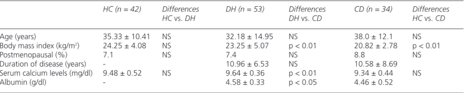
Table I. Main demographical and clinical parameters of patients with coeliac disease (CD), dermatitis herpetiformis (DH) and healthy control (HC), expressed as mean ± standard deviations
HC (n = 42) Differences DH (n = 53) Differences CD (n = 34) Differences
HC vs.DH DH vs.CD HC vs.CD
Age (years) 35.33 ± 10.41 NS 32.18 ± 14.95 NS 38.0 ± 12.1 NS
Body mass index (kg/m2) 24.25 ± 4.08 NS 23.25 ± 5.07 p < 0.01 20.82 ± 2.78 p < 0.01
Postmenopausal (%) 7.1 NS 7.4 NS 8.8 NS
Duration of disease (years) - 10.96 ± 6.53 NS 10.58 ± 8.69
Serum calcium levels (mg/dl) 9.48 ± 0.52 NS 9.64 ± 0.36 p < 0.01 9.34 ± 0.44 NS
Albumin (g/dl) - 4.58 ± 0.33 p < 0.05 4.46 ± 0.52
NS: Non significant differences.
Fig. 1. Bone mineral density of patients with coeliac disease (CD), der- matitis herpetiformis (DHD) and healthy controls (HC) at lumbar spine, represented as mean and standard deviations.
Fig. 2. Bone mineral density of patients with coeliac disease (CD), dermatitis herpetiformis (DHD) and healthy controls (HC) at the femoral neck, repre- sented as mean and standard deviations (NS: non-significant differences).
Fig. 3. Bone mineral density of patients with coeliac disease (CD), der- matitis herpetiformis (DHD) and healthy control (HC) at the radius (p:
significance; NS: non-significant differences).
Lumbar BMD
g/cm3
1.30 1.20 1.10 1.00 0.90 0.80 0.70 0.60
0.50 HC DHD CD
Lumbar BMD 1.06 0.99 0.88
p < 0.1 p < 0.1 p < 0.5
Radius BMD
g/cm3
0.80 0.75 0.70 0.65 0.60 0.55 0.50
p < 0.1
NS p = 0.1
HC DHD CD
0.67 0.62
Radius BMD 0.67
Femoral neck BMD
g/cm3
1.10 1.00 0.90 0.80 0.70 0.60 0.50
p < 0.1
NS p < 0.1
HC DHD CD
0.87 0.73
Femoral neck BMD 0.84
A higher ratio of patients had severe villous atrophy with- in the CD group compared to DH patients, according to Marsh-Oberhuber grading system (in CD group 3 % of patients presented stage 0, 9 % stage 1-2, 26 % stage 3a, 15 % stage 3b, and 47 % stage 3c; in DH group, 8 % of patients exhibited a stage 0, 24 % had stage 1-2, 38 % stage 3a, 13 % stage 3b, and stage 3c was present in 17 % of patients).
There was a tendency, but no significant difference, to present a higher BMD in DH patients with normal villous structure (Marsh-Oberhuber grade 0-2) compared to patients with villous atrophy (grade 3a-c), being at lumbar axis 0.985 ± 0,092 vs.0.997 ± 0,145 g/cm2(p = NS), at femoral neck 0.912 ± 0,034 vs.0.890 ± 0,123 g/cm2(p = NS); and at radius 0.757 ± 0,062 vs.0.686 ± 0,079 g/cm2 (p = NS).
Sixty-two percent of DH patients presented EMA pos- itive. BMD did not differ in EMA positive compared to EMA negative DH patients (lumbar axis: 0.795 ± 0.344 vs.
0.885 ± 0.320 g/cm2 [p = NS]; femoral neck: 0.553 ± 0.395 vs.0.698 ± 0.229 g/cm2[p = NS]; and radius: 0.551 ± 0.272 vs.0.608 ± 0.204 g/cm2[p = NS], respectively).
Correlations between BMD, EMA status and Marsh stage for enteropathy were not observed.
Serum calcium levels (Ca) were corrected with serum albumin in every group of subjects. Corrected Ca values were not significantly different between the DH and CD groups (10.33 ± 0.48 vs.9.89 ± 0.71 mg/dl; p = NS). There was a higher albumin serum concentration in DH compared to CD patients (4.58 ± 0.33 vs.4.46 ± 0.52 g/dl, respectively;
p < 0.05), however serum calcium levels differed signifi- cantly (Table I). A higher phosphorous serum concentration was also observed in DH compared to CD patients (1.16 ± 0.16 vs.1.14 ± 0.12 mg/dl; p < 0.01). Patients with CD pre- sented a lower body mass index (BMI) than that with DH and HC (p < 0.01). Further parameters regarding the possible malabsorption status of patients are presented in table I.
Table II. Osteodensitometric parameters of the subjects with coeliac disease (CD), dermatitis herpetiformis (DH) and healthy control (HC), expressed as mean ± standard deviation
Bone mineral density (g/cm2) T-score Z-score
Lumbar region
CD 0.880 ± 0.155 -1.848 ± 1.398 -1.490 ± 1.263
p < 0.01 p < 0.01 p < 0.01
DH 0.993 ± 0.136 -0.855 ± 1.227 -0.509 ± 1.216
p < 0.05 p < 0.05 NS
HC 1.056 ± 0.126 -0.269 ± 1.151 -0.060 ± 1.173
Left femoral neck
CD 0.733 ± 0.151 -1.165 ± 1.295 -0.784 ± 1.131
p < 0.01 p < 0.01 p < 0.01
DH 0.870 ± 0.128 -0.182 ± 1.183 0.224 ± 0.890
NS NS NS
HC 0.841 ± 0.119 -0.211 ± 1.053 0.009 ± 0.984
Non-dominant radius
CD 0.618 ± 0.068 -1.551 ± 1.329 -1.226 ± 1.358
p = 0.01 NS NS
DH 0.670 ± 0.098 -1.342 ± 1.378 -0.751 ± 1.114
NS p < 0.01 NS
HC 0.667 ± 0.072 -0.822 ± 1.106 -0.478 ± 1.122
NS: Non significant differences.
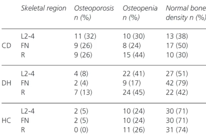
Table III. Distribution of bone mineral density of patients with coeliac disease (CD), dermatitis herpetiformis (DH)
and healthy controls (HC) enrolled in our study Skeletal region Osteoporosis Osteopenia Normal bone
n (%) n (%) density n (%) L2-4 11 (32) 10 (30) 13 (38) CD FN 9 (26) 8 (24) 17 (50) R 9 (26) 15 (44) 10 (30) L2-4 4 (8) 22 (41) 27 (51) DH FN 2 (4) 9 (17) 42 (79) R 7 (13) 24 (45) 22 (42) L2-4 2 (5) 10 (24) 30 (71) HC FN 2 (5) 10 (24) 30 (71) R 0 (0) 11 (26) 31 (74) L2-4: lumbar axis 2-4; FN: left femoral neck; R: non-dominant radius.
Thirty three percent of DH patients were not on a GFD.
BMD parameters of non-adherent DH patients did not differ from those who followed a GFD (lumbar: 0.995 ± 0.152 vs.1.025 ± 0.111 g/cm2; p = NS; femoral neck: 0.833 ± 0.186 vs.0.888 ± 0.155 g/cm2; p = NS; and radius: 0.679
± 0.146 vs.0.670 ± 0.088 g/cm2; p = NS). Mean parathyroid hormone serum concentrations were within normal ranges in all groups of subjects.
DISCUSSION
CD and one of its predominantly cutaneous manifesta- tions, DH are both gluten-sensitive disorders. CD manifests with intestinal and extraintestinal symptoms with a same frequency (20,21). Both conditions are mediated by IgA- class autoantibodies, and an underlying genetic predispo- sition and environmental factors also play an important pathophysiological role.
The vast majority of DH patients do not have any abdom- inal complaint, even when more than 75-90 % of all cases present histological changes in their small bowel mucosa similar to those in CD patients: partial or total villous atro- phy and lymphocytic infiltration of the bowel’s mucous membrane (2,22).
Although osteopenia as a common complication of CD is well recognized, only a few data on BMD in DH were available; up to the present day, the association between DH and low BMD had been investigated only in three pub- lished studies (23-25).
Abuzakouk et al. (23) investigated the bone metabolism of 25 DH patients. The mean duration of the disease was 9 years. Neither BMD nor bone biomarkers were significantly different in patient and controls. One year long GFD prospec- tively controlled did not influence the BMD in a subgroup of patients. Di Stefano et al. (24) included 16 newly diagnosed DH and sufficient number of CD patients and healthy controls in their study. BMD was found to be lower in DH compared to HC, but it was higher than in CD patients. BMD correlated with BMI and nutritional status, but neither with age at diag- nosis nor with age at onset of symptoms. BMD was signifi- cantly higher in DH patients exhibiting partial in contrast to those with subtotal villous atrophy. Lewis et al. identified 846 DH patients from the General Practice Research Database to evaluate the risk of bone fracture, malignancy and mortality among these patients: an increased risk of fracture at hip, fore- arm or any other bones were not found (25).
We have documented a higher frequency of low bone mass in patients with DH compared to healthy subjects;
however it was observed to be lower than in CD. There is no definitive data in the literature regarding the origin of low BMD in DH. In contrast, the origin of a lowered bone mass in CD has been show to be multifactorial, being the most plausible explanations related to enteropathy and a concomitant malabsorption. In fact, the frequency of symp- tomatic enteropathy was reported to be lower in DH com- pared to CD patients (2).
Bone loss of patients suffering gluten sensitive diseases may depend of the grade of the enteropathy; we observed an association between the grade of villous atrophy and the BMD in DH patients, but not reaching statistical signifi- cance. Villous atrophy correlated to the degree of bone loss in this Hungarian cohort and also in a recently published Spanish research: Garcia-Manzanares et al. (26) document- ed that the degree of bone loss in the lumbar spine correlated to Marsh stage, moreover, significant differences in major fracture risk were observed depending on Marsh stage.
However, in a recent study by Abuzakouk et al. (23) this association between BMD and the severity of enteropathy graded by the Marsh classification was not observed. Rodri- go-Sáez et al. have described that a severe villous atrophy was more frequent in coeliac patients with a younger age at diagnosis (27). In contrast, an opposite relationship was documented in a Hungarian cohort, in which the severity of histology at diagnosis was higher in adults than in chil- dren (Mars-Oberhuber 3 was presented in 92 % of adults and 57 % of children) (28). Based on these observations, one should remind that demographical and geographical characteristics of the studied populations should be con- sidered while judging the relationship of enteropathy and its consequences.
Taking into consideration the previously mentioned con- flicting results (23,24) additional multicentre studies enrolling many patients would be needed to clearly deter- mine how relevant the grade of enteropathy is when assess- ing BMD in patients with DH.
In our study BMD in DH patients who were not on a GFD did not differ from those who were on it. Confusing results on the relationship between GFD and metabolism have been provided: most of studies demonstrated a lower prevalence of low BMD in coeliac patients who are on GDF (16,29), as well as GFD exert a beneficial effect on bone metabolism of coeliac patients (30-32). A similar prevalence of low BMD have been documented after one (33) and three years (34) on GFD, which shows that the main recovery in bone mass is reached in the first year of following the diet.
Lewis et al. (35) observed that years of exposure to gluten before diagnosis of CD do not appear to influence BMD significantly. The relationship between GFD and bone restoration seems to be more pronounced in paediatric pop- ulation (36,37): one year long GFD did not improve BMD in adult population according to the previously mentioned Irish study (23).
Calcium malabsorption has been suspected to be the most plausible cause of low BMD in CD (38). In our study corrected Ca serum levels were not significantly different between DH and the CD patients, but serum calcium levels differed significantly. There was a higher albumin serum concentration in DH patients compared to the CD group.
Additionally, BMI was similar in HC and DH patients, while it was lower in the studied coeliac population.
Secondary hyperparathyroidism (39) and concomitant lactase deficiency (40) may also worsen Ca balance in CD.
In fact, reduction of cortical bone mass is very common
in hyperparathyroidism, which should be tested in all patients with CD. Parathyroid hormone concentrations were within the normal range in our patients and bone loss was prominent in those bones containing mainly trabecular component.
An increased receptor-associated nuclear factor kappa ligand/osteoprotegerin ratio has been also detected in newly diagnosed patients with CD (41); this dissociation causing uncoupled bone remodelling have been also proved by the elevation of markers of increased bone turnover in CD (42,43). Local factors may affect the bone remodelling in CD as well. Several pro-inflammatory cytokines (inter- leukin (IL)-1, IL-6, and tumour necrosis factor alpha) are produced in the intestinal mucosa in CD and result in an increased osteoclast activity (44). Low levels of other cytokines, such as IL-12 and IL-18, result in osteoclasto- genesis due to the lack of the inhibitory effects of these cytokines (45). Some data show increase serum pro-inflam- matory cytokine concentrations (IL-8, tumour necrosis fac- tor alpha) in DH patient (46). Milder degrees of bone loss in DH compared to CD were observed in our study. We are not able to explain this phenomenon based on our results.
A more patchy distribution of villous atrophy or a marker cytokine alteration in CD compared to DH may be hypoth- esized in order to explain this phenomenon; however, these comparisons have been never performed until now.
A loss of bone density in the peripheral skeleton may persist despite normalization of lumbar and femoral bone density after patients are on a GFD. The importance of an early diagnosed of metabolic bone disease is clear in CD.
The proper diagnostic workup and strict GFD is mandatory to prevent bone fractures, but timing of densitometry in the follow up has not been clarified, but to measure BMD after the first year on a GFD has been recommended proposed as useful.
Our results suggest the usefulness of measuring the bone density of DH patients, but doubts on the appropriate timing for the first bone density scan, the diagnostic workup and the most appropriate therapy for DH patients with a low BMD remain.
There are some limitations of our study. More complete and accurate information would have been achieved from in a greater number of patients, but we should remain that the incidence of DH has been estimated to be 8-16 times lower than that of CD (47,48). So, to the best of our knowl- edge, this study investigated BMD in the widest number of DH cases up to now. We did not examine more sophis- ticated laboratory parameters, as biomarkers related to bone formation and resorption, or cytokines. The exact role of these markers has not clarified in the diagnostic workup of gastrointestinal disorders involved with bone metabolism.
Further investigation on this biomarkers will help to under- stand the pathogenesis of DH-associated bone loss.
As a conclusion, bone mineral content in patients with DH was significantly lower than in healthy controls, but higher than in CD. Bone mineral loss was more prominent in bones containing more trabecular than cortical compo-
nent. This phenomenon suggests a similar pathogenetic ori- gin for DH and CD-associated osteopenia and osteoporosis, even when a relationship between the severity of villous atrophy and bone loss was not clearly shown. Further research will define a proper diagnostic and therapeutic algorithm for the management of DH.
ACKNOWLEDGEMENTS
We are grateful to William Gesztes for his constructive criticism and language lecturing our paper as a native Eng- lish speaker. We also thank to Miklós Tóth MD, DSc the evaluation of the densitometry measurements.
REFERENCES
1. Duhring LA. Dermatitis herpetiformis. JAMA 1983;250:212-16.
2. Rose C, Brocker EB, Zillikens D. Clinical, histological and immunpathological findings in 32 patients with dermatitis herpetiformis Duhring. J Dtsch Dermatol Ges 2010;8:265-71.
3. Smith JB, Tulloch JE, Meyer LJ, Zone JJ. The incidence and prevalence of der- matitis herpetiformis in Utah. Arch Dermatol 1992;128:1608-10.
4. Reunala TL. Dermatitis herpetiformis. Clin Dermatol 2001;19:728-36.
5. Marietta E, Black K, Camilleri M, Krause P, Rogers RS 3rd, David C, et al. A new model for dermatitis herpetiformis that uses HLA-DQ8 transgenic NOD mice. J Clin Invest 2004;114:1090-7.
6. Sardy M, Tietze J. Dermatitis herpetiformis. An update of the patho- genesis. Hautarzt 2009;60:627-30.
7. Piccini B, Vascotto M, Serracca L, Luddi A, Margollicci MA, Balestri P, et al. HLA-DQ typing in the diagnostic algorithm of celiac disease.
Rev Esp Enferm Dig 2012;104:248-54.
8. Caputo I, Barone MV, Martucciello S, Lepretti M, Esposito C. Tissue transglutaminase in celiac disease: Role of autoantibodies. Amino Acids 2009;36:693-9.
9. Karpati S. Dermatitis herpetiformis: Close to unravelling a disease. J Dermatol Sci 2004;34:83-90.
10. Sardy M, Karpati S, Merkl B, Paulsson M, Smyth N. Epidermal trans- glutaminase (TGase 3) is the autoantigen of dermatitis herpetiformis.
J Exp Med 2002;195:747-57.
11. Jaskowski TD, Hamblin T, Wilson AR, Hill HR, Book LS, Meyer LJ, et al. IgA anti-epidermal transglutaminase antibodies in dermatitis her- petiformis and pediatric celiac disease. J Invest Dermatol 2009;129:
2728-30.
12. Vasquez H, Mazure R, Gonzalez D, Flores D, Pedreira S, Niveloni S, et al. Risk of fractures in celiac disease patients: A cross-sectional, case-control study. Am J Gastroenterol 2000;95:183-9.
13. Jafri MR, Nordstrom CW, Murray JA, Van Dyke CT, Dierkhising RA, Zinsmeister AR, et al. Long-term fracture risk in patients with celiac disease: A population-based study in Olmsted County, Minnesota. Dig Dis Sci 2008;53:964-71.
14. Ludvigsson JF, Michaelsson K, Ekbom A, Montgomery SM. Coeliac disease and the risk of fractures - A general population-based cohort study. Aliment Pharmacol Ther 2007;25:273-85.
15. Zanini B, Caselani F, Magni A, Turini D, Ferraresi A, Lanzarotto F, et al. Celiac disease with mild enteropathy is not mild disease. Clin Gas- troenterol Hepatol 2013;11:253-8.
16. Corazza GR, Di Sario A, Cecchetti L, Jorizzo RA, Di Stefano M, Min- guzzi L, et al. Influence of pattern of clinical presentation and of gluten- free diet on bone mass and metabolism in adult coeliac disease. Bone 1996;18:525-30.
17. Olmos M, Antelo M, Vazquez H, Smecuol E, Maurino E, Bai JC. Sys- tematic review and meta-analysis of observational studies on the preva- lence of fractures in coeliac disease. Dig Liver Dis 2008;40:46-53.
18. Oberhuber G, Granditsch G, Vogelsang H. The histopathology of coeli- ac disease: Time for a standardized report scheme for pathologists. Eur J Gastroenterol Hepatol 1999;11:1185-94.
19. Kanis JA. Assessment of fracture risk and its application to screening for postmenopausal osteoporosis: Synopsis of a WHO report. WHO Study Group. Osteoporos Int 1994;4:368-81.
20. Fernandez A, Gonzalez L, de-la-Fuente J. Coeliac disease: Clinical features in adult populations. Rev Esp Enferm Dig 2010;102:466-71.
21. Fernandez Salazar LI, de la Torre Ferrera N, Velayos Jimenez B, Nocito Colon M, Gonzalez Hernandez JM, Garrote Adrados JA. Diag- nostic problems in adult celiac disease. Rev Esp Enferm Dig 2008;100:24-8.
22. Dahlbom I, Korponay-Szabo IR, Kovacs JB, Szalai Z, Maki M, Hans- son T. Prediction of clinical and mucosal severity of coeliac disease and dermatitis herpetiformis by quantification of IgA/IgG serum anti- bodies to tissue transglutaminase. J Pediatr Gastroenterol Nutr 2010;50:140-6.
23. Abuzakouk M, Barnes L, O’Gorman N, O’Grady A, Mohamed B, McKenna MJ, et al. Dermatitis herpetiformis: No evidence of bone dis- ease despite evidence of enteropathy. Dig Dis Sci 2007;52:659-64.
24. Di Stefano M, Jorizzo RA, Veneto G, Cecchetti L, Gasbarrini G, Coraz- za GR. Bone mass and metabolism in dermatitis herpetiformis. Dig Dis Sci 1999;44:2139-43.
25. Lewis NR, Logan RF, Hubbard RB, West J. No increase in risk of frac- ture, malignancy or mortality in dermatitis herpetiformis: A cohort study. Aliment Pharmacol Ther 2008;27:1140-7.
26. Garcia-Manzanares A, Tenias JM, Lucendo AJ. Bone mineral density directly correlates with duodenal Marsh stage in newly diagnosed adult celiac patients. Scand J Gastroenterol 2012;8-9:927-36.
27. Rodrigo-Saez L, Fuentes-Alvarez D, Perez-Martinez I, Alvarez-Mieres N, Nino-Garcia P, de-Francisco-Garcia R, et al. Differences between pediatric and adult celiac disease. Rev Esp Enferm Dig 2011;103:238- 44.
28. Juhasz M, Kocsis D, Zagoni T, Miheller P, Herszenyi L, Tulassay Z.
Retrospective evaluation of the ten-year experience of a single coeliac centre. Orv Hetil 2012;153:776-85.
29. McFarlane XA, Bhalla AK, Reeves DE, Morgan LM, Robertson DA.
Osteoporosis in treated adult coeliac disease. Gut 1995;36:710-4.
30. Mora S, Barera G, Beccio S, Proverbio MC, Weber G, Bianchi C, et al. Bone density and bone metabolism are normal after long-term gluten- free diet in young celiac patients. Am J Gastroenterol 1999;94:398- 403.
31. Heyman R, Guggenbuhl P, Corbel A, Bridoux-Henno L, Tourtelier Y, Balencon-Morival M, et al. Effect of a gluten-free diet on bone mineral density in children with celiac disease. Gastroenterol Clin Biol 2009;33:109-14.
32. Capriles VD, Martini LA, Areas JA. Metabolic osteopathy in celiac disease: Importance of a gluten-free diet. Nutr Rev 2009;67:599-606.
33. Mautalen C, Gonzalez D, Mazure R, Vazquez H, Lorenzetti MP, Mau- rino E, et al. Effect of treatment on bone mass, mineral metabolism, and body composition in untreated celiac disease patients. Am J Gas- troenterol 1997;92:313-8.
34. Bai JC, Gonzalez D, Mautalen C, Mazure R, Pedreira S, Vazquez H, et al. Long-term effect of gluten restriction on bone mineral density of patients with coeliac disease. Aliment Pharmacol Ther 1997;11:157- 64.
35. Lewis NR, Scott BB. Should patients with coeliac disease have their bone mineral density measured? Eur J Gastroenterol Hepatol 2005;17:1065-70.
36. Mora S, Barera G, Ricotti A, Weber G, Bianchi C, Chiumello G. Rever- sal of low bone density with a gluten-free diet in children and adoles- cents with celiac disease. Am J Clin Nutr 1998;67:477-81.
37. Mazure R, Vazquez H, Gonzalez D, Mautalen C, Pedreira S, Boerr L, et al. Bone mineral affection in asymptomatic adult patients with celiac disease. Am J Gastroenterol 1994;89:2130-4.
38. Szymczak J, Bohdanowicz-Pawlak A, Waszczuk E, Jakubowska J.
Low bone mineral density in adult patients with coeliac disease.
Endokrynol Pol 2012;63:270-6.
39. Keaveny AP, Freaney R, McKenna MJ, Masterson J, O’Donoghue DP.
Bone remodeling indices and secondary hyperparathyroidism in celiac disease. Am J Gastroenterol 1996;91:1226-31.
40. Di Stefano M, Veneto G, Malservisi S, Cecchetti L, Minguzzi L, Stroc- chi A, et al. Lactose malabsorption and intolerance and peak bone mass.
Gastroenterology 2002;122:1793-9.
41. Horwood NJ, Elliott J, Martin TJ, Gillespie MT. IL-12 alone and in synergy with IL-18 inhibits osteoclast formation in vitro. J Immunol 2001;166:4915-21.
42. Fiore CE, Pennisi P, Ferro G, Ximenes B, Privitelli L, Mangiafico RA, et al. Altered osteoprotegerin/RANKL ratio and low bone mineral den- sity in celiac patients on long-term treatment with gluten-free diet.
Horm Metab Res 2006;38:417-22.
43. Corazza GR, Di Sario A, Cecchetti L, Tarozzi C, Corrao G, Bernardi M, et al. Bone mass and metabolism in patients with celiac disease.
Gastroenterology 1995;109:122-8.
44. Taranta A, Fortunati D, Longo M, Rucci N, Iacomino E, Aliberti F, et al. Imbalance of osteoclastogenesis-regulating factors in patients with celiac disease. J Bone Miner Res 2004;19:1112-21.
45. Fornari MC, Pedreira S, Niveloni S, Gonzalez D, Diez RA, Vazquez H, et al. Pre- and post-treatment serum levels of cytokines IL-1beta, IL-6, and IL-1 receptor antagonist in celiac disease. Are they related to the associated osteopenia? Am J Gastroenterol 1998;93:413-8.
46. Hall RP 3rd, Takeuchi F, Benbenisty KM, Streilein RD. Cutaneous endothelial cell activation in normal skin of patients with dermatitis herpetiformis associated with increased serum levels of IL-8, sE- Selectin, and TNF-alpha. J Invest Dermatol 2006;126:1331-7.
47. Salmi TT, Hervonen K, Kautiainen H, Collin P, Reunala T. Prevalence and incidence of dermatitis herpetiformis: A 40-year prospective study from Finland. Br J Dermatol 2011;165:354-9.
48. Virta LJ, Kaukinen K, Collin P. Incidence and prevalence of diagnosed coeliac disease in Finland: Results of effective case finding in adults.
Scand J Gastroenterol 2009;44:933-8.