T cell responses to an HLA-A2-restricted
adipophilin peptide correlate with BMI in patients with atherosclerosis
K. KALANTAR
1, Z. FARZANEH
1, M. ESHKEVAR VAKILI
1,
M.H. KARIMI
3, M. ASADI
1, S. KHOSROPANAH
2and M. DOROUDCHI
1p1Department of Immunology, School of Medicine, Shiraz University of Medical Sciences, Shiraz, Islamic Republic of Iran
2Department of Cardiology, School of Medicine, Shiraz University of Medical Sciences, Shiraz, Islamic Republic of Iran
3Transplant Research Center, Nemazee Hospital, Shiraz University of Medical Sciences, Shiraz, Islamic Republic of Iran
Received: October 28, 2019 • Accepted: February 5, 2020 Published online: July 13, 2020
© 2020 Akademiai Kiado, Budapest
ABSTRACT
Introduction:Atherosclerosis is an inflammatory disease causing a vast array of cardiovascular diseases.
Adipophilin has been reported to be highly expressed in atherosclerotic lesions. This study investigated the possible existence of auto-reactive T cells against an HLA-A02-restricted adipophilin-derived peptide as well as peptides from Epstein-barr virus (EBV), Cytomegalovirus (CMV) and influenza (Flu) virus in patients with atherosclerosis. Methods: HLA-A02 expression on peripheral blood mononuclear cells (PBMCs) was examined by flow cytometry. PBMCs from HLA-A02 individuals were stimulated with adipophilin, CMV, EBV, and Flu peptides at a concentration of 10mM. Interferon (IFN)-gproduction was evaluated in the culture supernatant using a commercial ELISA test.Results:
The levels of IFN-gproduction against an HLA-A02-restricted adipophilin peptide and peptides from CMV, EBV, and Flu revealed no statistically significant differences between patients and healthy controls. However, we found a positive correlation between IFN-gproduction against adipophilin and Body mass index (BMI) of patients (R50.8,P50.003), whereas no significant correlation was found in healthy controls (R5 0.267, P 5 0.378). No correlation between BMI and IFN-g production
pCorresponding author. Memory T Cell Laboratory, Department of Immunology, School of Medicine, Shiraz University of Medical Sciences, P.O. Box: 71345-3119, Shiraz, Islamic Republic of Iran. Tel./Fax:þ98 (71) 3235 1575. E-mail: mdoroud@sums.ac.ir
against CMV, EBV, or Flu peptides was found.Discussion:Atherosclerotic patients with higher BMIs might have greater numbers of T cells against adipophilin that is highly expressed in atherosclerotic plaques. Therefore, autoimmune reactions may have a greater role in the development of athero- sclerosis in individuals with higher BMI.
KEYWORDS
atherosclerosis, adipophilin, HLA-A02, BMI
INTRODUCTION
Atherosclerosis is a chronic immune inflammatory disease of the arterial walls characterized by formation of atherosclerotic plaques [1]. The major clinical manifestations of the disease are peripheral arterial disease, ischemic heart pain, myocardial infarction and ischemic stroke which lead to premature death worldwide [2]. While the exact etiology of atherosclerosis is unknown, it is widely accepted that it is a multifactorial disease; for example with contribution of infections and the immune system [2–4].
The contribution of immune cells in the disease process is well acknowledged, and a large body of evidence suggests the underlying importance of the immune system in the devel- opment and progression of atherosclerosis [1, 5]. Various infiltrating immune cell types, including monocytes/foam cell macrophages and lymphocytes with their secreted cytokines and chemokines constitute key components of atherosclerotic plaque inflammation [1].
Monocytes can be recruited to the inflamed endothelium where they differentiate into macrophage phenotypes with highly phagocytic ability. They can scavenge modified low- density lipoprotein(mLDL) and eventually form foam cells, which along with other immune cells can increase the risk of the development of the unstable atherosclerotic plaque phenotype [5].
In addition, other immune cells including CD4þ T helper and CD8þ cytotoxic T lym- phocytes (CTLs) play a pivotal role in the development or instability of the plaque [6, 7]. It is suggested that CD8þT cells represent about 29% of all lymphocytes in early lesions, increasing up to 50% in advanced plaques [6]. They play critical roles in the development of vulnerable atherosclerotic plaques by recognition of oxidized low-density lipoprotein (Ox-LDL)-derived peptides and other probable auto-antigens presented via MHC class I molecules [8]. The cytotoxic effects of CD8þ T cells happen via granule exocytosis and release of key effector molecules, perforin and granzymes by which they induce apoptosis in different target cells including smooth muscle cells, endothelial cells and macrophages, and eventually lead to necrotic core and vulnerable atherosclerotic plaques formation [7]. Moreover, several cytokines produced by CD8þT cells, like tumor-necrosis factor (TNF)-aand interferon (IFN)-gcan exert cytotoxic action when secreted in the proximity of target cells [9]. The central role of IFN-gin pathogenesis of atherosclerosis is shown in IFN-g-deficient mice which develop smaller and more stable plaques [10].
This cytokine is highly expressed in atherosclerotic lesions and exerts its detrimental roles in atherosclerosis via different mechanisms; IFN-g activates macrophage and
endothelial cells to produce chemokines and adhesion molecules, respectively, by which it provokes monocyte/lymphocyte recruitment and infiltration into the sub-endothelium, thus accelerating plaque growth. IFN-gpromotes foam cell formation by increasing the uptake of mLDL by scavenger receptors on macrophages. IFN-gcan also help formation of vulnerable plaques by inducing the formation and release of reactive oxygen species and matrix met- alloproteinases (MMPs) [11].
Numerous atherosclerosis-related T cell antigens are reported which include both microbial and self-antigens. Among bacterial and viral pathogens, Chlamydia pneumoniae, Helicobacter pylori, Porphyromonas gingivalis, Cytomegalovirus (CMV), Epstein-barr virus (EBV) and influenza (Flu) virus [12] are documented. The proposed mechanisms in microbial pathogenesis in atherosclerosis include direct infection of cells in the arterial wall, molecular mimicry, acti- vation of pattern recognition receptors (PRR; e.g. Toll-like receptor; TLR) and subsequent bystander activation of auto-reactive T-cells and epitope spreading. Another mechanism is based on “danger hypothesis” in which damage-associated molecular patterns (DAMPs) released during cell death act as endogenous self-adjuvants and induce inflammation and auto- reactive T cell responses [13].
Potential candidate T cell auto-antigens in atherosclerosis lie within altered self-structures, such as modified lipoproteins and other proteins includingb2-glycoprotein I and heat shock proteins [14].
Adipophilin, or adipose differentiation-related protein (ADRP) is a 50 kDa protein expressed in adipocytes, which controls the structure and formation of lipid droplets. It is also expressed in macrophage foam cells and its expression is up-regulated by mLDL [15, 16]. This protein is expressed at higher levels in atherosclerotic lesions compared to healthy arterial intima, and the relative expression of adipophilin is higher in symptomatic compared to asymptomatic carotid plaques [15]. In addition, studies have shown that adipophilin is highly overexpressed in some tumors and is a target of CD8þT cell responses [17]. In a study conducted by Weinschenk et al.
a HLA-A*0201-binding peptide, SVASTITGV, derived from the adipophilin protein was identified in renal cell carcinoma (RCC) tumor samples [18] which was further confirmed by other studies [17]. As adipophilin is highly expressed in atherosclerotic lesions, and previous experimental data have revealed the “SVASTITGV” peptide as a CD8þ T cell target, we examined the possible potential of this peptide as a CD8þT-cell epitope that can induce an- tigen-specific CD8þT cells in HLA-A2 atherosclerosis patients.
The existence of CD8þ T-cells specific to adipophilin-derived peptides in patients with cancers has already been shown [17]. Therefore, we hypothesized that adipophilin could be a potential target for T cells in atherosclerosis [16, 19]. Thus the purpose of the present study was to investigate the possible existence of auto-reactive T cells against an HLA-A02-restricted adipophilin-derived peptide as well as peptides from EBV, CMV and Flu virus in patients with atherosclerosis.
MATERIALS AND METHODS
Patients and controls
Thirty-two patients with atherosclerosis from the Diabetes Clinic of Shiraz University of Medical Sciences, Iran entered the study. Forty age- and sex-matched healthy blood donors
were also enrolled in the study. Informed consent was obtained from all the subjects and the study was approved by the relevant Ethics Committees. Coronary angiography (CAG) was performed by a team of expert interventional cardiologists to estimate the coronary lesions.
The inclusion and exclusion criteria were as follows: patients who were diagnosed based on the results of coronary angiography were included in the present study. We excluded pa- tients with chronic diseases, including diabetes, hepatic or renal diseases or active infection.
Patients with autoimmune diseases, malignancies and histories of taking any immunosup- pressive drugs during last 3 months were also excluded from this study. For healthy subjects, the exclusion criteria were current smoking, hypertension, hypercholesterolemia, diabetes, obesity, active infection, autoimmune disease. The patients were grouped into single vessel disease (SVD), double vessel disease (DVD), and triple vessel disease (TVD) according to the number of major epicardial coronary arteries involved. Blood samples from both patients and controls were collected in heparin tubes and kept at room temperature (RT) until processing.
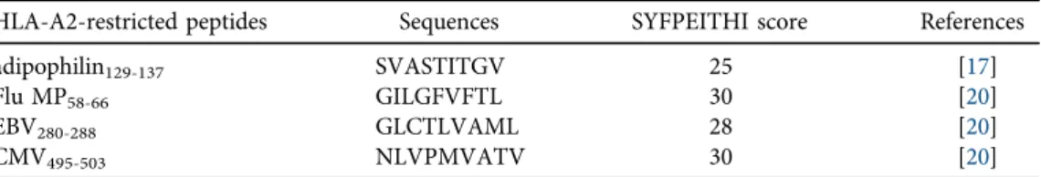
Peptides
HLA-A2-restricted peptides used in this study were >80% pure (China Peptides, Shanghai, China) (Table 1). Peptides were added at a final concentration of 10
m
M.Isolation of peripheral blood mononuclear cells (PBMCs)
Ficoll-Hypaque solution (Lymphodex, Germany) was placed into a 50-mL conical centrifuge tube using a sterile pipette. Heparinized blood was mixed with an equal volume of phosphate buffered saline (PBS) and the diluted blood was slowly layered over the Ficoll-Hypaque solution.
The tube was centrifuged for 20 min at 4003g, 22 8C. The ring of mononuclear cells was carefully collected and transferred into a 15-mL conical tube, 10 mL complete Roswell Park Memorial Institute (RPMI) medium was added and mixed thoroughly. The tube was centrifuged for 10 min at 4003g, 48C. The supernatant was discarded and peripheral blood mononuclear cells (PBMCs) were used for further experiments.
HLA-A02 typing of PBMCs by flow cytometry
106PBMCs were re-suspended in 1 mL ice-cold PBS containing 10% fetal calf serum (FCS) and 1% sodium azide to prevent the modulation and internalization of surface antigens which can
Table 1.HLA-A02-restricted peptides used in the stimulation of cultured PBMCs
HLA-A2-restricted peptides Sequences SYFPEITHI score References
adipophilin129-137 SVASTITGV 25 [17]
Flu MP58-66 GILGFVFTL 30 [20]
EBV280-288 GLCTLVAML 28 [20]
CMV495-503 NLVPMVATV 30 [20]
Flu MP, Influenza Matrix protein; EBV, Epstein-Barr virus; CMV, Cytomegalovirus; SYFPEITHI (a database for MHC ligands and peptide motifs).
decrease thefluorescence intensity. 100
m
L of cell suspension was added to each eppendorf tube, 2m
g of anti HLA-A02 monoclonal antibody (mouse IgG2b, Abcam, USA) was added to the cell suspension (final concentration 20m
g/mL), and cells were incubated for at least 30 min at 48C in the dark. Anti HLA-Class I antibody (IgG2a, Abcam, USA) was used as positive control. Cells were washed three times by centrifugation for 5 min at 4003gand re-suspended in ice-cold PBS.10
m
L of fluorescent isothiocyanate (FITC)-conjugated anti-mouse IgG secondary antibody (IgG2a, Abcam, USA) was added and incubation was continued for 30 min at 4˚C in the dark.Cells stained only with the secondary antibody were used as negative control. In the next step, cells were washed 3 times by centrifugation for 5 min at 4003g and re-suspended in ice-cold PBS containing 3% bovine serum albumin (BSA) and 1% sodium azide. The cell suspension was analyzed in a BD FACSCalibur flow cytometer. Analysis was performed by FlowJo version 7 software (TreeStar, USA).
PBMCs culture and stimulation with specific peptides
PBMCs from HLA-A02 individuals (n514) were seeded in 96-well plates (Nunc, Denmark) at a concentration of 2 3 105 cells/well. PBMCs cultured alone were used as negative control. Phytohemagglutinin (PHA) (Invitrogen, USA) at afinal concentration of 2.5
m
g/mLwas used as positive control of the kit. Flu, CMV, and EBV peptides at a concentration of 10
m
M/well/200m
L were used as positive controls for the subjects [20]. Adipophilin peptide was also used at the mentioned concentration to determine the responsiveness of PBMCs [17].Plates were incubated at 5% CO2in a humidified incubator for 24 h. Cell culture media were next collected and centrifuged (1,500 rpm, 48C for 10 min). Supernatants were kept at80 8C until use for further processing. The sequences of selected peptides used in the present study were listed inTable 1.
IFN-g detection in the supernatant of cultured PBMCs using ELISA
IFN-gin the culture supernatant was assessed by using a commercial ELISA kit (Human IFN gamma ELISA Ready-SET-Go, ebioscience, USA) (sensitivity: 4 pg/mL) based on the manufacturer’s instructions. Briefly, the ELISA plate was coated with 100
m
L/well ofcapture antibody in coating buffer. The plate was sealed and incubated overnight at 48C.
Wells were aspirated and washed 5 times with >250
m
L/well wash buffer (13PBS, 0.05%Tween-20) and allowed to soak for about 1 minute during each wash step to increase the effectiveness of the washes. The plate was blotted on absorbent paper to remove any re- sidual buffer. 1 part of 5X concentrated assay diluent was diluted with 4 parts Deionized (DI) water and blocking was done using 200
m
L/well of fetal bovine serum (FBS) 2%. The plate was incubated at room temperature for 1 hour. Wells were aspirated/washed as in step 3 (5 washes). Standards were diluted using 1X assay diluent. 100m
L/well of standard was added to the appropriate wells. Two-fold serial dilutions of the top standards were made to make the standard curve. 100m
L/well of the cell culture supernatant was added to the appropriate wells, and the plate was covered or sealed and incubated overnight at 48C for maximal sensitivity. The next day, wells were aspirated/washed as in step 3 (5 washes).100
m
L/well of detection antibody diluted in 1X assay diluent was added, and the plate was sealed and incubated at room temperature for 1 hour. Wells were aspirated/washed as in step 3 (5 washes). 100m
L/well of Avidin-HRP diluted in 1X assay diluent was added, andthe plate was sealed and incubated at room temperature for 30 minutes. Wells were aspirated, washed, and soaked (7 washes). 100
m
L/well of substrate solution was added to each well, and the plate was incubated at room temperature for 15 min. 50m
L of stopsolution (2N H2SO4) was added to each well. The plate was read at 450 nm and a 630 nm filter as a reference wavelength. The levels of IFN-gwithin the sample were determined by setting up a standard curve of known target protein concentrations which was provided in the kit.
Statistical analysis
Data presented in the text and Figures are mean±SEM of at least three independent experi- ments. Unpaired Mann-Whitney U test and Kruskal-Wallis test were used to test the probability of significant differences between groups. For testing the correlation between the levels of IFN-g and clinical manifestation, Pearson’s correlation test was used. A Pvalue less than 0.05 was considered statistically significant.
RESULTS
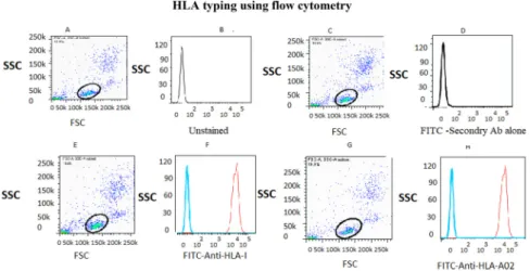
HLA typing of patients and healthy individuals
HLA typing using flow cytometry revealed that 14 out of the 32 recruited patients with atherosclerosis and 16 out of the 40 healthy individuals were positive for HLA-A02 (Fig. 1). Angiography indicated that five out of the 14 HLA-A02 patients had SVD, three had DVD and six had three-vessel disease (3VD). All HLA-A02 individuals were used for
Fig. 1.HLA typing usingflow cytometry. Dot plot and histogram plot obtained from flow cytometry analysis of PBMCs isolated from patients and healthy individuals. (A) Unstained; (B) Negative control; (C)
Positive control; (D) HLA-A02-positive individual.
further experiments. The demographic characteristics of the two groups are listed in Table 2.
ELISA on PBMCs supernatant
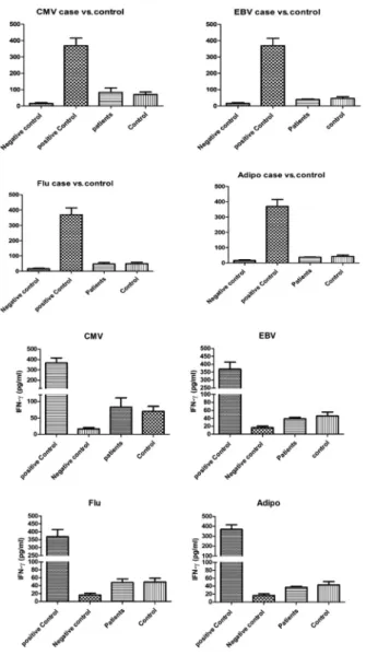
The levels of IFN-g (pg/mL) for HLA-A2-positive patients and controls after stimulation with CMV, EBV, Flu- and adipophilin-derived peptides are shown in Table 3. PBMCs stimulation with the mentioned peptides revealed no statistically significant differences in the levels of IFN-g production between HLA-A2-positive patients and healthy controls (Fig. 2).
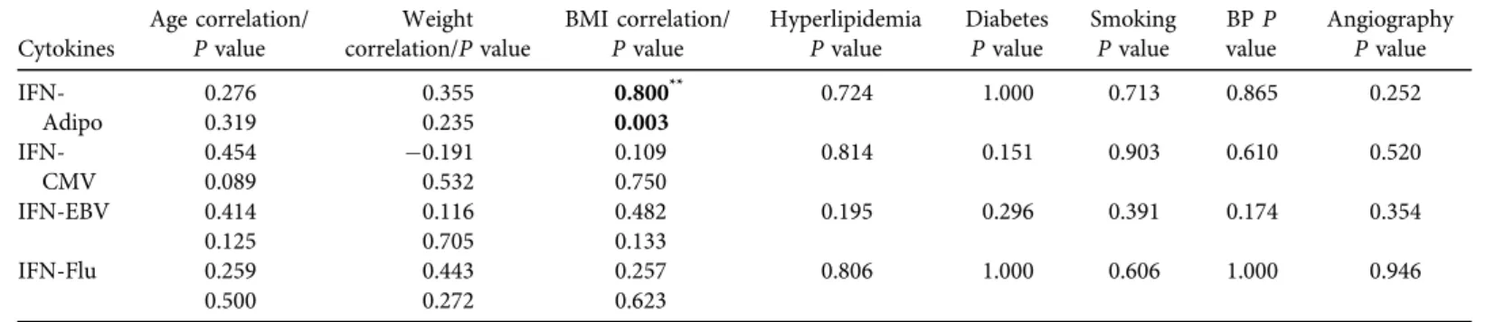
Correlation between IFN-g and clinicopathologic manifestations of atherosclerosis in patients group
The results of the correlation analysis are shown in Table 4. Interestingly, a positive cor- relation was found between levels of IFN-gproduction against adipophilin and Body Mass Index (BMI) of patients (R50.8,P50.003). No significant differences were found between the levels of IFN-g against adipophilin and other clinicopathologic manifestations. In respect to the other selected peptides, no significant correlations were found between IFN-g production and clinical manifestations. In addition, the same analysis was done in healthy controls to predict the correlation between IFN-gproductions against adipophilin and BMI.
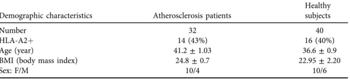
No statistically significant correlation was seen (R50.267,P50.378). The same results Table 2.Demographic characteristics of atherosclerosis patients and healthy controls
Demographic characteristics Atherosclerosis patients
Healthy subjects
Number 32 40
HLA-A2þ 14 (43%) 16 (40%)
Age (year) 41.2±1.03 36.6±0.9
BMI (body mass index) 24.8±0.7 22.95±2.20
Sex: F/M 10/4 10/6
Data represent mean±SEM of age and BMI.
Table 3.The levels of IFN-g(pg/mL) for HLA-A2-positive patients and healthy controls after PBMCs stimulation with CMV-, EBV-, Flu- and adipophilin-derived peptides
Peptides Patients (n514) Controls (n516)
P value
CMV495-503 83.3±27.6 70.6±14.8 0.68
EBV280-288 39.0±3.3 46.1±9.7 0.5
Flu MP58-66 47.8±8.9 49.2±9.5 0.92
adipophilin129-137 36.9±2.4 42.7±8.8 0.53
Flu MP, Influenza (matrix protein); EBV, Epstein-Barr virus; CMV, Cytomegalovirus.
were obtained when we pooled the results of IFN-gof healthy subjects and atherosclerosis patients together. In this case no correlation was seen between IFN-gand BMI (R50.140, P50.514).
Fig. 2.IFN-gELISA results in PBMCs cultured with CMV-, EBV-, Flu- and Adipo-derived peptides.
As shown, the levels of IFN-gbetween HLA-A2-positive patients and healthy blood donors exhibit no significant differences
Table 4.Correlations between amounts of IFN-gproduction against selected peptides and demographic and clinical manifestations of patients
Cytokines
Age correlation/
Pvalue
Weight correlation/Pvalue
BMI correlation/
Pvalue
Hyperlipidemia Pvalue
Diabetes Pvalue
Smoking Pvalue
BPP value
Angiography Pvalue IFN-
Adipo
0.276 0.355 0.800** 0.724 1.000 0.713 0.865 0.252
0.319 0.235 0.003
IFN- CMV
0.454 0.191 0.109 0.814 0.151 0.903 0.610 0.520
0.089 0.532 0.750
IFN-EBV 0.414 0.116 0.482 0.195 0.296 0.391 0.174 0.354
0.125 0.705 0.133
IFN-Flu 0.259 0.443 0.257 0.806 1.000 0.606 1.000 0.946
0.500 0.272 0.623
BMI, Body mass index; BP, Blood pressure; IFN, Interferon-gamma; Adipo, adipophilin; CMV, Cytomegalovirus; EBV, Epstein-Barr virus; Flu, Influenza The bold values show the statistically significant correlation between IFN gamma levels against adipophilin and the BMI of the patients.
PhysiologyInternational107(2020)2,280–293
DISCUSSION
In the current study we found that HLA-A2-restricted adipophilin129-137is recognized by T cells from patients with atherosclerosis as well as control individuals. The magnitude of IFN-g response was comparable to that of EBV280-288 and Flu MP58-66 (Fig. 2). Both patients and controls produced notable amounts of IFN-gin response to CMV495-503.
Adipophilin is expressed in a broad variety of human malignancies and the HLA-A*02- binding peptide derived from this protein was identified in tumors of RCC patients [17]. A previous study reported that CTLs can lyse different cancer cells which endogenously express the adipophilin-derived peptides in a HLA-A*0201-restricted manner [16]. CTLs in RCC, malignant melanoma, breast cancer, and multiple myeloma cells can recognize and lyse tumor cells which endogenously express adipophilin in an antigen-specific and HLA-A2-restricted manner [17]. Adipophilin is also shown to be highly expressed in the atherosclerotic plaque [15].
Since the T cells in the circulation home to different tissues as well as pathologic sites, we asked if T (CD8þ) cell responses towards adipophilin129-137 exist in our patients and if so, how different these responses would be from that of healthy controls. While such responses in the form of IFN-gsecretion existed in both groups, we did not find any significant difference be- tween the two groups. However, we found a positive correlation between the extent of IFN-g production upon adipophilin peptide stimulation and BMI only among patients. Interestingly, such a correlation did not exist in healthy controls or when both groups were considered. Our result supports the relationship between inflammation induced by obesity and atherosclerosis.
Studies have shown that obesity is associated with the production of inflammatory molecules, including inflammatory cytokines. The inflammatory cytokines, by affecting the artery wall, lead to the production of different mediators and surface proteins, followed by accumulation of inflammatory cells, endothelial dysfunction, substantial structural change in the artery and ul- timately to atherosclerosis [21]. Earlier studies have demonstrated that the risk of cardiovascular disease (CVD) significantly increases in obese individuals in comparison to those with normal BMI [22, 23]. As noted above, high adipophilin expression in atherosclerotic plaques has already been reported [15]. Furthermore, association of total plaque area with obesity is demonstrated in a recent study [24]. Hence, we assume that atherosclerotic patients with higher BMI might have a greater number of T cells against this protein. Adipophilin plays a very important role in the formation of fat droplets, and the expression of this protein is very high in the adipose tissue of obese people [25]. Accordingly, we suggest that our study provides new information on the auto-reactive responses in general and in atherosclerosis in particular [17]. In our study both patients and controls responded to peptides derived from CMV, EBV and Flu viruses. As already mentioned, several evidences have indicated the relationship between pathogens and athero- sclerosis [4, 12, 26]. The proposed mechanisms include direct presence of pathogens in the plaques, molecular mimicry, activation of PRRs, bystander activation of auto-reactive T-cells and epitope spreading [13].
We found no statistically significant differences in the levels of IFN-g against the CMV peptide between HLA-A2-positive patients and healthy controls. Several studies indicate that CMV infection may contribute to accelerated atherosclerosis [4, 27–31]. CMV infection has been associated with accelerated heart transplant vasculopathy, cardiac allograft rejection and atherosclerosis [32, 33]. A recent report has shown an association between CVD mortality and CMV antibody titers [34]. Although there have been a large number of reports of the presence of
CMV in human plaques [35–39], a number of studies failed to document the presence of CMV in human lesions [40, 41]. The complex role of CMV in atherogenesis and the complex nature of the disease make it difficult to draw any conclusion at this point.
In addition to the CMV, EBV is another herpesvirus that is related to the pathogenesis of atherosclerosis [30]. A previous study indicated that EBV-specific cytotoxic T-cells and EBV DNA could be frequently observed in human atherosclerotic plaques, therefore T-cell response against EBV could contribute to plaque inflammation [42]. Ourfindings, however, did not show any differences with respect to CD8þT-Cell IFN-g responses against a common HLA-A2- restricted EBV peptide between patients and controls. Also, there have been both positive and negative reports of an association between Flu infection or seropositivity for Flu antibodies and coronary artery disease [43–45]. However, IFN-gresponses to FluMP58-66, while present, did not differ between our studied groups. Obviously by using only one peptide we cannot rule out the differences between patients and controls; however, the production of comparable amounts of IFN-ginduced by adipophilin, EBV and FLU peptides in our study is noteworthy.
This study set out for the first time to explore the presence of specific CD8þT-cells against adipophilin as an autoantigen in patients with atherosclerosis compared to healthy individuals.
Our results showed a high number of cells producing IFN-gagainst adipophilin in patients with higher BMI. However, the results of this investigation show no significant differences between the two groups; i.e. adipophilin129-137is recognized by CD8þT-cells from similar numbers of patients and healthy blood donors as are EBV and Flu peptides. Therefore, auto-reactive re- sponses may be more common than previously thought. It is possible that studying other adipophilin-derived peptides would clarify the role of this molecule and specifically that of CD8þ T-cells in atherosclerosis and other diseases. In addition, culturing and stimulating plaque-derived CD8þ T-cells instead of PBMCs is a more accurate option for finding the candidate antigenic peptides in atherosclerosis. Finally, using more sensitive techniques such as ELISPOT and flow cytometry to investigate the production and secretion of IFN-g, or using peptide tetramers could help us tofind out the role of self and foreign antigens in the patho- genesis of atherosclerosis.
Conflict of interest:The authors declare no conflicts of interest.
ACKNOWLEDGMENTS
This article was supported by Shiraz University of Medical Sciences, under the grant number 10108.
REFERENCES
1. Galkina E, Ley K. Immune and inflammatory mechanisms of atherosclerosis. Annu Rev Immunol 2009; 27:
165–97.
2. Herrington W, Lacey B, Sherliker P, Armitage J, Lewington S. Epidemiology of atherosclerosis and the po- tential to reduce the global burden of atherothrombotic disease. Circ Res 2016; 118: 535–46.
3. Andersson J, Libby P, Hansson GK. Adaptive immunity and atherosclerosis. Clin Immunol 2010; 134: 33–46.
4. Rosenfeld M, Campbell LA. Pathogens and atherosclerosis: update on the potential contribution of multiple infectious organisms to the pathogenesis of atherosclerosis. Thromb Haemost 2011; 106: 858–67.
5. Woollard KJ. Immunological aspects of atherosclerosis. Clin Sci (Lond) 2013; 125: 221–35.
6. Gewaltig J, Kummer M, Koella C, Cathomas G, Biedermann BC. Requirements for CD8 T-cell migration into the human arterial wall. Hum Pathol 2008; 39: 1756–62.
7. Kyaw T, Winship A, Tay C, Kanellakis P, Hosseini H, Cao A, et al. Cytotoxic and proinflammatory CD8þT lymphocytes promote development of vulnerable atherosclerotic plaques in ApoE-deficient mice. Circulation 2013; 127: 1028–39.
8. Wu R, Giscombe R, Holm G, Lefvert AK. Induction of human cytotoxic T lymphocytes by oxidized low density lipoproteins. Scand J Immunol 1996; 43: 381–4.
9. Barry M, Bleackley RC. Cytotoxic T lymphocytes: all roads lead to death. Nat Rev Immunol 2002; 2: 401–9.
10. Gupta S, Pablo AM, Jiang Xc, Wang N, Tall AR, Schindler C. IFN-gamma potentiates atherosclerosis in ApoE knock-out mice. J Clin Invest 1997; 99: 2752–61.
11. Voloshyna I, Littlefield MJ, Reiss AB. Atherosclerosis and interferon-g: new insights and therapeutic targets.
Trends Cardiovasc Med 2014; 24: 45–51.
12. Sessa R, Pietro MD, Filardo S, Turriziani O. Infectious burden and atherosclerosis: a clinical issue. World J Clin Cases 2014; 2: 240–9.
13. Pedicino D, Giglio AF, Galiffa VA, Cialdella P, Trotta F, Graziani F, et al. Infections, immunity and atherosclerosis: pathogenic mechanisms and unsolved questions. Int J Cardiol 2013; 166: 572–83.
14. Mallat Z, Taleb S, Ait-Oufella H, Tedgui A. The role of adaptive T cell immunity in atherosclerosis. J Lipid Res 2009; 50: S364–9.
15. Nuotio K, Isoviita PM, Saksi J, Ijäs P, Pitkäniemi J, Sonninen R, et al. Adipophilin expression is increased in symptomatic carotid atherosclerosis. Stroke 2007; 38: 1791–8.
16. Wang X, Reape TJ, Li X, Rayner K, Webb CL, Burnand KG, et al. Induced expression of adipophilin mRNA in human macrophages stimulated with oxidized low-density lipoprotein and in atherosclerotic lesions. FEBS Lett 1999; 462: 145–50.
17. Schmidt SM, Schag K, M€uller MR, Weinschenk T, Appel S, Schoor O, et al. Induction of adipophilin-specific cytotoxic T lymphocytes using a novel HLA-A2-binding peptide that mediates tumor cell lysis. Cancer Res 2004; 64: 1164–70.
18. Weinschenk T, Gouttefangeas C, Schirle M, Obermayr F, Walter S, Schoor O, et al. Integrated functional genomics approach for the design of patient-individual antitumor vaccines. Cancer Res 2002; 62: 5818–27.
19. Heid HW, Moll R, Schwetlick I, Rackwitz H-R, Keenan TW. Adipophilin is a specific marker of lipid accumulation in diverse cell types and diseases. Cell Tissue Res 1998; 294: 309–21.
20. Martinuzzi E, Scotto M,En ee E, Brezar V, Ribeil J-A, van Endert P, et al. Serum-free culture medium and IL-7 costimulation increase the sensitivity of ELISpot detection. J Immunol Methods 2008; 333: 61–70.
21. Wang Z, Nakayama T. Inflammation, a link between obesity and cardiovascular disease. Mediators Inflamm 2010; 2010: 535918.
22. Irace C, Scavelli F, Carallo C, Serra R, Cortese C, Gnasso A. Body mass index, metabolic syndrome and carotid atherosclerosis. Coron Artery Dis 2009; 20: 94–9.
23. Khan SS, Ning H, Wilkins JT, Allen N, Carnethon M, Berry JD, et al. Association of body mass index with lifetime risk of cardiovascular disease and compression of morbidity. JAMA Cardiol 2018; 3: 280–7.
24. Imahori Y, Mathiesen EB, Leon DA, Hopstock LA, Hughes AD, Johnsen SH, et al. The contribution of obesity to carotid atherosclerotic plaque burden in a general population sample in Norway: The Tromsø Study.
Atherosclerosis 2018; 273: 15–20.
25. Straub BK, Gyoengyoesi B, Koenig M, Hashani M, Pawella LM, Herpel E, et al. Adipophilin/perilipin-2 as a lipid droplet-specific marker for metabolically active cells and diseases associated with metabolic dysregu- lation. Histopathology 2013; 62: 617–31.
26. Campbell LA, Rosenfeld ME. Infection and atherosclerosis development. Arch Med Res 2015; 46: 339–50.
27. Hendrix MG, Salimans MM, van Boven CP, Bruggeman CA. High prevalence of latently present cytomeg- alovirus in arterial walls of patients suffering from grade III atherosclerosis. Am J Pathol 1990; 136: 23–8.
28. Jonasson L, Tompa A, Wikby A. Expansion of peripheral CD8þT cells in patients with coronary artery disease: relation to cytomegalovirus infection. J Intern Med 2003; 254: 472–8.
29. Melnick JL, Adam E, Debakey ME. Cytomegalovirus and atherosclerosis. Bioessays 1995; 17: 899–903.
30. Shi Y, Tokunaga O. Herpesvirus (HSV-1, EBV and CMV) infections in atherosclerotic compared with non- atherosclerotic aortic tissue. Pathol Int 2002; 52: 31–9.
31. Yi L, Wang D-X, Feng Z-J. Detection of human cytomegalovirus in atherosclerotic carotid arteries in humans.
J Formos Med Assoc 2008; 107: 774–81.
32. Fateh-Moghadam S, Bocksch W, Wessely R, J€ager G, Hetzer R, Gawaz M. Cytomegalovirus infection status predicts progression of heart-transplant vasculopathy. Transplantation 2003; 76: 1470–4.
33. Grattan MT. Cytomegalovirus infection is associated with cardiac allograft rejection and atherosclerosis.
JAMA 1989; 261: 3561–6.
34. Roberts ET, Haan MN, Dowd JB, Aiello AE. Cytomegalovirus antibody levels, inflammation, and mortality among elderly Latinos over 9 years of follow-up. Am J Epidemiol 2010; 172: 363–71.
35. Chen R, Xiong S, Yang Y, Fu W, Wang Y, Ge J. The relationship between human cytomegalovirus infection and atherosclerosis development. Mol Cell Biochem 2003; 249: 91–6.
36. Ibrahim AI, Obeid MT, Jouma MJ, Moasis GA, Al-Richane WL, Kindermann I, et al. Detection of herpes simplex virus, cytomegalovirus and Epstein-Barr virus DNA in atherosclerotic plaques and in unaffected bypass grafts. J Clin Virol 2005; 32: 29–32.
37. Latsios G, Saetta A, Michalopoulos NV, Agapitos E, Patsouris E. Detection of cytomegalovirus, Helicobacter pylori and Chlamydia pneumoniae DNA in carotid atherosclerotic plaques by the polymerase chain reaction.
Acta Cardiol 2004; 59: 652–7.
38. Lin TM, Chen Wj, Chen HY, Wang PW, Eng HL. Increased incidence of cytomegalovirus but not Chlamydia pneumoniae in atherosclerotic lesions of arteries of lower extremities from patients with diabetes mellitus undergoing amputation. J Clin Pathol 2003; 56: 429–32.
39. Reszka E, Jegier B, Wasowicz W, Lelonek M, Banach M, Jaszewski R. Detection of infectious agents by polymerase chain reaction in human aortic wall. Cardiovasc Pathol 2008; 17: 297–302.
40. Bartels C, Maass M, Bein G, Malisius R, Brill N, Bechtel JFM, et al. Detection of Chlamydia pneumoniae but not cytomegalovirus in occluded saphenous vein coronary artery bypass grafts. Circulation 1999; 99: 879–82.
41. Daus H,Ozbek C, Saage D, Scheller B, Schieffer H, Pfreundschuh M, et al. Lack of evidence for a pathogenic€ role ofChlamydia pneumoniaeand cytomegalovirus infection in coronary atheroma formation. Cardiology 1998; 90: 83–8.
42. de Boer OJ, Teeling P, Idu MM, Becker AE, Wal ACvd. Epstein Barr virus specific T-cells generated from unstable human atherosclerotic lesions: Implications for plaque inflammation. Atherosclerosis 2006; 184:
322–9.
43. Auer J, Leitinger M, Berent R, Prammer W, Weber T, Lassnig E, et al. Influenza A and B IgG seropositivity and coronary atherosclerosis assessed by angiography. Heart Dis 2002; 4: 349–54.
44. Guan X-R, Li X, Xin X-M, Jiang L-X, Cui L-Y, Wang L-F, et al. Influenza virus infection and risk of acute myocardial infarction. Inflammation 2008; 31: 266–72.
45. Keller TT, van der Meer JJ, Teeling P, van der Sluijs K, Idu MM, Rimmelzwaan GF, et al. Selective expansion of influenza A virus–specific T cells in symptomatic human carotid artery atherosclerotic plaques. Stroke 2008; 39: 174–9.