BINDING OF SYNTHETIC CHEMOTACTIC PEPTIDES AS A MODEL OF LIGAND-RECEPTOR INTERACTION
Edward J. Fudman Ralph Snyderman
INTRODUCTION
Certain synthetic N-formylated peptides are potent chemo- tactic factors for phagocytic cells (1,2). W-Formylated pep- tides initiate chemotaxis of macrophages as well as polymorpho- nuclear leukocytes by combining with a high-affinity cell sur- face receptor (3-6). The characteristics of this receptor, such as number per cell, dissociation constant, and specifici- ty, can be quantified by a radioligand binding assay (7). A radiolabeled chemotactic peptide is incubated with cells and the association of peptide with the cells is measured by liq- uid scintillation spectrophotometry. The portion of total binding that is due to a specific receptor is determined by subtracting nonspecific binding, the amount of binding not in- hibited by a vast excess of unlabeled peptide.
The macrophage chemotactic factor receptor may be useful as a marker for investigating potential heterogeneity and dif- ferentiation of mononuclear phagocytes. The radioligand bind-
METHODS FOR STUDYING Copyright © 1981 by Academic Press, Inc.
MONONUCLEAR PHAGOCYTES 9 3 3 All rights of reproduction in any form reserved.
ISBN 0-12-044220-5
ing assay described here uses guinea pig peritoneal macro- phages; other mononuclear phagocytes as well as polymorpho- nuclear leukocytes can be studied using similar methods. How- ever, the ligand binds very poorly to murine macrophages.
II. REAGENTS
Incubation buffer stock (5 x concentration): 0.7 M NaCl, 9.5 mM KH2P04, 25.5 mM Na2HP04. To prepare 1 liter of 5 x stock, use 40.9 gm NaCl, 1.3 gm KH2P04, and 6.85 gm Na2HP04. Store at 0°C.
Stock minerals: 0.5 M MgCl2, 0.15 M CaCl2· To prepare 100 ml stock minerals, use 10.16 gm MgCl2 and 1.66 gm CaCl2· Store at 4°C.
Chemotactic peptides: Tritiated iV-formylmethionly- leucyl-phenylalanine (fMet-Leu-[^H]Phe), 57 Ci/mmol (New Eng- land Nuclear, Boston, Massachusetts). Store at -20°C.
Unlabeled fMet-Leu-Phe (Peninsula Laboratories, San Carlos, California). Prepare stock solution of 10" 2 M in water and store at -20°C. Less concentrated solutions are unstable and should be prepared daily as needed. Other unlabeled N-formy- lated chemotactic peptides are available from Sigma Chemicals, St. Louis, Missouri, Andrulis Research, Bethesda, Maryland, and Miles Laboratories, Elkhart, Indiana. Both labeled and unlabeled peptides should be kept in an ice bath at all times while working with them.
Sodium azide, 0.1 Ai.
III. PROCEDURES
A. Buffer Preparation
Prepare a mininum of 1000 ml fresh buffer daily by making a 1:5 dilution of concentrated buffer stock in water. Add 1 ml stock minerals per liter of buffer and adjust pH to 7.2.
The final incubation buffer concentration is 0.14 M NaCl, 1.9 mM KH2P04, 5.1 mM Na2HP04, 0.15 mM CaCl2, and 0.5 mM MgCl2- Keep buffer at 0°-4°C intil used.
B. Cell Preparations
Resident or inflammatory macrophages are obtained from the peritoneal cavities of adult male Hartley guinea pigs by la- vage with 0.01 M phosphate-buffered isotonic saline, pH 7.2,
with 10 U heparin/ml. After centrifugation at 350 g for 7 min at 0°C, wash the cells in incubation buffer and resuspend in incubation buffer at the concentration of 1.5 x 10^ cells/ml.
Keep the cells at 0°C. About 20 min before starting the in- cubation with peptide, add 1 mM sodium azide to the cells as 10 yl of 0.1 M sodium azide per milliliter of cells. Azide minimizes internalization of the peptides. Vortex the cells vigorously immediately upon adding sodium azide. Allow the cells to come to room temperature.
C. Incubation of Cells and Peptide
Incubations are done in 12 x 75 mm polypropylene tubes (Falcon Plastics, Oxnard, California) with a reaction volume of 150 yl. The reaction mixture consists of 100 yl of the cell suspension, 25 yl of labeled fMet-Leu-[3H]Phe, and 25 yl of unlabeled fMet-Leu-Phe (for nonspecific binding determina- tion) , buffer (for total binding determination), or other pep- tide (for specificity and inhibition experiments).
Dilute the tritiated peptide fMet-Leu-[*^H]Phe in incubation buffer to the desired concentrations (usually 1-20 nM range) and keep at 0°C until use. Dilute with incubation buffer the unlabeled fMet-Leu-Phe from 10"2 M stock to 6 x 10""5 M. The final concentration of unlabeled peptide when 25 y1 of 6 x 10""5 Af is added as part of the 150 yl reaction volume will be 10""^ M, or 1000 times the concentration of labeled peptide.
After bringing the reagents to room temperature (or the de- sired temperature for a particular study), pipette 25 yl of fMet-Leu-[3H]Phe and 25 yl of buffer, unlabeled fMet-Leu-Phe, or other agent into the 12 x 75 mm tubes. Carefully add 100 yl of the macrophage suspension (1.5 x 10^/ml) to each tube and vortex the tube. For saturation experiments, incubate the cells
for 45 min with constant shaking at room temperature. For ki- netic studies, appropriately vary the incubation time.
D. Determination of Cellular Binding
Cellular binding is determined following suitable incuba- tion by rapidly filtering the cells and measuring the radio- activity of the filter paper. A suction filtration device
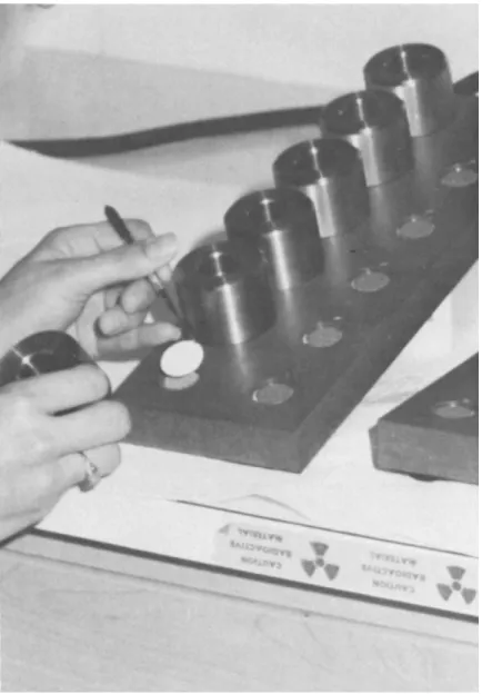
(Duke University Physiological Instrument Shop, Durham, North Carolina) is used in which the filter rests on a stainless steel grid and is held in place by a weighted stainless steel wall (Fig. 1) (7).
Prepare the filtration apparatus for use by rinsing the metal grids and place a Whatman GFC glass fiber filter on each,
Fig. 1. Suction filtration device used for radioligand binding assay. The glass fiber filter rests on a stainless steel grid and is held in place by the metal weight that serves as a reservoir above the filter. [From Williams and Lefkowitz (7).]
with the cross-hatched side of the filter facing upward. Po- sition the steel wall around each filter and fill with 2 ml of incubation buffer. Terminate the incubation of cells with
the radioligand after the appropriate time by adding 4 ml of ice-cold incubation buffer to the tube, using a Cornwall sy- ringe, and quickly pour the contents of the tube through a filter on the suction filtration apparatus. Wash the tube twice, emptying the contents onto the filter, and the filter once with an additional 4 ml volume of buffer. Place the filters in scintillation vials with 10 ml Aquasol (New England Nuclear) and vigorously shake. Wait at least 1 hr before mea- suring radioactivity by liquid scintillation spectrometry.
IV. CALCULATION OF DATA
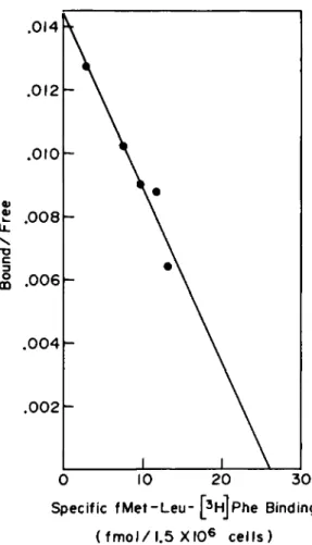
The dissociation constant (2Cd) and number of binding sites per cell can be determined from a saturation curve in which the specific fMet-Leu-[3H]Phe binding is plotted as a function of the concentration of labeled peptide (Fig. 2). Specific binding is determined by subtracting the amount of radioligand that binds in the presence of 10"5 M unlabeled fMet-Leu-Phe from the total binding. Theoretically, the number of receptors
4 6 8 10 [fMet-Leu-[3H]Phe] (nM)
12 14
Fig. 2. Specific binding of fMet-Leu-[3H]Phe to inflam- matory macrophages as a function of fMet-Leu-[3H]Phe concentra-
tion. Guinea pigs were injected intraperitoneally with 25 ml of 0.5% glycogen in saline 3 days prior to harvest of the cells.
[From Snyderman and Fudman (3).]
per cell (or fmol receptor/1.5 x 106 cells as shown) can be calculated from the amount of specific binding present at the point where the curve becomes horizontal. The K^ is the con- centration of fMet-Leu-[^H]Phe needed for half-maximal satura- tion. In practice, these values are more accurately determined by Scatchard analysis (8), an algebraic manipulation of the saturation curve which makes the data linear. A plot of the bound ligand/free ligand ratio as a function of the specific binding is a linear relationship with the slope equal to -1/Kà and the x intercept equal to the number of receptors
per cell (Fig. 3). Total binding rather than nonspecific bind- ing is used in calculating the amount of free ligand. For a more detailed explanation of Scatchard analysis in radioligand binding assays, see Chapter 4 of Williams and Lefkowitz (7).
The affinity of other ligands for the fMet-Leu-Phe receptor can be determined indirectly by measuring their ability to in- hibit fMet-Leu-[^H]Phe binding. The concentration of unlabeled peptide that causes half-maximal inhibition of fMet-Leu-[^H]Phe binding (EC50 binding) is a reflection of its affinity for the receptor. Nonspecific binding, the amount of fMet-Leu-[3H]Phe binding measured in the presence of 10"5 M unlabeled fMet-Leu- Phe, is defined as 0% specific binding or 100% inhibition.
V. CRITICAL COMMENTS
The assay described here demonstrates that N-formylated peptides bind to a specific receptor on macrophages. The bind- ing site may be classified as a receptor because it meets the following criteria (7):
1. The binding is saturable, reversible, and of high affinity.
2. The concentration range over which the binding sites are occupied is comparable to the range over which the ligands elicit a response.
3. The binding sites have specificity for ligands similar to the order of potency with which the ligands elicit a bio- logical response.
Endocytosis of the ligands by mononuclear phagocytes compli- cates the measurement of cell surface receptors. Internaliza- tion of receptor-ligand complexes may prove to be a necessary step in eliciting a chemotactic response, however, it inter- feres with studying whether or not binding is saturable and reversible. Endocytosis of free ligand may produce unaccept- ably high nonspecific binding. We use 0.1 M sodium azide in the incubation mixture to arrest the metabolic functions of
0 10 20 30 Specific fMet-Leu- [3HjPhe Binding
( f m o l / 1 . 5 XIO6 cells)
Fig. 3. Scatchard plot derived from the data in Fig. 1.
Free ligand is the amount of fMet-Leu-[3HJPhe added minus the total fMet-Leu-[3H]Phe binding. The equilibrium dissociation constant (Kd) and number of binding sites per cell are calcu-
lated from the slope and x intercept, respectively. [From Snyderman and Fudman, (3).]
the cell during the binding assay. In the presence of sodium azide we get a nearly linear Scatchard plot and nonspecific binding of only 10-15% on the rapidly rising portion of the saturation curve.
Using this assay, we have demonstrated that iV-formylated peptide receptors on both resident and inflammatory guinea pig peritoneal macrophages have a Κ<$ of about 10 nM and that there are about 10,000 receptors per cell (3).
The radioligand binding assay described here was initially developed for studying human polymorphonuclear leukocytes (4) and was modified for guinea pig peritoneal macrophages by changing the cell concentrations and the incubation time. In adapting this assay for other mononuclear phagocytes, the in- vestigator should determine the optimal cell concentration and incubation time for the particular cell type. One must avoid cell concentrations or conditions that lead to cell agglutina- tion or adherence of the cells to the surface of the tube used for the incubation assay. Sufficient washing of the tubes and filters must be used to obtain good reproducibility and to minimize nonspecific binding.
REFERENCES
E. Schiffmann, B. A. Corcoran, and S. M. Wahl. iV-For- mylated peptides as chemoattractants for leukocytes.
Proc. Natl. Acad. Sei. USA 72.-1059-1062, 1975.
H. S. Showeil, F. J. Freer, S. H. Zigmond, E. Schiffmann, S. Aswanikumar, B. Corcoran, and E. L. Becker. The structure-activity relations of synthetic peptides as chemotactic factors and inducers of lysosomal enzyme secretions for neutrophils. J. Exp. Med. 143:1154-1169, 1976.
R. Snyderman and E. J. Fudman. Demonstration of a chemo- tactic factor receptor on macrophages. J. Immunol. 124:
2754-2757, 1980.
L. T. Williams, R. Snyderman, M. C. Pike, and R. J.
Lefkowitz. Specific receptor sites for chemotactic pep- tides on human polymorphonuclear leukocytes. Proc. Natl.
Acad. Sei. USA 74:1204-1208, 1977.
S. Aswanikumar, B. Corcoran, E. Schiffmann, A. R. Day, R. J. Freer, H. J. Showell, E. L. Becker, and C. B. Pert.
Demonstration of a receptor on rabbit neutrophils for chemotactic peptides. Biochem. Biophys. Res. Commun. 74:
810-817, 1977.
J. Niedel, S. Wilkinson, and P. Cuatrecases. Receptor- mediated uptake and degradation of 125i-chemotactic pep- tide by human neutrophils. J. Biol. Chem. 254:10700- 10706, 1979.
L. T. Williams and R. J. Lefkowitz. "Receptor Binding Studies in Adrenergic Pharmacology." Raven Press, New York, 1978.
8. G. Scatchard. The attraction of proteins for small
molecules and ions. Ann. NY Acad. Sei. 51:660-612, 1949.
![Fig. 2. Specific binding of fMet-Leu-[ 3 H]Phe to inflam- inflam-matory macrophages as a function of fMet-Leu-[ 3 H]Phe](https://thumb-eu.123doks.com/thumbv2/9dokorg/1153060.82998/5.648.68.561.497.792/fig-specific-binding-inflam-inflam-matory-macrophages-function.webp)