in Cardiac Resynchronization Therapy. One Year Experience in a High Volume Center
I. NEUHOFF1, S. SZILÁGYI 2, L. MOLNÁR2, I. OSZTHEIMER2, E. ZIMA2, G.A. DAN1,3, B. MERKELY2, L. GELLÉR2
1“Carol Davila” University of Medicine and Pharmacy, Bucharest, Romania
2Semmelweis University, Heart Center, Budapest, Hungary
3“Colentina” Clinical Hospital, Bucharest, Romania
Introduction. In patients receiving cardiac resynchronization therapy (CRT), failure rate to implant the left ventricular (LV) lead by the traditional trans-venous approach is 4-8%. Surgical epicardial implantation is considered as an alternative, but this technique is not without morbidity.
Evidence from case documentation and from small trial batches demonstrated the viability of endocardial LV lead implantation where surgical epicardial lead placement is not applicable.
Material and Methods. Four patients were implanted with endocardial LV lead using the transseptal atrial approach after unsuccessful transvenous implantation. Implantation of an endocardial active fixation LV leads was successful in all patients with stable electrical parameters immediately after implantation and over the follow-up period. All patients received anticoagulation therapy in order to target the international normalized ratio of 2.5-3.5 and have not experienced any thromboembolic, hemorrhagic events, or infection.
Results. Follow-up echocardiography indicated significant improvement of LV systolic function (24 + 4.9 to 32 + 5.1 %, P = 0.023) with a notable improvement of the functional status.
Conclusions. Endocardial left ventricular lead implantation can be a valuable and safe alternative technique to enable LV stimulation in high surgical risk patients where standard coronary sinus implant is unsuccessful.
Key words: biventricular pacing, transseptal lead, transseptal pacing, endocardial stimulation, cardiac resynchronization therapy.
INTRODUCTION
Cardiac resynchronization therapy (CRT), with atrio-biventricular pacing has been a tremendous advance in the treatment of symptomatic congestive heart failure patients presenting with severely impaired left ventricular function associated with ventricular dyssynchrony and intraventricular con- duction disturbances [1-3]. The standard approach indicates the necessity for the implantation of an LV pacing lead transvenously into one of the tributary veins of the coronary sinus (CS). However, even with the improvement in dedicated devices and increased operator experience, failure to implant a CS lead has been reported in 4-8% of the cases [4, 5]. Unfavorable CS or vein anatomy, such as CS dissection, occlusion, or abnormal ostium of the CS or focal coronary vein stenosis are the most frequent reasons. Post-implant dislocation of the lead, high pacing threshold or phrenic nerve stimulation also diminishes the effectiveness of LV stimulation [6-8]. In cases of unsuccessful LV lead placement
through the CS, surgical epicardial lead implantation via a lateral thoracotomy or minimal-invasive thoracoscopy is considered the standard alternative method that requires general anesthesia [9-11]. In patients with advanced heart failure, this approach may be linked with a high morbidity and mortality.
Endocardial LV lead placement through an atrial transseptal approach has been described previously as an alternative technique to provide CRT in cases where epicardial LV lead implantation is contra- indicated or is at increased risk [12-18]. In the following paper we describe the technique that we used and the outcomes of the transseptal endocardial LV implant in patients for whom transvenous CS lead placement had failed and are not eligible for surgical epicardial implantation and have no contraindication for lifelong oral anticoagulation.
Patients
From October 2014 to October 2015, at the Electrophysiology and PM department of Semmel- weis University Heart Center, four patients (3 males;
ROM. J. INTERN. MED., 2016, 54, 2, 121–127 DOI: 10.1515/rjim-2016-0020
mean age 62.5 years) had undergone endocardial LV lead implantation where LV pacing could not be achieved via the coronary sinus despite the application of interventional techniques including venoplasty. Patients had New York Heart Association III to IV symptoms and had compelling indication for life long systemic anticoagulation. All patients had left bundle branch block (LBBB). Pre-implant- ation echocardiography revealed severely depressed LV function. Patient’s characteristic features are listed in Table 1. Surgical lead implantation was
contraindicated in three patients because of a very high anesthetic and surgical risk. Patient 2 did not sign the consent for epicardial surgical LV lead implantation. In patients 1, 3 and 4, the CRT devices together with the right atrial and ventricular lead were implanted in the first procedure having the port of the LV lead sealed with an IS-1 plug.
The transseptal LV lead implantation was performed on a later procedure. In patient 2 the transseptal LV lead implantation was implanted during the first procedure.
Table 1
Characteristic features of patients
Pt no. Age Gender Etiology of cardiomyopathy
NYHA Functional
Class
QRS (ms)
LVEF (%)
Follow- up months
MR grade before implantation (I-IV)
ACT before implantation
1 60 M Ischemic III 186 28 11 III Yes (for AF)
2 45 M non-ischemic Iva 193 21 10 III Yes (IVT)
3 70 M non-ischemic III-IV 205 27 7 III-IV Yes (for AF)
4 75 F Ischemic III 210 24 6 III Yes (for AF)
Pt, patient, NYHA, New York Heart Association Functional Class, LV, Left Ventricular, LVEF, Left Ventricular Ejection Fraction, MR, Mitral, regurgitation, ACT, Anticoagulation Therapy, IVT, Interventricular Thrombosis, AF, Atrial fibrillation
MATERIAL AND METHODS
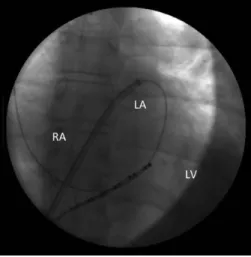
The placement of the LV endocardial lead was done via a combined femoral and subclavian approach. The implantation starts with a standard transseptal puncture from the right femoral vein.
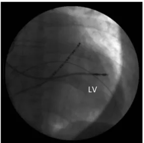
The puncture was performed with the guidance of fluoroscopy and with continuous monitoring of the arterial pressure. Following the successful trans- septal puncture, confirmed by the left atrial pressure curve, 5000 IU of intravenous heparin was administered. A guide wire (0.035 inch × 260 cm) was advanced into one of the pulmonary veins and the dilator was removed and the sheath was with- drawn into the right atrium (Figure 1). An angioplasty balloon (8 mm × 40 mm Fox Plus Abbott Vascular) was positioned across the septal puncture site and was inflated using contrast agent. In a CS guiding sheath a steerable electrophysiological catheter is advanced from the left subclavian area towards the septum of the right atrium. The balloon was deflated then it was withdrawn into the transseptal sheath and the guide wire was maintained in the left atrium (Figure 2). The steerable catheter is pushed on transseptally into the left atrium and then to the left ventricle. Maintaining the electrophysiological catheter in position the guiding sheath is advanced to the left atrium. In cases where the sheath did not advance through the site of the puncture, the angio- plasty balloon was again placed at the puncture site and it was inflated and deflated as per the need
(Figure 3). When we identified the optimal pacing site, the electrophysiological catheter was with- drawn into the sheath and the sheath was pushed against the LV wall to ensure stable position.
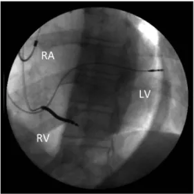
Subsequently, a standard active fixation bipolar lead was screwed into the desired location of the postero-lateral area of the left ventricle (Figure 4).
After measuring the pacing and sensing thresholds, the guiding sheath had been removed using a longitu- dinal slitter tool. Maintaining a sufficient slack in the lead, the proximal end was secured in the pre- pectoral region and connected to the CRT device.In all patients, the atrial lead has been implanted in the right atrial appendage and the right ventricular lead was placed in the right ventricular septum.
Figure 1. A guide wire in the Left Atrium after successful transseptal puncture.
Figure 2 A. Balloon dilatation of the trans- septal puncture site.
Figure 2B. Advancement of the EP catheter to the right atrium.
Figure 2C. The balloon is deflated and withdrawn into the right atrium.
Figure 3A. The EP catheter is advanced trans- septally into the left atrium.
Figure 3B. The EP catheter and the sheath are advanced into the left ventricle.
Figure 4A. The active fixation lead is screwed in the left ventricle.
Figure 4B. Final lead position.
RESULTS
All patients had successfully been implanted with LV endocardial leads. Three of our patients
received CRT-D devices, while one patient received a CRT-pacemaker. A sum-up of the device implantation and follow-up parameters is available in Table 2.
There was no phrenic nerve stimulation observed at stimulation with 10 V, 0.4 msec. During the follow-up period, no lead dysfunction or dislocation was observed. All patients have been on long-term anti- coagulation and the international normalized ratio was maintained between 2.5 and 3.5. There were no hematoma or serious post-procedural bleeding observed due to the use of full anticoagulation during the procedure. None of the patients developed any thromboembolic events on last follow-up. All patients had improvement in the functional status thus symptoms had ameliorated at least one NYHA class. There was a significant improvement in the left ventricular systolic function, from a mean LV ejection fraction of 24 + 4.9 to 32 + 5.1 % (p = 0.023). We did not observe significant changes in the grade of mitral regurgitation throughout the follow-up period (p = 0.18).
Table 2
Implant and Follow-up data of the patients
Pt 1 2 3 4
Device Incepta Boston Scientific, CRT-D
Miami, USA Incepta Boston Scientific, CRT-D
Miami, USA Entovis, HF-T, CRT-P,
BiotronikGmbH&Co, Berlin, Germany Protecta XT, CRT-D, Medtronic;
Minnesota, USA
LV lead Medtronic 5076-65cm Medtronic 5076-65cm Medtronic 5076-65cm Medtronic 5076-65cm
Success Yes Yes Yes Yes
R wave
(mV) Threshold
(Volts/ms) Impedance (Ohms) R wave
(mV) Threshold
(Volts/ms) Impedance (Ohms) R wave
(mV) Threshold
(Volts/ms) Impedance (Ohms) R wave
(mV) Threshold
(Volts/ms) Impedance (Ohms)
Implant 8.5 0.6/0.4 495 15.5 0.7/0.4 754 20 0.8/0.4 678 7.2 0.75/0.4 754
1 Week postimplant 9.1 0.7/0.4 537 14.7 0.7/0.4 461 21 0.9/0.4 658 7.4 1/0.4 713
3 months postimplant 9 0.7/0.4 430 15 0.7/0.4 490 20 0.8/0.4 589 7.3 0.75/0.4 710
6 months postimplant 9.3 0.7/0.4 483 15.3 0.7/0.4 710 22 0.8/0.4 630 7.2 0.75/0.4 720
Complications None none none none
Follow-up NYHA
Class II II-III II II
LVEF at follow-up 38% 29% 35% 27%
Grade of MR at
follow-up III II-III III III
Thromboembolism None none none none
Pt, patient, NYHA, New York Heart Association Functional Class, LV, Left Ventricular, LVEF, Left Ventricular Ejection Fraction, MR, Mitral regurgitation
DISCUSSION
For patients in whom transvenous left ventricular lead placement in the tributary veins of the CS is unsuccessful and are ineligible for surgical epicardial implantation, transseptal endocardial LV lead im- plantation is an alternative method for providing resynchronization therapy. Evidence from case documentation and from small trial batches proved the safety and feasibility of this alternative technique.
In 1998 Jaïs et al. described the first successful implant of endocardial LV lead using a combined approach with a transseptal puncture from the femoral vein and a snare technique through the right jugular vein [12]. Since then, Leclercq et al.
have described several cases of successful LV lead implantation using a direct transseptal approach via the right internal jugular vein [14]. Another group has described a method using direct TS puncture from the left axillary vein [18]. In 2007 Van Gelder et al. and Nuta et al. described successful endocardial LV lead placement from the subclavian vein into the LV cavity via the interatrial septum, following transseptal puncture through the femoral vein to allow entry of the LV lead from above [15, 16]. In our patients, we used the latter approach.
One of the advantages of this interventional alternative technique is that there is no need for general anesthesia and there is minimum post- operative recovery. In comparison to epicardial
pacing, endocardial LV stimulation has some potential advantages. The transseptal access allows a broader choice for the pacing site inside the LV cavity and is not limited by the anatomy of the tributary veins of the CS. In case of a high pacing threshold, the lead can be repositioned to a different site inside the LV cavity, this is often difficult when maneuvering the lead in the cardiac venous system. The risk for phrenic nerve stimulation is low. In our small study population no phrenic nerve stimulation occurred at high energy stimulation and optimal implantation and follow-up electrical para- meters were observed (Table 2). Experimental and clinical observations also indicated that LV endo- cardial stimulation enables a more physiological electrical activation of the left ventricle, with a transmural activation spreading from the endocardium to the epicardium [19, 20]. Better hemodynamic results with higher aortic and mitral time velocity integral, improvement of LV fractional shortening and less ventricular dyssynchrony were reported with endocardial than with epicardial LV stimulation [21, 22]. During follow-up in our patients, we observed that the totally endocardial pacing was correlated with a marked improvement of the LV systolic function and improvement in the functional status of the patients.
The disadvantages of this alternative technique include the risk associated with the transseptal catheterization, the unknown long-term thrombo- embolic risk, the need for lifetime anticoagulation, risks associated with position of the lead across the mitral valve, and the need of complex procedures to manage device related infections. The transseptal approach via the interatrial septum has been used for various interventional and electrophysiological procedures. Therefore, without any doubt, experienced operators have less risk with this procedure. The use of intraoperative anticoagulation and the need of anticoagulation right after the implantation lead to the increase of the risk of periprocedural bleeding complications. In our small study cohort there has not been any complications related to the procedure nor major hematoma was observed. The main concern is regarding the long-term risks of thromboembolic complications associated with endocardial LV stimulation. It is mandatory to use
anticoagulant therapy in these patients and the recommended target INR is between 2.5 and 3.5 [23, 24]. However, the use of anticoagulation is common in the majority of these patients due to the presence of atrial fibrillation. In all of our patients there was an indication for the use anticoagulant therapy, before the implantation of a transseptal system – three patients for paroxysmal atrial fibrillation and one for intraventricular thrombosis.
Long-term follow-up data of the thromboembolic complications in patients who had a transseptal endocardial LV lead implanted are scarce. So far no thromboembolic complications where reported as long as anticoagulant therapy was followed [13, 25-27]. During follow-up no thromboembolic events occurred in our study population. In patients with transseptal CRT with an endocardial LV lead crossing the interatrial septum and the mitral valve, there is a theoretically increased risk of worsening the mitral regurgitation and of mitral valve endo- carditis. No significant increase in the grade of mitral regurgitation has been reported with LV endocardial pacing [13-16, 26, 28]. We did not document any significant deterioration of mitral regurgitation in our study patients. To date there is no record of more frequent endocarditis associated with this technique. Throughout the follow-up period we did not have any lead infection.
CONCLUSIONS
Transseptalendocardial left ventricular lead implantation appears to be a valuable and safe second alternative with the benefit of an endo- cardial pacing site. The disadvantages of this technique are the need for permanent anti- coagulation and the lead interaction with the mitral valve, though any significant complications as a result of this phenomena have not been reported until now. Nevertheless, more data is required to evaluate the safety and long-term efficacy of this alternative technique.
Acknowledgment: This paper is supported by the Sectorial Operational Programme Human Resources Develop- ment (SOP HRD), financed from the European Social Fund and by the Romanian Government under the contract number POSDRU/159/1.5/S/137390.
Conflicts of interest: None declared.
Introducere. Implantarea clasică prin abord epicardic transvenos a electrodului de ventricul stâng (VS) pentru a realiza terapia de resincronizare cardiacă (TRC) nu este posibilă în 4-8% din cazuri. La acei pacienţi, la care plasarea epicardică chirurgicală a electrodului de stimulare al VS nu este posibilă datorită unui risc
anestezico-chirurgical inacceptabil, implantarea endocardică a electrodului de stimulare al VS poate fi o alternative viabilă.
Metode. S-a efectuat TRC în cazul a patru pacienţi, cu plasarea endocardică prin abord transseptal atrial a sondei de VS, după ce implantarea prin abord epicardic transvenos a fost lipsită de succes. La toţi pacienţii s-a realizat poziţionarea cu succes a unui electrod de stimulare cu fixare activă la nivelul endocardului VS, cu parametrii electrici stabili la momentul implantului şi pe perioada de urmărire.
Toţi pacienţii au primit tratament anticoagulant oral cu o ţintă a INR de 2,5-3,5.
Pe perioada de urmărire nu am observant complicaţii tromboembolice, hemo- ragice sau infecţioase.
Rezultate. Toţi pacienţii au avut un răspuns clinic şi structural adecvat cu ameliorarea semnificativă a funcţiei sistolice a VS (24 + 4,9 to 32 + 5.1 % P = 0.023) şi a statusului funcţional.
Concluzii. Plasarea endocardică prin abord transseptal atrial a electrodului de stimulare al VS, poate fi o tehnică intervenţională alternativă, sigură şi valoroasă la acei pacienţi la care riscul anestezico-chirurgical este ridicat şi poziţionarea clasică prin abord transvenos al electrodului de stimulare al VS nu este posibilă.
Correspondence to: Neuhoff István, MD, PhD student, “Colentina” Clinical Hospital
19-21 Ştefan cel Mare, Bucharest, District 2, 020125, Romania, Phone: + 40 751 240 566 E-mail: ineuhoff@yahoo.com
REFERENCES
1. ABRAHAM WT, FISHER WG, SMITH AL, DELURGIO DB, LEON AR, LOH E et al. Cardiac resynchronization in chronic heart failure. N Engl J Med. 2002; 346:1845-53.
2. CAZEAU S, LECLERCQ C, LAVERGNE T, WALKER S, VARMA C, LINDE C et al. Effects of multisite biventricular pacing in patients with heart failure and intraventricular conduction delay. N Engl J Med. 2001; 344:873-80.
3. CLELAND JGF, DAUBERT J-C, ERDMANN E, FREEMANTLE N, GRAS D, KAPPENBERGER L et al. The effect of cardiac resynchronization on morbidity and mortality in heart failure. N Engl J Med. 2005; 352:1539-49.
4. BRISTOW MR, SAXON LA, BOEHMERJ, KRUEGER S, KASSDA, DE MARCO T et al. Cardiac-resynchronization therapy with or without an implantable defibrillator in advanced chronic heart failure. N Engl J Med 2004; 350:2140-50.
5. GRAS D, BOCKER D, LUNATI M,WELLENSHJ, CALVERT M, FREEMANTLE N et al. Implantation of cardiac resynchronization therapy systems in the CARE-HF trial: procedural success rate and safety. Europace 2007; 9:516-22.
6. MEISEL E, PFEIFFER D, ENGELMANN L, TEBBENJOHANNSJ, SCHUERT B, HAHN S et al. Investigation of coronary venous anatomy by retrograde venography in patients with malignant ventricular tachycardia. Circulation 2001; 104:442-7.
7. HIGGINS SL, HUMMEL JD, NIAZIIK, GIUDICI MC, WORLEY SJ, SAXON LA et al. Cardiac resynchronization therapy for the treatment of heart failure in patients with intra-ventricular conduction delay and malignant ventricular tachy- arrhythmias. J Am Coll Cardiol 2003; 42:1454-9.
8. LEÓN AR, ABRAHAM WT, CURTIS AB, DAUBERT JP, FISHER WG, GURLEY J et al. Safety of transvenous cardiac resynchronization system implantation in patients with chronic heart failure: combined results of over 2000 patients from a multicenter study program. J Am Coll Cardiol. 2005; 46:2348-56.
9. MAIR H, JANSENSJL, LATTOUF OM, REICHART B, DABRITZS. Epicardial lead implantation techniques for biventricular pacing via left lateral mini-thoracotomy, video-assisted thoracoscopy, and robotic approach. Heart Surg Forum 2003; 6:412-7.
10. GABOR S, PRENNER G, WASLER A, SCHWEIGER M, TSCHELIESSNIGGKH, SMOLLE -JUTTNERFM. A simplified technique for implantation of left ventricular epicardial leads for biventricular re-synchronization using video-assisted thoracoscopy (VATS). Eur J Cardiothorac Surg 2005; 28:797-800.
11. KOOS R,SINHA AM, MARKUS K, BREITHARDT OA, MISCHKE K, ZARSEM et al. Comparison of left ventricular lead placement via the coronary venous approach versus lateral thoracotomy in patients receiving cardiac resynchronization therapy. Am J Cardiol 2004; 94:59-63.
12. JAÏS P, DOUARD H, SHAH DC, BAROLD S, BARAT JL, CLÉMENTY J. Endocardial biventricular pacing. Pacing ClinElectrophysiol 1998; 21: 2128-31.
13. JAÏS P, TAKAHASHI A, GARRIGUE S, YAMANE T, HOCINI M, SHAH DC et al. Mid-term follow-up of endocardial biventricular pacing. Pacing ClinElectrophysiol 2000; 23: 1744-7.
14. LECLERCQ F, HAGER FX, MACIA JC, MARIOTTINI CJ, PASQUIÉ JL, GROLLEAU R. Left ventricular lead insertion using a modified transseptal catheterization technique: a totally endocardial approach for permanent biventricular pacing in end-stage heart failure. Pacing Clin Electrophysiol 1999; 22:1570-5.
15. VAN GELDER BM, SCHEFFER MG, MEIJER A, BRACKE FA. Transseptal endocardial left ventricular pacing: an alternative technique for coronary sinus lead placement in cardiac resynchronization therapy. Heart Rhythm 2007; 4:454-60.
16. NUTA B, LINES I, MACINTYRE I, HAYWOOD GA. Biventricular ICD implant using endocardial LV lead placement from the left subclavian vein approach and transseptal puncture via the transfemoral route. Europace 2007;9:1038-40.
17. MORGAN JM, SCOTT PA, TURNER NG, YUE AM, ROBERTS PR. Targeted left ventricular endocardial pacing using a steerable introducing guide catheter and active fixation pacing lead. Europace 2009; 11:502-6.
18. JI S, CESARIO DA, SWERDLOW CD, SHIVKUMAR K. Left ventricular endocardial lead placement using a modified transseptal approach. J Cardiovasc Electrophysiol 2004; 15:234-66.
19. FISH JM, DI DIEGO JM, NESTERENKO V, ANTZELEVITCH C. Epicardial activation of left ventricular wall prolongs QT interval and transmural dispersion of repolarization: implications for biventricular pacing. Circulation 2004; 109:2136-42.
20. FISH JM, BRUGADA J, ANTZELEVITCH C. Potential proar rhythmic effects of biventricular pacing. J Am CollCardiol 2005; 46:2340-47.
21. GARRIGUE S, JAIS P, ESPIL G, LABEQUEJN, HOCINI M, SHAH DC et al. Comparison of chronic biventricular pacing between epicardial and endocardial left ventricular stimulation using Doppler tissue imaging in patients with heart failure. Am J Cardiol 2001; 88:858-62.
22. JAISPSF, LABORDERIE J, REUTER S, BORDACHAR P, HSU LF, SANDERS P et al. Tailored endocardial left ventricular pacing is superior to coronary sinus in heart failure patients needing cardiac resynchronization. Heart Rhythm 2006; 3:S247.
23. VAN GELDER BM, BRACKE FA, OTO A, YILDIRIR A, HAAS PC, SEGERJ J et al. Diagnosis and management of inadvertently placed pacing and ICD leads in the left ventricle: a multicenter experience and review of the literature. Pacing Clin Electrophysiol. 2000; 23:877-83.
24. SHARIFI M, SORKIN R, SHARIFI V, LAKIER JB. Inadvertent malposition of a transvenous-inserted pacing lead in the left ventricular chamber. Am J Cardiol 1995; 76:92-95.
25. BRACKE FA, HOUTHUIZEN P, RAHEL BM, VAN GELDER BM. Left ventricular endocardial pacing improves the clinical efficacy in a non- responder to cardiac resynchronization therapy: role of acute haemodynamic testing. Europace. 2010;
12:1032–4.
26. PASQUIEJL, MASSIN F, MACIAJC, GERVASONI R, BORTONE A, CAYLA G et al. Long-term follow-up of biventricular pacing using a totally endocardial approach in patients with end-stage cardiac failure. Pacing Clin Electrophysiol. 2007;
30Suppl 1:S31-3.
27. BORDACHAR P, DERVAL N, PLOUX S, GARRIGUE S, RITTER P, HAISSAGUERRE M, et al. Left ventricular endocardial stimulation for severe heart failure. J Am Coll Cardiol 2010; 56:747-753.
28. KUTYIFA V, MERKELY B, SZILAGYI S, ZIMA E, ROKA A, KIRALY A, OSZTHEIMERI, et al. Usefulness of electroanatomical mapping during transseptal endocardial left ventricular lead implantation. Europace 2012; 14:599-604.
Received November 1, 2015