Reprint
www.chemeurj.org
A Journal of
& Bioinorganic Chemistry
C-terminal Cysteines of CueR Act as Auxiliary Metal Site Ligands upon Hg
IIBinding—A Mechanism To Prevent Transcriptional Activation by Divalent Metal Ions?
Ria K. Balogh,
[a]B8la Gyurcsik,
[a]Pva Hunyadi-Guly#s,
[b]Juliana Schell,
[c]Peter W. Thulstrup,
[d]Lars Hemmingsen,*
[d]and Attila Jancsj*
[a]Abstract: Intracellular CuI is controlled by the transcrip- tional regulator CueR, which effectively discriminates be- tween monovalent and divalent metal ions. It is intriguing that HgII does not activate transcription, as bis-thiolate metal sites exhibit high affinity for HgII. Here the binding of HgIIto CueR and a truncated variant, DC7-CueR, with- out the last 7 amino acids at the C-terminus including a conserved CCHH motif is explored. ESI-MS demonstrates that up to two HgIIbind to CueR, whileDC7-CueR accom- modates only one HgII. 199mHg PAC and UV absorption spectroscopy indicate HgS2structure at both the function- al and the CCHH metal site. However, at sub-equimolar concentrations of HgII at pH 8.0, the metal binding site displays an equilibrium between HgS2and HgS3, involving cysteines from both sites. We hypothesize that the C-ter- minal CCHH motif provides auxiliary ligands that coordi- nate to HgII and thereby prevents activation of transcrip- tion.
The CueR metalloregulatory protein controls cellular copper homeostasis by activating the transcription ofcueO andcopA genes in prokaryotes and some eukaryotes.[1] CueR responds to CuI, AgI and AuI, but not to the divalent ions HgII or ZnII.[2]
SC-XRD studies onEscherichia coliCueR and EXAFS in solution revealed that the inducer metal ions are coordinated by C112 and C120 residues in a linear, bis-cysteinate fashion.[2,3]These two cysteines are essential to the protein function, as shown by mutation studies (C112S and/or C120S) both in vitro[3]and in vivo.[4]
CueR proteins from various bacteria contain two additional well conserved cysteines at the C-terminal, disordered segment of the protein (Figure 1).[2] Crystal structures of the activator
and the repressor forms of the DNA-bound CueR dimer sug- gest that a two-turn helix between the metal binding loop and the CCHH motif may have a key role in the protein function.[5]
Upon AgI binding, the activator conformation is stabilized by the docking of the C-terminal helix (via residues I122, I123, L126) into an opened, hydrophobic pocket, formed by residues of the dimerization helix and the DNA-binding domain. This re- sults in a small “scissoring” movement and bending of the DNA chain allowing the transcription to be carried out by the RNA polymerase. The allosteric role of the C-terminal helix was confirmed by constructing the CuI-independent constitutive activator (T84V/N125L/C112S/C120S) and the constitutive re- pressor (truncation from I122) mutants of CueR.[5]
Several representative examples can be found in the litera- ture where non-cognate metal ions bind to a metalloprotein with the same or even higher affinity than the inducer metal ion. However, despite the high affinity binding of non-cognate metal ions, they cannot trigger the functional structural change of the protein, because the coordination number or geometry differ.[6–9] Thus, studying the interaction of metallo- regulatory proteins with non-cognate metal ions may provide a deeper insight into the mechanism of metal ion selection and the regulation of the transcription.[8]
Although CueR is one of the most thoroughly characterized proteins in the MerR family, the mechanism of discrimination between mono- and divalent metal ions is still not fully under- stood. Surprisingly, HgIIdoes not trigger the activation of tran- scription by CueR,[2] despite its well-known preference for a bis-thiolate coordination environment.[10] O’Halloran et al. de- termined a CuI-binding sensitivity of the CueR protein (1–2 V 10@21m) based on an in vitro transcriptional assay.[2]Our previ- ous studies on model peptides of the metal binding loop of CueR also showed that these fragments bind CuIwith a high Figure 1.Structure of CueR (E.coli) (PDB id.: 1Q05-modified) showing the po- tential metal binding sites (top). Sequence alignment of CueR proteins from various organisms (bottom). Conserved cysteine residues are highlighted in yellow.
[a]R. K. Balogh, Prof. B. Gyurcsik, Prof. A. Jancsj Department of Inorganic and Analytical Chemistry University of Szeged
Djm t8r 7, 6720 Szeged (Hungary) E-mail: jancso@chem.u-szeged.hu [b]Dr. P. Hunyadi-Guly#s
Laboratory of Proteomics Research, Institute of Biochemistry Biological Research Centre of the Hungarian Academy of Sciences Temesv#ri krt. 62, 6726 Szeged (Hungary)
[c] Dr. J. Schell
Institute for Materials Science and Center for Nanointegration Duisburg- Essen (CENIDE)
University of Duisburg-Essen, 45141 Essen (Germany) European Organization for Nuclear Research (CERN) 1211 Geneva (Switzerland)
[d]Prof. P. W. Thulstrup, Prof. L. Hemmingsen Department of Chemistry, University of Copenhagen Universitetsparken 5, 2100 Copenhagen (Denmark) E-mail: lhe@chem.ku.dk
Supporting information and the ORCID identification number(s) for the author(s) of this article can be found under:
https://doi.org/10.1002/chem.201902940.
T 2019 The Authors. Published by Wiley-VCH Verlag GmbH & Co. KGaA.
This is an open access article under the terms of the Creative Commons At- tribution License, which permits use, distribution and reproduction in any medium, provided the original work is properly cited.
Chem. Eur. J.2019,25, 15030 – 15035 www.chemeurj.org 15031 T 2019The Authors. Published by Wiley-VCH Verlag GmbH & Co. KGaA, Weinheim
Communication
affinity.[11] However, according to model peptide studies[12,13]
and QM/MM calculations,[14]HgIIions may be coordinated even more efficiently. Moreover, HgIIis also able to bind to a CC se- quence,[15]and therefore coordination of HgIIion by the CCHH motif is also highly probable.
With the present work we aim to explore the role of the C- terminal CCHH motif with a particular focus on the binding of HgIIto CueR. To achieve this, we studied the HgII-interaction of E. coliCueR and its truncated variant, lacking seven C-terminal residues (including the CCHH motif), DC7-CueR. The integrity of this variant was confirmed by CD spectroscopy and electro- phoretic mobility shift assay, see Figure S3.
A series of ESI-MS spectra were recorded with the two pro- tein variants, see Figures 2, S4 and S5. The disappearance of
the signals of the apo-form in the presence of 1.0 equivalent of HgIIimplies that HgIIions display high affinity to both pro- teins. The spectra obtained at twofold HgII-excess per protein clearly demonstrate the availability of two binding sites for HgII ions in the Wild-type (WT) CueR. These are most likely the metal ion binding loop formed by C112 and C120, and the C- terminal CCHH motif. Participation of the latter CCHH se- quence motif in HgIIbinding is supported by the lack of signals corresponding to a Hg2-DC7-CueR complex, even at twofold HgII-excess over the truncated protein. Both the Hg-CueR and Hg2-CueR species are observed at 1.0 equivalent HgII, suggest- ing that there is no significant difference in the HgII-binding af- finities of the two sites.
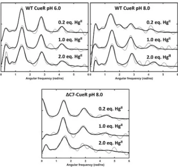
199mHg-perturbed angular correlation (PAC) spectrosco-
py[12,13,16–20]was used to elucidate the metal site structures and
dynamics at the nanosecond timescale, see Figure 3 and Sup- porting Information Figure S6. At pH 6.0 and HgII:CueR of 0.2 and 1.0, the signals agree well with a HgS2coordination geom- etry, that is, coordination of HgII by two cysteinates.[18] This is also the case at HgII:CueR of 2.0, although a slightly larger line- width is observed, in particular for the first peak at around 1.4 radns@1. This line broadening presumably reflects the occu- pation of two HgS2 sites, and it can originate either from minor differences in structure of the two sites, or from metal site dynamics at the nanosecond time scale becoming more pronounced upon binding of the second HgII(Figure 3).
The spectrum recorded with 0.2 equivalent of HgIIper CueR at pH 8.0 is more complex than at pH 6.0. Qualitatively, the first peak is shifted to slightly lower frequency and exhibits considerable broadening, and the second peak (ca.
2.8 radns@1) is significantly attenuated, to the extent that it barely rises above the noise level. A reliable analysis of the data requires the inclusion of two nuclear quadrupole interac- tions (NQIs). One of these NQIs is very similar to that observed in the spectra at pH 6.0, most likely reflecting a HgS2structure.
The other NQI has a higher asymmetry parameter and a lower frequency, see Table S1, indicating a higher coordination number than 2. The lower frequency agrees well with an ideal trigonal planar HgS3structure, but the relatively high asymme- try parameter rules out this possibility. However, in the simple angular overlap model (AOM),[21]a T-shaped HgS3coordination geometry gives the same frequency as a trigonal planar struc- ture, but an asymmetry parameter of 1. Thus, a HgS3structure in between trigonal planar and T-shaped, with the third ligand in a slightly longer Hg@S distance seems to be a plausible structural interpretation of the low frequency signal. It is also Figure 2.Deconvoluted native ESI-MS spectra of the WT and truncated CueR
in the absence and presence of 0.2, 0.5, 1.0 and 2.0 equivalents of HgIIions.
Individual samples contained 20mmprotein in a 10 mmNH4HCO3buffer, 0.5 mmTCEP, pH 7.5.
Figure 3.Experimental (grey) and fitted (black)199mHg PAC spectra of WT and truncated CueR in the presence of DNA with 0.2, 1.0 and 2.0 equivalents of HgII. Top left: WT at pH 6.0; top right: WT at pH 8.0cWT CueR=12mm, 0.5 equiv. DNA, and bottom:DC7-CueR at pH 8.0cDC7-CueR=8.4mm, 0.5 equiv.
DNA.
possible that the PAC data reflect a trigonal planar HgS2N structure, with a histidine coordinating, as this would give an asymmetry parameter different from zero. However, this seems less likely, given the thiophilicity of HgII, and the UV absorption data, vide infra. Finally, it is conceivable that the spectrum re- flects intermediate (nanosecond) exchange between HgS2and HgS3 structures. Notice that this entails a flip of principal axis of the electric field gradient tensor, which has Vzz along the axis of HgS2 but perpendicular to the HgS3 plane, and there- fore the asymmetry parameter will depend on the dynamics in a non-trivial manner. It cannot be excluded that the data re- corded at 1.0 equivalent of HgIIalso contain signals reflecting both of these species, but the reduced chi-square does not im- prove significantly upon including a second NQI. Consequently, we have only included the high frequency NQI (HgS2) in the analysis. For the experiment with 2.0 equivalents of HgII the signal may be satisfactorily fitted with just one (high frequen- cy) NQI, presumably reflecting HgS2 structure for both HgII bound to CueR (Figure 3).
Most interestingly, the 199mHg PAC spectrum recorded at pH 8.0 with 0.2 equivalents HgIIforDC7-CueR exhibits a signal reflecting only HgS2structure (Figure 3). The fact that theDC7- CueR HgII site exhibits a HgS2 structure strongly supports the interpretation presented above for the WT CueR: if HgS3 is formed by occupation of the functional site, a third thiolate is recruited from the CCHH motif, or vice versa, HgIIbinds to the CCHH motif and recruits one of the cysteines from the func- tional binding site. With 2.0 equivalents of HgII perDC7-CueR at pH 8.0, the signal changes as compared to experiments with ,1 equivalent HgII, presumably because the functional metal site is filled, and the additional HgIIaccommodates a coordina- tion geometry other than linear HgS2due to weak or non-spe- cific HgIIadducts. This agrees well with the ESI-MS data, where no Hg2-DC7-CueR was observed. Thus it is likely that the signal includes more than one NQI. Surprisingly, the signal shifts to slightly higher frequency, which is difficult to account for, except if a positive charge appears in the equatorial plane of HgS2, vide infra.
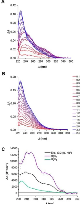
To further characterize the metal site coordination geome- tries, we applied UV absorption spectroscopy (Figure 4). HgII- thiolate complexes possess characteristic charge transfer (CT) bands in the region of 230–300 nm. Moreover, features of the absorption spectrum reflect the coordination geometry of the complexes. Using Hg(SEt)2 and [Et4N][Hg(SBut)3] model com- pounds, the UV-absorption spectra of linearly and trigonal planar coordinated HgII, respectively, were characterized.[24]Lin- early coordinated HgII-thiolate species display a transition at around 230 nm.[22] The increase of the coordination number shifts the absorption bands towards longer wavelengths.[23,25]
The spectrum of a trigonal HgII-thiolate complex has a charac- teristic absorption maximum at 245 nm with a distinct should- er at around 290 nm.[22]Qualitatively, the absorption difference spectra at sub-equimolar HgII:WT CueR ratios exhibit a charac- teristic absorption at around 290 nm reflecting the presence of HgS3structure (Figure 4), in agreement with the PAC data, vide supra. The PAC data indicate 40 % HgS3 and 60 % HgS2 at 0.2 equivalents HgII. We used the recorded spectrum with
2.0 equivalents HgIIper WT CueR (Figure 4A) to determine the molar absorption of the HgS2 species (green curve in Fig- ure 4C). Next, we predicted the pure HgS3 molar absorption spectrum (Figure 4C, purple curve) by assuming that the ex- perimentally determined spectrum is given by 0.6 HgS2+0.4 HgS3. The UV absorption spectra derived in this manner for HgS2 and HgS3 agree well with those reported in the litera- ture,[23]strongly supporting the interpretation of the PAC data presented above. We present molar absorption data at select- ed wavelength values in Table 1. The UV absorption spectra re- corded forDC7-CueR exclusively exhibit the signature of HgS2
Figure 4.UV absorption difference spectra of WT CueR (A) andDC7-CueR (B) titrated with HgIIions (0.1–2.4 equivalents). Spectra recorded in the pres- ence of 1.0 and 2.0 equivalents of HgIIare shown with dashed lines. pH 7.5, cWT CueR=14mm,cDC7-CueR=12mm. (C) Estimated molar absorbance for the HgS2and HgS3species derived from the WT CueR UV absorption spectrum recorded with 2.0 equiv. HgIIand 0.2 equiv. HgIIcombined with the relative population of the two species derived from199mHg PAC data, see the text for details.
Chem. Eur. J.2019,25, 15030 – 15035 www.chemeurj.org 15033 T 2019The Authors. Published by Wiley-VCH Verlag GmbH & Co. KGaA, Weinheim
Communication
structure, corroborating the interpretation of other experimen- tal data. Surprisingly, the absorbance for DC7-CueR continues to increase beyond 1.0 equivalent HgIIand saturates at ca. 2:1 HgII:DC7-CueR, indicating that the truncated protein can ac- commodate two HgIIions in a HgS2coordination environment.
This may be realized if a dinuclear Hg2S2 site is formed with the two thiolates as bridging ligands. Interestingly, this agrees with the unexpectedly high frequency observed by PAC spec- troscopy, which can be explained by the presence of a positive charge (the second HgII) in the Hg2S2structure, vide supra. The fact that the species with two HgIIbound per CueR monomer is not observed in ESI-MS implies that the binding of the second HgIIis relatively weak.
In Figure 5, we present model structures which agree with all the experimental data presented in this work. At pH 8.0
with 0.2 and 1.0 equivalent HgII, two species co-exist, most likely the linear HgS2and a HgS3 structure with the equatorial Hg-S bond being longer than the other two. Such structures have also been observed in small, HgII containing inorganic compounds.[26] The increased availability of deprotonated cys- teines with increasing pH agrees well with this change in spe- ciation observed from pH 6.0 to pH 8.0, that is, a change from HgS2towards HgS3coordination mode, and a similar trend has been observed for de novo designed proteins by Iranzo et al.[18]The additional thiolate is most likely recruited from the CCHH motif, or vice versa, and may thus prevent the docking of the C-terminal helix into the hydrophobic pocket, and con- sequently inhibit activation of transcription. The net negative charge of HgS3may be stabilized due to the presence of lysine or arginine in the C-terminal fragment of CueR in almost all the organisms listed in Figure 1. That is, we hypothesize that the CCHH motif is not involved in the function of CueR when sensing the monovalent coinage metals, but it does take part in binding of divalent metal ions, a mechanism that would ac- count for the selectivity of CueR.
It may seem intriguing that with 1.0 equivalent HgIIboth the PAC and UV absorption spectra differ significantly from those recorded with 0.2 equivalent HgII. However, a simple probabil- istic model qualitatively accounts for this change, assuming that the two sites are independent (i.e. distributing HgII ran- domly among the 4 metal sites of a protein dimer), and that population of two adjacent sites (the functional site and the C- terminal site) leads to formation of HgS2, because there are no more cysteines locally available to form HgS3, see the Support- ing Information for details. This very simple interpretation is to some extent supported by the ESI-MS data, which display pop- ulation of the Hg2-CueR species when HgIIand CueR are pres- ent in equimolar amounts. Obviously, the model is too simple because formation of HgS3 requires that cysteines from both metal binding sites are involved, but the alternative, that is, that one binding site (either the functional site or the C-termi- nal site) binds HgII with significantly higher affinity than the other, does not agree with the spectroscopic data, because this would imply that the HgS2/HgS3ratio should be the same at 0.2 and at 1.0 equivalent, nor with the ESI-MS data, which indicate the presence of Hg2-CueR already at 1.0 equivalent HgII. At 2.0 equivalents HgII, of course, there is no more possi- bility to form HgS3, because the protein is saturated with HgII in HgS2structures. Similar geometrical rearrangement was ob- served in metallothioneins (by UV absorption) upon saturating the protein by the metal ion in a titration with HgII.[27,28] The function of the CCHH motif has also been studied by Stoyanov and Brown, using an in vivo assay to monitor the CueR con- trolled transcription.[4] The double mutation of histidine (H131N/H132N) or cysteine residues (C129S/C130S) and trunca- tion from G128 in E. coliCueR resulted in an only slightly al- tered induction of the transcription by cognate metal ions. Al- though experimental data were not presented, Stoyanov and Brown indicated that the selectivity of reaction with other, un- specified metal ions was not affected. To further explore this issue, a series of in vitro and in vivo transcriptional assays should be conducted.
Figure 5.Model structures of HgIIbound to WT CueR at pH 8.0. Binding of HgIIto CueR gives rise to an equilibrium between HgS2and HgS3when HgII:CueR<2, and to pure HgS2coordination upon addition of 2 HgIIions per protein monomer. This can only be accounted for if the CCHH C-termi- nal motif participates in the coordination of HgII, see the text for details.
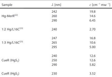
Table 1.Spectroscopic properties of the HgS2and HgS3 species com- pared to HgII/MerR and Hg/L16C complexes. The two entries for CueR are from this work, see Figure 4C.
Sample l[nm] e[cm@1mm@1]
Hg-MerR[22] 242 19.8
260 14.6
290 6.45
1:2 Hg/L16C[23] 240 2.70
1:3 Hg/L16C[23] 247 16.8
265 10.6
295 5.00
CueR (HgS3) 240 12.6
250 12.6
290 5.82
CueR (HgS2) 230 3.52
In summary, we have demonstrated that up to two HgIIions bind with high affinity to WT CueR, one at the functional (C112 and C120) metal binding site, and the other at the C-terminal CCHH motif. Moreover, under conditions where the protein is not saturated by HgII, a higher coordination number (presuma- bly HgS3) is observed for WT CueR but not forDC7-CueR, indi- cating that side chains from the CCHH motif may be recruited as auxiliary ligands at the functional metal site (or vice versa).
This implies a mechanism where the specificity of CueR for monovalent coinage metal ions and against divalent metal ions is achieved by coordination to divalent metal ions by the CCHH motif, preventing the docking of the C-terminal helix into the hydrophobic pocket,[5]and consequently inhibiting ac- tivation of transcription. Indeed, the CCHH motif provides a se- lection of ligands that may participate in coordination of both soft and intermediate metal ions. As the findings presented here on HgIIdo represent a special case, the generalization to other divalent metal ions should be considered carefully.
Acknowledgements
We acknowledge the financial support received from the Fed- eral Ministry of Education and Research (BMBF) through grant 05K16PGA and from the European Union’s Horizon 2020 Framework research and innovation program under grant agreement no. 654002 (ENSAR2). We further thank J.G. Correia (C2TN-DECN-IST-UL) and project CERN-FIS-PAR-0005–2017 FCT- Portugal for technical assistance during the beam time and Jens-Christian Navarro Poulsen and Morten J. Bjerrum (Univer- sity of Copenhagen) for their support during expression and purification of the DC7-CueR variant. We thank ISOLDE/CERN for beam time, EURONS and NICE for financial support. Finan- cial support from the Hungarian National Research, Develop- ment and Innovation Office (GINOP-2.3.2-15-2016-00038, GINOP-2.3.2-15-2016-0001 and K 16/120130) is also acknowl- edged.
Conflict of interest
The authors declare no conflict of interest.
Keywords: coordination modes · CueR metalloregulatory protein · mercury · metal ion selectivity · perturbed angular correlation (PAC) spectroscopy
[1] J. V. Stoyanov, J. L. Hobman, N. L. Brown,Mol. Microbiol.2001,39, 502 – 512.
[2] A. Changela, K. Chen, Y. Xue, J. Holschen, C. E. Outten, T. V. Halloran, A.
Mondragjn,Science2003,301, 1383.
[3] K. Chen, S. Yuldasheva, J. E. Penner-Hahn, T. V. O’Halloran,J. Am. Chem.
Soc.2003,125, 12088– 12089.
[4] J. V. Stoyanov, N. L. Brown,J. Biol. Chem.2003,278, 1407 – 1410.
[5] S. J. Philips, M. Canalizo-Hernandez, I. Yildirim, G. C. Schatz, A. Mondra- gjn, T. V. O’Halloran,Science2015,349, 877–881.
[6] M. V. Golynskiy, W. A. Gunderson, M. P. Hendrich, S. M. Cohen,Biochem- istry2006,45, 15359 –15372.
[7] J. S. Cavet, W. Meng, M. A. Pennella, R. J. Appelhoff, D. P. Giedroc, N. J.
Robinson,J. Biol. Chem.2002,277, 38441–38448.
[8] Z. Ma, D. M. Cowart, R. A. Scott, D. P. Giedroc,Biochemistry2009,48, 3325 –3334.
[9] C. M. Phillips, E. R. Schreiter, Y. Guo, S. C. Wang, D. B. Zamble, C. L.
Drennan,Biochemistry2008,47, 1938 –1946.
[10] D. C. Bebout, Encyclopedia of Inorganic and Bioinorganic Chemistry 2011.
[11] E. Mesterh#zy, B. Boff, C. Lebrun, P. Delangle, A. Jancsj, Inorg. Chim.
Acta2018,472, 192 –198.
[12] D. Szunyogh, H. Szokolai, P. W. Thulstrup, F. H. Larsen, B. Gyurcsik, N. J.
Christensen, M. Stachura, L. Hemmingsen, A. Jancsj,Angew. Chem. Int.
Ed.2015,54, 15756–15761;Angew. Chem.2015,127, 15982– 15987.
[13] D. Szunyogh, B. Gyurcsik, F. H. Larsen, M. Stachura, P. W. Thulstrup, L.
Hemmingsen, A. Jancsj,Dalton Trans.2015,44, 12576–12588.
[14] L. Rao, Q. Cui, X. Xu,J. Am. Chem. Soc.2010,132, 18092– 18102.
[15] T. M. DeSilva, G. Veglia, F. Porcelli, A. M. Prantner, S. J. Opella,Biopoly- mers2002,64, 189– 197.
[16] S. Chakraborty, S. Pallada, J. T. Pedersen, A. Jancso, J. G. Correia, L. Hem- mingsen,Acc. Chem. Res.2017,50, 2225 –2232.
[17] A. Jancso, J. G. Correia, A. Gottberg, J. Schell, M. Stachura, D. Szunyogh, S. Pallada, D. C. Lupascu, M. Kowalska, L. Hemmingsen,J. Phys. G2017, 44, 064003.
[18] O. Iranzo, P. W. Thulstrup, S.-b. Ryu, L. Hemmingsen, V. L. Pecoraro, Chem. Eur. J.2007,13, 9178 –9190.
[19] L. Hemmingsen, K. N. Sas, E. Danielsen, Chem. Rev. 2004, 104, 4027 – 4062.
[20] T. Butz, S. Saibene, T. Fraenzke, M. Weber,Nuclear Instruments and Meth- ods in Physics Research Section A: Accelerators, Spectrometers, Detectors and Associated Equipment1989,284, 417 –421.
[21] R. Bauer, S. J. Jensen, B. Schmidt-Nielsen,Hyperfine Interact. 1988,39, 203– 234.
[22] S. P. Watton, J. G. Wright, F. M. MacDonnell, J. W. Bryson, M. Sabat, T. V.
O’Halloran,J. Am. Chem. Soc.1990,112, 2824– 2826.
[23] G. R. Dieckmann, D. K. McRorie, D. L. Tierney, L. M. Utschig, C. P. Singer, T. V. O’Halloran, J. E. Penner-Hahn, W. F. DeGrado, V. L. Pecoraro,J. Am.
Chem. Soc.1997,119, 6195 –6196.
[24] J. G. Wright, M. J. Natan, F. M. MacDonnel, D. M. Ralston, T. V. O’Halloran, Progress in Inorg. Chem.1990,38, 323– 412.
[25] F. Jalilehvand, B. O. Leung, M. Izadifard, E. Damian,Inorg. Chem.2006, 45, 66– 73.
[26] A. Manceau, K. L. Nagy,Dalton Trans.2008, 1421– 1425.
[27] E. Freisinger,Inorg. Chim. Acta2007,360, 369–380.
[28] O. Schicht, E. Freisinger,Inorg. Chim. Acta2009,362, 714– 724.
Manuscript received: June 26, 2019 Accepted manuscript online: July 31, 2019 Version of record online: October 15, 2019
Chem. Eur. J.2019,25, 15030 – 15035 www.chemeurj.org 15035 T 2019The Authors. Published by Wiley-VCH Verlag GmbH & Co. KGaA, Weinheim