I N T R O D U C T O R Y L E C T U R E D . S H U G A R
Institute of Biochemistry & Biophysics, Academy of Sciences, Warsaw
Interest in the photochemistry of nucleic acids has long stemmed from the established involvement of these important biological polymers in the biological effects of irradiation in the quartz u.v. The phenomenon of photoreactivation (PR), as well as the subsequent elucidation of additional cellular processes which are capable of repairing or reversing u.v. lesions in nucleic acids, provided an additional incentive to such studies, the more so in that reversible reactions are more readily unravelled experimentally.
The rate of growth of work in this and related fields since the last Congress in Copenhagen in i960 has, however, been truly phenomenal and accompanied by numerous important advances. It should be recognized that this is due not only to the biological significance of the subject, but is related in part to the general revival of interest in photo- chemical reactions, with increased emphasis on photoproduct isolation and identification as compared to the earlier widespread use of kinetics as an indirect tool for this purpose.
Since the accompanying rapporteur lecture is devoted exclusively to biological aspects, we shall attempt to present a brief general outline of several features of the photochemistry of nucleic acids and their derivatives, with some reference to their biological significance.
M O N O C H R O M A T I C S O U R C E S
It is, in my opinion, neither mundane nor superfluous to emphasize at the outset that the use of monochromatic radiation of different wavelengths has become a very important requirement not only for studies on the photochemistry of isolated nucleic acids, but also in photobiological work in general. The hitherto widespread use of resonance lamps, although still of value for many purposes, is more and more becoming a limiting factor in experimental work as our knowledge of the basic processes increases. This can be overcome in almost all biological studies, as well as in some aspects of nucleic acid
37
38 P H O T O C H E M I S T R Y O F N U C L E I C A C I D S
photochemistry, with the aid of new commercially available mono- chromators or of laboratory models specifically designed for this purpose. In many experiments band-pass and cut-off filters may often replace monochromators, but all too often at the expense of quantita- tive measurements. On the other hand, for photoproduct isolation on a preparative scale, the use of commercial or laboratory designed reactors with suitable band-pass or cut-off filters frequently offers more advantages than a monochromator.
P H O T O H Y D R A T I O N O F U R A C I L A N D C Y T O S I N E
By comparison with the complex effects of ionizing radiations, the photochemistry of pyrimidines and nucleic acids is considerably simplified by the fact that the naturally occurring purines are almost one hundred-fold more radiation resistant than the commonly occurring pyrimidines. All the available evidence points to the validity of this relationship when the various bases are incorporated in polymer chains. Hence, in the absence of evidence to the contrary, our attention need be confined mainly to the pyrimidines in the interpretation of biological experiments, if we exclude photosensitization, which will be briefly discussed later on.
The first important photochemical transformation of a pyrimidine to be elucidated was that of uracil and its glycosides, which undergo photohydration at the 5,6 bond, the location of the water hydroxy 1 on the 6-position of the uracil ring being confirmed by a number of methods, including direct chemical synthesis. The water molecule is readily eliminated, to regenerate the parent compound, by acid, heat at neutral pH, and to some extent in alkali.
Evidence has also been advanced for an analogous photohydration of the 5,6 bond in cytosine and its glycosides. These are, however, much more labile than their uracil analogues, thus effectively pre- cluding their isolation. The evidence for the hydration reaction in this instance is consequently indirect, the most convincing being the similarity of the u.v. absorption spectra of the photoproducts to those of the corresponding dihydroderivatives of cytosine and its glycosides, as well as to the dihydroderivatives of uracil and its glycosides in alkaline medium.
Objections have been raised against the above interpretation on the grounds that the absorption spectra of the photoproducts of cytosine and i-methylcytosine are not fully in accord with the above. An alter- native proposal was advanced to the effect that the photoproducts of
39 cytosine and its glycosides are the result of an intramolecular isomer- ization. Since this has led to some doubts, which are repeatedly cropping up in the literature, as to the nature of cytosine photo- products, a few words on this subject are called for.
It was pointed out some time ago that the difference in behaviour between cytosine and ι-methylcytosine on the one hand, and cytosine glycosides on the other, was due to the much more rapid rate of
80
60
.40
20h
R^H or alky I R2=H oralkyl
R3=alkyl orribose or ribosephosphate
2200 2400 2600 2800 Wavelength ( a ) 3000 FIG. Ι. Photochemical transformation of i-methyl-4-methylamino- cytosine, i.e. Rx = H, R2 = C H3, R3 = C H3. Irradiation was at 254 nm and longer wavelengths at neutral pH :
a—Absorption spectrum of solution prior to irradiation.
b to e—Absorption spectra following increasing periods of irradiation to about 85 per cent photoproduct formation for curve e. T h e spectral changes resulting from irradiation are almost identical when R3 = ribose.
f—Absorption spectrum following heating of photoproduct at neutral pH. Reversal is not quantitative due to partial destruction of the primary, hydrated, photoproduct by irradiation (from FIKUS et al, 1962).
40 P H O T O C H E M I S T R Y O F N U C L E I C A C I D S
dehydration of the primary photoproducts of the former, the reaction being a function of the ionic strength of the medium. This has since been confirmed: (a) by the late A . M . M O O R E (1963), who showed that the u.v. absorption spectrum of cytosine photoproduct in unbuffered medium at pH 7 is indeed similar to that for cytosine glycoside photo- products, and (b) by an examination of the photoproducts of mono- and di-methylamino substituted cytosines and their glycosides. The photoproducts of these derivatives, although formed in low quantum yield, are remarkably stable ; and this has made it possible to show that the photoproduct of 1-methyl-4-methylaminocytosine (see Fig. 1) is practically identical with that of the corresponding methylamino glycoside (FIKUS et al, 1962). It should be added that these photo- products are sufficiently stable to permit of n.m.r. measurements, and it would be highly desirable that this be done in order to verify directly whether the water hydroxyl is on the 6-position of the cytosine ring.
S O M E M O D E L O L I G O - A N D P O L Y N U C L E O T I D E S
The foregoing should not be taken to imply that hydration is neces- sarily the sole reaction of cytosine residues in irradiated R N A and DNA, and it is instructive to re-examine some of the available data bearing on this question.
When a cytosine residue is adjacent to a purine, as in ApCp or GpCp, it can be shown that photohydration is the only reaction, the purine being unaffected; furthermore, the reaction is quantitatively reversible in the dark. For a cytosine residue sandwiched between two purines, as in GpCpGpCp, the foregoing also applies (WIERZCHOWSKI and SHUGAR, i960, 1962).
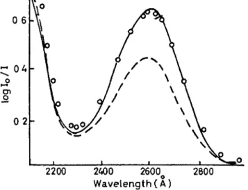
The experimental data are not quite so unequivocal when a cytosine residue neighbours on another pyrimidine. For CpU, for example, dark reversibility following almost complete photolysis is only about 90 per cent. For CpCp it is also about 90 per cent, with evidence that at least part of the non-reversible reaction occurs during the initial stage of irradiation. For poly-C, dark thermal reversibility is only about 85 per cent. All this leads to the inference that, while cytosine hydration is the predominant reaction of cytosine residues in poly- nucleotides, some other reactions appear to be involved which require further investigation. This is also underlined by HAUG'S (1964b) study on T p C where, apart from hydration of cytosine residues, three other photoproducts accounted for 20 per cent of the total; these were isolated chromatographically, but not identified. Figure 2, from
FIG. 2. Photoproduct formation from T p C by irradiation in neutral, aqueous medium at 280 nm. Continuous line is absorption spectrum prior to irradiation, dashed line following irradiation to a stationary state. Note in the latter the maximum at about 245 nm corresponding largely to hydrated cytosine, and the maximum at about 270 nm corresponding to unchanged thymine. That hydrated cytosine is not the only photoproduct is testified to by the fact that thermal reversal gives back only 80 per cent of the original T p C . T h e increased absorption of the photoproduct above 300 nm is also indicative of formation of photoproducts other than cytosine hydrates (from
HA U G , 1964a).
It has also been reported that cytosine may dimerize with other pyrimidines, and then undergo deamination, but the evidence in this case is not convincing. Finally, reference should be made to the finding of RUPERT (1963) that poly-dGC exhibits slight susceptibility to the photoreactivating enzyme; although puzzling, this may well imply that a few cytosine residues do indeed dimerize.
HAUG'S paper, exhibits the absorption spectrum of T p C prior to, and following, photochemical transformation to a stationary state. The dashed curve is the absorption spectrum following irradiation and corresponds roughly with that to be expected. But dark thermal reversibility was only 80 per cent, in agreement with the isolation of other photoproducts.
42 P H O T O C H E M I S T R Y O F N U C L E I C A C I D S
P Y R I M I D I N E D I M E R I Z A T I O N
A good deal of work had already been done on photohydration of pyrimidines at the time BEUKERS and BERENDS (i960) first isolated the thymine photodimer, although the formation of such dimers was independently postulated from the results of work on poly-U and on D N A films. It was soon demonstrated that, under appropriate conditions, dimer formation could occur between a variety of free and substituted uracil and thymine derivatives.
Dimer formation immediately aroused wide interest because of the remarkable stability of the dimers and their ability to dissociate photo- chemically to release the original pyrimidine rings. The acid stability of the dimers made possible their direct isolation from hydrolysates of natural nucleic acids and hence an examination of their biological role.
This, in turn, stimulated intensive investigations on the kinetics of formation and dissociation of thymine dimers, the latest and most elegant of which is the study of JOHNS et al (1964) on T p T . Four photoproducts were isolated chromatographically, formed according to the following scheme :
T p T T p T
I
«04«02
«20 T p T2
T p T4
31311m Ä43 Ä34 240 nm T p T3
Two of these, as seen from the diagram, are formed reversibly from T p T and are isomeric dimers. Another photoproduct, T p T4, is produced in low yield irreversibly from T p T . T p T4 absorbs to the red of 300 nm and most likely bears some relationship to a similar photoproduct observed in irradiated D N A or frozen solutions of thymine ; it is converted by irradiation at 313 nm to another product T p T3 which, in turn, reverts to T p T4 on irradiation at 240 nm. The nature of these two interconvertible photoproducts, as well as the two isomeric dimers, remains to be established.
In natural nucleic acids steric considerations would, of course, lead
one to expect only one isomeric dimer for intrastrand dimerization, viz. a eis head-to-head dimer. For interstrand dimerization, that is cross-linking, additional possibilities exist. At the moment the chances of definite identification of the isomeric nature of dimer photoproducts from irradiated nucleic acids do not look promising.
B I O L O G I C A L R O L E O F D I M E R I Z A T I O N
Qualitative demonstrations of dimer formation in irradiated nucleic acids and micro-organisms were rapidly obtained in several labora- tories. It remained for W U L F F and RUPERT (see RUPERT, 1963), however, first to pin down the biological significance of this reaction when they showed not only that u.v. inactivation of transforming D N A was accompanied by thymine photodimerization, but that subsequent treatment of the irradiated D N A with the PR enzyme in the presence of visible light resulted in the disappearance of the photodimers with concomitant reactivation of the D N A .
This biological evidence was rapidly supplemented by the elegant demonstration by SETLOW and SETLOW (1962) that inactivation of transforming D N A at 280 nm is partially reversed by subsequent irradiation at 230 nm, in accordance with the expected photochemical behaviour of such dimers from studies on model polynucleotides. It was again shown by BOLLUM and SETLOW (1963) that thymine photo- dimerization is likewise involved in the u.v. inactivation of D N A primer activity in the calf thymus polymerase system.
The available evidence points to these dimers as being largely intrastrand in origin. However, the possibility of partial involvement of interstrand dimers cannot be fully excluded. Detailed investigations of this latter phenomenon, in particular that of WILSON and GROSSMAN (1964) on model polymers, which included the alternating twin- stranded poly-dAT where intrastrand dimerization cannot occur, suggest that such cross-linking proceeds with much lower yield and is of lesser importance in biological inactivation.
Summing up, it will be seen that there is sound evidence for the postulate that thymine dimerization is involved in D N A inactivation and that a function of the PR enzyme is to dissociate these dimers.
There is one experiment in apparent contradiction with this con- clusion. KLECZKOWSKI and KLECZKOWSKI (1964, personal communica- tion) have examined the inactivation of a Rhizobium bacteriophage at different wavelengths and find that, following inactivation at 285 nm, irradiation at 230 nm gives no reversal but additional inactivation.
44 P H O T O C H E M I S T R Y O F N U C L E I C A C I D S
But it seems unlikely that this result invalidates the above findings since the high absorption of the phage protein component at 230 nm may effectively reduce the dose directed at the nucleic acid component.
Furthermore the protein component may conceivably cross-link with the nucleic acid component (SMITH, 1963a), particularly at 230 nm;
such cross-linking of protein to nucleic acid has been demonstrated in u.v. irradiated T M V , the cross-links being non-covalent. This may also be related to the failure of irradiated intact T M V to exhibit photo- reactivation, and undoubtedly merits closer examination.
R O L E OF P H O T O H Y D R A T I O N
Thymine dimerization is not the only reaction in irradiated nucleic acids. Cytosine hydration proceeds with a comparable yield. In fact, in a rigid twin-stranded D N A helix, where adjacent thymine rings are rotated out of line with respect to each other by 360, it is difficult to see how dimerization could be the primary event. It is worth recalling that in twin-stranded poly-(A + U) the sole initial reaction on irradiation is hydration; only following partial collapse of the twin-stranded structure, as a result of hydration, is dimerization initiated. This is even more strikingly illustrated by some unpublished experiments in our laboratories by Dr K.L.WIERZCHOWSKI on twin-stranded poly- (A + rT), where hydration is excluded. This twin helix is remarkably radiation resistant when compared with the rapid rate of dimerization in poly-rT alone.
It consequently appears likely that thymine dimerization is preceded and accompanied by cytosine hydration in irradiated D N A . We shall ignore at this point other possible reactions, for example between thymine and cytosine residues as in T p C (HAUG, 1964b), between cytosine residues as in CpCp and poly-C, etc. Any modification of one or more bases may lead to loss in activity of a given biological marker. But, in terms of present concepts of coding, dimerization between two adjacent bases must necessarily lead to an interruption of the message contained in the polymer, resulting in lethality (or latent lethality, since the effect can be reversed). In the case of cross-linking by H N 02 this has recently been directly demonstrated (ZIMMERMAN and GEIDUSCHEK, 6th Intern. Biochem. Cong.).
A quite different situation prevails when we are dealing with a reaction which modifies an individual base. One such reaction is hydration, although it may prove to be not the only one. Let us now examine this in more detail.
I N T R O D U C T O R Y L E C T U R E 45
2200 2400 2600o 2800 Wavelength (A)
FIG. 3. Absorption spectrum at neutral pH of A p U p : prior to irradiation, following irradiation at 254 nm to a photostationary state, O O O O following removal of irradiation source and heating the solution for 3 h at 8o° C. Note that thermal reversal is quantitative.
and no modification of the A p component. Note that the photoproduct ApUp H20 must be heated at pH 7 for 3 h at 8o° C to fully eliminate the water molecule. Even at 50° C dehydration is so slow as to be barely measurable in this time interval. The foregoing example was selected for purposes of illustration since both the forward and dark reactions are fully quantitative. But when we examine other uracil- containing model oligo- and polynucleotides, it is found in all instances that uracil hydration products are stable for many hours at 370 C.
Hydrated cytosine residues are, of course, much more labile. For example, in ApCp where photohydration and dark reversal are
Following discovery of pyrimidine photodimers, a tendency arose to discount the potential role of hydration on the grounds of the relative instability of photohydrated pyrimidines. Looked at from the point of view of the chemist, this is sound reasoning. But are hydrated pyri- midines really so ( unstable ' ? Let us first examine the case of hydrated uracil residues at physiological pH.
Figure 3 presents the behaviour of a dinucleotide ApUp, irradiation of which at 254 nm leads to quantitative hydration of the uracil ring
φ P H O T O C H E M I S T R Y O F N U C L E I C A C I D S
quantitative, the dark reaction requires 3 h at 300 C and pH 7 to go to completion. For GpCp it is about 20 min at 8o° C. For the cytosine residue in C p U the ti/2 for dehydration at 30°C is about 90 min.
Approximately the same value prevails for some of the cytosine resi- dues in poly-C. Extensive data for dehydration rates of cytosine residues, both in the free state and when incorporated in model polymers, are available (WIERZCHOWSKI and SHUGAR, 1961, 1962).
It is clear, however, that even at 370 C we may expect a number of hydrated cytosine residues to be present in irradiated nucleic acids even after a period of an hour in the dark, which is certainly long enough to produce some biological effects during post-irradiation metabolism.
I M P L I C A T I O N O F P H O T O H Y D R A T I O N IN B I O L O G I C A L E F F E C T S
It was reported some time ago by KAPLAN et al (i960) that, while PR for lethality of the χ-phage of Serratia marcescens proceeds only following adsorption of the phage to the host cells, photoreversion of mutations (actually pre-mutations) occurred readily on warming the extracellular phage for 1 h at 400 C, and it was suggested that this was due to cytosine dehydration. It is unfortunate that these experiments have not been repeated, since it is the sole example hitherto reported for extracellular temperature reversal of a u.v.-induced biological effect. Experiments in our laboratories on a number of coli T-phages have failed to find extracellular thermal reactivation, although such reactivation is appreciable for irradiated T2 and T4 phages when multiply adsorbed to the host cells.
Although T M V is not susceptible to PR, the isolated infectious RNA from this virus is, the degree of PR attainable following irradia- tion being independent of the wavelength used. KLECZKOWSKI (1964) has carried out on T M V - R N A an experiment similar to that of SETLOW and SETLOW (1962) on transforming D N A , that is the R N A was first inactivated by irradiation at 285 nm and then submitted to irradiation at 230 nm. The assumption was that, if irradiation at 285 nm produces more dimers than at 230 nm, these should undergo partial reversal at the shorter wavelength. Hence if dimerization is mainly responsible for loss of activity, irradiation at 230 nm should result in at least partial reactivation. Not only was no reactivation observed on exposure to 230 nm, but the sample suffered additional inactivation. It is, of course, conceivable that irradiation at 230 nm did dissociate some uracil dimers, releasing excited uracil residues
which may have hydrated. But the evidence is rather against this, since measurements of quantum yields for uracil dimerization in poly-U at wavelengths to the red of 280 nm gave values about one- tenth that for hydration (SWENSON and SETLOW, 1963). It follows that inactivation of T M V - R N A is due principally to hydration. Additional confirmation of this finding is to be desired. Several possible experi- ments suggest themselves, for example the use of labelled uracil and isolation of any dimers formed by irradiation at both 230 nm (where dimerization should be appreciable) and 280 nm. Another possible indirect approach would involve an examination of the isotope effect for inactivation in heavy water at 230 nm and 280 nm, since this would be expected to show up for hydration but not dimerization. A n additional possibility is a study of the behaviour of R N A containing incorporated fluorouracil residues, which appear to undergo only hydration at wavelengths to the red of 265 nm (FIKUS et al, 1964).
Experiments similar to the above have been performed with unfractionated s-RNA, the results being equally negative as regards dimer involvement (SWENSON and NISHIMURA, 1964). On the alter- native assumption that inactivation was due to photohydration, the inactivated s-RNA was heated at 85°C to effect dehydration, again with negative results; this may not be conclusive, however, since heating of control non-irradiated samples resulted in loss of up to 10 per cent activity. It was concluded that neither uracil hydration or dimerization were involved in s-RNA inactivation.
More decisive evidence is, however, forthcoming from experiments using the in vitro amino acid incorporating system and the R N A polymerase system with the aid of synthetic polyribonucleotides as templates.
When poly-U is employed as a messenger in the in vitro amino acid incorporating system, under conditions where it codes for phenyl- alanine, it loses this ability on irradiation with u.v. Simultaneously, it begins to catalyse the incorporation of serine. GROSSMAN (1963) demonstrated, by an examination of the effects of various wavelengths, that the loss in activity towards phenylalanine is related to the forma- tion of uracil dimers, while the new activity towards serine is due to hydrated residues. This finding is by no means illogical if it is recalled that a triplet for phenylalanine is U U U , while serine is expressed by U C U and U C C amongst others. While it is not immediately obvious how U - H20 can replace cytosine, it is not at all difficult to visualize the possibility of a hydrated pyrimidine making some 'sense'. On the
48 P H O T O C H E M I S T R Y OF N U C L E I C A C I D S
other hand, these findings have been regarded with some sceptism in view of the fact that the extent of serine incorporation obtained following irradiation is relatively low, while conflicting results have been reported with regard to the coding ability of poly-U itself for amino acids other than phenylalanine. It would clearly be desirable to
100 90 80 70 60 50 40
*~ϋν"*0 30 60 1 4 x 1 0A Time (min) at 37°
e r a s / c m2
FIG. 4. Effect of 254 nm irradiation on the template activity of poly-C ( ) and the influence of temperature on the activity of the irradi- ated polymer ( ). Note that thermal treatment restores about 90 per cent of the activity, in agreement with other physico-chemical observations (from ΟΝ Ο and GR O S S M A N , 1964).
extend the above to a study of the behaviour of poly-rT, where hydration is excluded, and to poly-FU where hydration appears to be the principal reaction at wavelengths longer than 265 nm. Both of these polymers are known to be active as messengers.
Much more striking are the findings with the R N A polymerase system carried out by O N O and GROSSMAN (1964) with the use of poly-C as template and the enzyme system from M . lysodeikticus.
49 This enzyme is a key one in the expression of genetic information since it is concerned with the transmission of genetic information from D N A to RNA. The replication of poly-C in this system leads to the polymerization of the complementary G T P to give acid insoluble poly-G.
Irradiation of poly-C leads to a loss in ability to promote G T P incorporation. When the irradiated poly-C is first heated, there is a recovery of G T P incorporation, as can be seen from Fig. 4, to a level of about 90 per cent of the initial activity. There is little doubt in this instance that the loss of G T P incorporation as a result of irradiation is due to hydration of cytosine residues, the sequential coding property of the polymer being interrupted wherever a hydrated residue appears.
It should nonetheless be noted that recovery on heating is only 90 per cent, in agreement with physico-chemical measurements of thermal dehydration of irradiated poly-C.
Now if a hydrated residue codes for some other nucleoside tri- phosphate, addition of this latter should result in a renewal of the replicating process. This was, in fact, found to be the case; addition of A T P partially restored the loss in G T P incorporation, with simul- taneous A T P incorporation (see Fig. 5), the net result being a mixed polymer, poly-AG. Independent evidence for actual incorporation of A T P by irradiated poly-C was obtained by isolation of the synthesized polymer and a nearest-neighbour analysis according to standard procedures.
Summing up, it will be seen that hydration and dimerization both lead to a loss of initial biological properties, but that hydration is capable of accounting for at least some of the mutagenic effects of u.v.
through base-pairing modifications in accordance with accepted views.
For loss of biological activity via dimerization we can state un- equivocally that biological photoreactivation proceeds in large part via a light-activated enzyme-catalysed dimer dissociation. If we accept the findings of KLECZKOWSKI (1963) for T M V - R N A indicating that hydration is largely responsible for inactivation, it may be concluded that there also exists some light-activated enzyme system which catalyses pyrimidine dehydration at ambient temperatures. But direct proof of this may be difficult until isolation of the PR enzyme from tobacco leaves has been achieved, so that appropriate experiments may be carried out in vitro. Such a result is, however, to be expected since u.v. induced pre-mutants are photoreactivable. There also exists a
4
50 P H O T O C H E M I S T R Y O F N U C L E I C A C I D S
One is tempted at this point to ask what is the function of photo- reactivating enzymes, which appeared to be quite puzzling when the existence of a PR enzyme was first established. It is much less so now in the light of the numerous mechanisms at present known to exist in the living cell for the reversal or repair of either radiation or other types of damage to essential nucleic acid molecules. The survival of chromo- somes and the information they contain is, after all, essential for the survival of a given organism. Particularly striking is the demonstration by SETLOW and his collaborators of the existence of a cellular repair good deal of indirect evidence from biological experiments that the u.v. lesions leading to mutagenesis differ in some respects from those provoking only loss in biological activity; for example W I T K I N (1963) has shown that post-irradiation treatment with acriflavine of a tyrosine-requiring mutant of E. colt B/r is without effect on PR of lethality, but considerably reduces the efficiency of PR of induced prototrophy.
mechanism involving the enzymatic excision of thymine dimers from irradiated D N A ; the cellular apparatus then intervenes in some way to fill in the gap with the correct sequences. The implications of this finding extend far beyond the realm of photobiology itself.
If the thymine in the D N A of mammalian cells, bacteria or bacterio- phage is replaced by 5-halogeno (Cl, Br, I) uracils, a marked increase in apparent photosensitivity is observed, with a loss in ability to undergo photo- or other forms of biological reactivation. This interesting field has been extensively reviewed by WACKER (1963) and SMITH (1963a), and two recent publications by SMITH (1963b, 1964) have provided a wealth of experimental data on the photochemical behaviour of bromouracil in vitro and in vivo. It appears that free bromouracil, which is itself relatively photoresistant, readily reacts with other favourably orientated pyrimidines, dehalogenation usually accompanying these reactions.
While the problem of photoproduct identification in these instances may at one time have appeared formidable, an investigation by HAUG (1964a) of a model dinucleotide, TpBrU, led to the isolation of what appears to be the principal photoproduct, the structure of which appears as follows :
It will be noted that the proposed photoproduct results from the formation of a cyclobutene ring between the 5,6 bonds of the two bases, accompanied by dehalogenation. While final proof for this structure remains to be established, it is fully consistent with the experimental data, including absence of reversibility and absorption spectra at neutral and alkaline pH. The latter point is perhaps best exemplified by an examination of the calculated absorption spectra at neutral and alkaline pH of an equimolar solution of dihydrouracil and thymine shown in Fig. 6, which agrees remarkably well with the observed spectrum. The proposed structure is also in accord with the
H A L O G E N O S U B S T I T U T E D N U C L E I C A C I D S
ο ο
R — Ρ — R D ρ
52 P H O T O C H E M I S T R Y O F N U C L E I C A C I D S
Wavelength (nm)
FIG. 6. Evidence for cyclobutewe dimer formation in irradiated T p B r U . Tn is absorption spectrum of dihydrothymidine at neutral pH, Un that of uridine at neutral pH and P„ that of an equimolar mixture of the two. Ta, U0 and Pa are the corresponding spectra at pH 12. P„ and Pa should agree with the neutral and alkaline spectra of T p B r U photoproduct ; and a comparison of the data given by HAUG (1964b) shows that this is indeed so.
that suitably orientated 5-halogeno residues likewise react readily with each other, leading to the formation of somewhat similar photo- products (FIKUS et al, 1964).
Apart from the increased apparent sensitivity of halogenated D N A in the quartz u.V., these polymers have long been known to exhibit earlier observation of RUPERT (1963) that u.v. irradiated Br-DNA competes for the P R enzyme. Our own observations on the photo- chemical behaviour of polymers of BrU and particularly C1U suggest
I N T R O D U C T O R Y L E C T U R E 53
Irradiation (hours)
FIG. 7. Photoinactivation at wavelengths longer than 310 nm of bacteriophage : Ο Ο ο Ο normal phage ; χ χ χ χ hybrid phage with only one of strands containing incorporated BrU ; · · · · fully bromin- ated phage (Fox and MESELSON, 1963).
experiment designed to test the function of the twin strands of bacteriophage λ. A comparison was made of the photosensitivity of normal phage and phage samples in which one or both strands were labelled with BrU, using wavelengths longer than 300 nm. Figure 7 shows that while normal phage is unaffected, as is to be expected, and photosensitivity in the region to the red of 300 nm, where BrU and C1U exhibit appreciable absorption (BERENS and SHUGAR, 1963). This fact was utilized by Fox and MESELSON (1963) in a very ingenious
54 P H O T O C H E M I S T R Y O F N U C L E I C A C I D S
the fully brominated phage completely inactivated, the hybrid con- taining one brominated strand suffers only 50 per cent inactivation. It follows that there is some vital function which only one of the two D N A strands is able to perform; evidence since obtained from other sources suggest that one such function is the transmission of informa- tion via messenger RNA. An additional bit of useful information emerges from a closer examination of Fig. 7. It will be noted that the degree of inactivation of the hybrid phage does not exceed 50 per cent even at high doses ; it follows from this that whatever energy transfer occurs between the two strands it is photochemically ineffective (cf.
WIERZCHOWSKI and SHUGAR, 1962).
In contrast to the above, fluorouracil is known to be incorporated into the R N A of plant and mammalian viruses, with an analogous increase in photosensitivity. The nature of the primary photoproduct of fluorouracil, both in the free state and when incorporated into a polymer, has now been partially elucidated (FIKUS et al, 1964) and leads to potential genetic applications of irradiated polymers con- taining fluorouracil residues.
P H O T O S E N S I T I Z E D I N A C T I V A T I O N
The dye-mediated action of visible light and near u.v. on nucleic acids in the presence of oxygen has been sporadically investigated for a number of years in relation to the phenomenon of photodynamic action. A recent development in this field is deserving of comment because of the new avenues of research which it opens up.
It was long ago pointed out by BURNETT (1963) that serologically related phages exhibit similar photodynamic sensitivity, with marked differences between individual serological groups. This important observation was subsequently repeatedly confirmed and extended, with attempts to apply it to practical use, for example to free live polio vaccines from other potentially harmful organisms. A variety of observations suggest that this differential sensitivity is dictated by differences in permeability of the phage protein coat through which the dye must diffuse to attain the nucleic acid component. That the latter is the principal site of attack is strongly supported by the fact that infectivity may be destroyed with only negligible loss of antigenicity.
The photosensitized inactivation of isolated nucleic acids, for example transforming D N A or infectious RNA, has likewise been demonstrated, and it has now been shown that this effect may be highly specific. SIMON and V A N VUNAKIS (1962) examined the photo-
12 16 Tîme(hr)
FIG. 8. Destruction of the various bases in DNA treated with methyl- ene blue under the action of visible light : guanine ; χ χ χ χ cytosine; ο 0 0 ο adenine; Α Α Α Α thymine (from SIMON and VAN V u N A K i s , 1962).
are photo-oxidized without perceptibly affecting the other con- stituents. The specificity of methylene blue in this reaction was testified to by the fact that the action of acridine orange on the free nucleosides was not confined to a selective attack on guanosine alone.
It remains to determine the nature of the photo-oxidation product of guanine. A start in this direction has been made by SUSSENBACH and BERENDS (1963), who found that lumichrome (which was selected as photosensitizing dye because it is itself inert to visible light) exhibits a similar specificity towards guanine, the reaction involving the elimina- tion of carbon C 8 as C 02, in agreement with the observation that the glycosidic linkage is unaffected. The photoproduct was isolated and, dynamic action of methylene blue on all the commonly occurring nucleic acid constituents as well as on intact and denatured bacterial and viral D N A . Figure 8 illustrates the essence of their findings. It will be observed that cytosine and adenine residues in D N A are unaffected, thymine only slightly so, while guanine residues react rapidly. The results are fully consistent with those for the free nucleotides. It will be noted from the figure that the reaction may be carried to the point where 50 per cent of the guanine residues in D N A
56 P H O T O C H E M I S T R Y O F N U C L E I C A C I D S
it is to be expected, will shortly be identified. Further work from the same laboratory has now shown that more than one photoproduct results from the action of lumichrome on guanine in the presence of visible light.
The foregoing brings some order into a domain of nucleic acid photochemistry which has important biological ramifications, extend- ing to genetic analysis and sequence studies in nucleic acids. It is worth noting that this approach is quite similar to that which had already been initiated and applied with considerable success some years earlier to proteins. It is to be anticipated that further work in this field will be extended to biologically active molecules with a view to correlating chemical and physico-chemical modifications with biological activity, as well as to a search for dyes which may provoke preferential attack of other nucleic acid constituents.
In conclusion it should be pointed out that the above may well be referred to generally as the descriptive aspects of nucleic acid photo- chemistry, in which the main emphasis is on the isolation and identi- fication of specific photoproducts, together with a study of their properties and the various biological effects they provoke. This is not only where the emphasis lies at the present time, but we may expect it to continue to do so for a long time yet. There is nothing unusual about this. After all we want to know what structural and chemical changes are responsible for such effects as lethality, mutagenicity, etc.
It must nonetheless be emphasized that little is as yet known about the fundamental mechanisms involved in the photochemical trans- formations described or about the intermediate excited states. There is no doubt that the initiation of serious investigations in this field are urgently required and are an essential prerequisite for a more adequate understanding not only of the photochemistry of nucleic acid deriva- tives and of the natural nucleic acids themselves, but also of the biological significance of these reactions. This is a fertile area of research which one may expect to be stimulated by the results already achieved.
R E F E R E N C E S
BERENS K. and SHUGAR D. (1963) Acta biochim., Pol. 1 0 , 25.
BEUKERS R. and BERENDS W . (i960) Biochim. biophys. Acta 4 1 , 550.
BURNETT F . M . (1933)^. Path. Bact. 37, 179.
BOLLUM F J . and SETLOW R . B . (1963) Biochim. biophys. Acta 68, 599.
FIKUS M . , WIERZCHOWSKI K.L. and SHUGAR D. (1962) Photochem. Photobiol.
ι, 325.
57
FIKUS M . , WIERZCHOWSKI K . L . and SHUGAR D . (1964) Biochem. Biophys.
Res. Comm. (in press).
Fox E . and MESELSON M . (1963)^. molec. Biol. 7 , 583.
GROSSMAN L . (1963) Proc. nat. Acad. Sei., Wash. 5 0 , 657.
HAUG A. (1964a) Z. Naturf. 1 9 b , 143.
HAUG A. (1964b) Photochem. Photobiol. 3 , 207.
JOHNS Η . Ε . , PEARSON M . L . , LEBLANC J.C. and HELLEINER C.W. (1964) (in
press).
KAPLAN R.W., WINKLER U. and WOLF-ELLMAUER H . (i960) Nature, Lond.
1 8 6 , 330.
KLECZKOWSKI A. (1963) Photochem. Photobiol. 2 , 497.
MOORE A. M . (1963) Can. J. Chem. 4 1 , 1937.
ONO J. and GROSSMAN L . (1964) (in press).
RUPERT C. (1963) In Photophysiology (A.C.Giese, ed.), Vol. II, Academic Press, New York.
SETLOW R . B . and SETLOW J . K . (1962) Proc. nat. Acad. Sei., Wash. 4 8 , 1250.
SIMON M. I . and VAN VUNAKIS H . (1962)^. molec. Biol. 4 , 488.
SMITH K. C . (1963a) In Photophysiology (A.C.Giese, ed.), Vol. II, Academic Press, New York.
SMITH K. C . (1963b) Photochem. Photobiol. 2 , 503.
SMITH K. C . (1964) Photochem. Photobiol. 3 , 1.
SUSSENBACH J.S. and BERENDS W. (1963) Biochim. biophys. Acta 7 6 , 154.
SWENSON P.A. and NISHIMURA S. (1964) Photochem. Photobiol. 3 , 85.
SWENSON P.A. and SETLOW R . B . (1963) Photochem. Photobiol. 2 , 419.
WIERZCHOWSKI K . L . and SHUGAR D . (i960) Acta biochim., Pol. 7 , 377.
WIERZCHOWSKI K . L . and SHUGAR D . (1961) Acta biochim., Pol. 8 , 219.
WIERZCHOWSKI K . L . and SHUGAR D . (1962) Photochem. Photobiol. 1 , 21.
WILSON R.G. and GROSSMAN L . (1964)^. molec. Biol, (in press).
WiTKiN Ε . Μ . (1963) Proc. nat. Acad. Sei., Wash. 5 0 , 425.