DISCRETE GEOMETRIC AND FUSION BASED TECHNIQUES FOR OBJECT DETECTION AND DECISION SUPPORT
Andr´ as Hajdu
Department of Computer Graphics and Image Processing Faculty of Informatics, University of Debrecen
Dissertation for the Doctoral Degree of the Hungarian Academy of Sciences
2015
To my family
I gratefully acknowledge the contributions of my MSc and PhD students from University of Debrecen, Hungary: B´alint Antal, Henrietta Tom´an, Bal´azs Harangi, L´aszl´o Kov´acs, J´anos T´oth, Istv´an L´az´ar, Brigitta Nagy, Roland Harangoz´o, Ign´ac Cs˝osz, L´aszl´o Szak´acs, J´ozsef Szak´acs, Ren´at´o Besenczi, Krist´of Szitha, D´avid Hornyik, L´aszl´o Farkas, L´aszl´o Dancsi; as well as Stylianos Asteriadis, Charalambos Giamas, Athanasios Roubies from Aristotle University of Thessaloniki, Greece. My colleagues at various institutions also provided valuable contributions: Ioannis Pitas and Nikos Nikolaidis from Aristotle University of Thessaloniki, Greece; Lajos Hajdu, J´anos Kormos†, Attila Fazekas, Gy¨orgy Kov´acs, Attila Szalkay and Adrienne Csutak from University of Debrecen, Hungary; Robert Tijdeman from Leiden University, The Netherlands; Rashid Jalal Qureshi from Islamia College Peshawar, Pakistan; T¨unde Pet˝o, Moorfields Eye Hospital London, UK; Fabric´e M´eriaudeau and D´esir´e Sidib´e, University of Bourgogne, France; Adri´an Cristal and Osman ¨Unsal, Barcelona Supercomputing Center; Spain.
Thermal videos were captured and provided by the Fire Department of Dortmund, Germany within the confines of the project FP6-004218, SHARE: Mobile Support for Rescue Forces, In- tegrating Multiple Modes of Interaction. Retinal images were collected and annotated by the Moorfields Eye Hospital Londok, UK and the Ophthalmology Clinics of the University of Debre- cen, Hungary.
Contents
List of Figures v
List of Tables ix
Introduction 1
1 Basic concepts and notations 7
1.1 General formalism . . . 8
1.2 Graph theory . . . 8
1.3 Information fusion . . . 8
1.3.1 Error measurement . . . 10
1.4 Clinical concepts . . . 12
1.5 Databases . . . 12
1.5.1 Retinopathy Online Challenge (ROC) database . . . 13
1.5.2 DiaretDB0 database . . . 13
1.5.3 DiaretDB1 database . . . 13
1.5.4 Database provided by the Moorfields Eye Hospital, London . . . 13
1.5.5 Messidor database . . . 13
1.5.6 DRIVE database . . . 14
1.6 Algorithms to create ensembles . . . 14
1.6.1 Preprocessing methods . . . 14
1.6.2 Single object (optic disc) candidate extractors . . . 15
1.6.3 Single object (macula) candidate extractors . . . 16
1.6.4 Multiple objects candidate extractors . . . 17
2 Optimal approach for fast object-template matching 19 2.1 Chamfer matching . . . 20
2.2 Reducing the number of object points involved in chamfer matching . . . 22
2.3 Experimental results . . . 28
2.3.1 Simplifying object contour representations . . . 28
2.3.2 Simplifying object region representations . . . 35
2.3.3 Subsampling strategies in learning-based vessel segmentation . . . 39
3 Piecewise linear digital curve representation and compression 45 3.1 Tracing curves using graph theory . . . 46
3.1.1 Assignment of a graph to a digital curve . . . 46
3.2 Optimized tracing for compression . . . 49
3.3 Curve compression by an alphabet of line segments . . . 50
3.4 Comparative analysis . . . 52
3.4.1 Comparing with JBEAM . . . 52
3.4.2 MPEG-4 contour-based shape coding . . . 52
3.5 Alternative compression approaches . . . 54
3.5.1 Compressing edges separately . . . 54
3.5.2 Curve compression without partitioning . . . 55
3.6 Discussion on curve compression . . . 57
3.7 Thickness-based binary morphological improvement of line intersections . . . 58
4 Combining algorithms for automatic detection of optic disc (OD) and macula in fundus images 63 4.1 Combination of single candidates of member algorithms . . . 65
4.1.1 Computing shift vectors for the output of detectors . . . 65
4.1.2 Detecting the OD and macula separately . . . 66
4.1.3 Detection based on mutual information . . . 67
4.1.4 Defining the object center as the weighted combination of candidates . . . . 69
4.2 Results for single candidates of members . . . 70
4.2.1 OD detection results . . . 70
4.2.2 Macula detection results . . . 71
4.2.3 Joint OD-macula detection results . . . 72
4.3 Improving detection accuracy by using multiple candidates of members . . . 72
4.3.1 Extraction of multiple candidates . . . 73
4.3.2 Finding a clique with maximal total weights . . . 75
4.3.3 Results for multiple candidates of members . . . 76
4.4 Detection of the OD by combining probability models . . . 76
4.4.1 Fusion of probability maps for the OD center and region . . . 77
4.4.2 Databases used for training and evaluation . . . 84
4.4.3 Experimental results . . . 84
4.4.4 Discussion on the combination of probability models . . . 87
5 Generalizing the majority voting scheme to spatially constrained voting 89 5.1 Generalization to constrained voting . . . 91
5.1.1 The independent case . . . 92
5.2 The dependent case . . . 95
5.2.1 Pattern of success and pattern of failure . . . 95
5.2.2 Extremal accuracies by linear programming . . . 98
5.3 Extending the ensemble by adding a new classifier . . . 99
5.4 Application – OD detection . . . 101
5.4.1 Constraining by shape characteristics . . . 101
5.4.2 An ensemble-based OD detector . . . 103
5.4.3 Characterizing and comparing OD-ensemble accuracies . . . 103
5.4.4 On adding algorithms to the detector . . . 104
5.4.5 Further testing and macula detection . . . 107
5.5 Generalizations to weighted voting systems . . . 107
5.5.1 Classic weighted voting system . . . 108
5.5.2 Assigning weights by adopting shape constraint . . . 108
5.5.3 The weighted majority voting in OD detection . . . 109
5.5.4 Experimental results . . . 109
5.6 Diversity measures for majority voting in the spatial domain . . . 111
5.6.1 Diversity measures in classic voting theory . . . 111
5.6.2 Generalized diversity measures for the spatial domain . . . 112
6 Creating ensembles for the automatic detection of microaneurysms (MAs) 115
6.1 Combining preprocessing methods (PPs) and candidate extractors (CEs) . . . 117
6.1.1 Methodology forhPP, CEi ensembles . . . 119
6.1.2 Energy functions to optimize ensemble performance . . . 120
6.1.3 Results and discussion . . . 122
6.2 An ensemble-based MA detector . . . 124
6.2.1 Ensemble selection for MA detection . . . 124
6.2.2 Results . . . 124
6.2.3 Discussion on the MA detector . . . 127
6.3 Improving MA detection by a context-aware approach . . . 129
6.3.1 Context-aware selection ofhPP, CEi pairs . . . 132
6.3.2 Adaptive weighting . . . 133
6.3.3 Methodology for the context-aware approach . . . 134
6.3.4 Results . . . 134
7 An ensemble-based system for the automatic screening of diabetic retinopathy (DR) 137 7.1 Components of an automatic system for DR screening . . . 139
7.1.1 Image-level components . . . 139
7.1.2 Lesion-specific components . . . 140
7.1.3 Anatomical components . . . 140
7.2 Ensemble selection . . . 141
7.3 Methodology for the automatic screening approach . . . 142
7.3.1 Features to be considered in classification . . . 142
7.3.2 Classifiers and energy functions . . . 143
7.3.3 Training and evaluation . . . 144
7.4 Results . . . 145
7.4.1 Ensemble selection . . . 145
7.4.2 Comparison with other automatic DR screening systems . . . 148
8 Related results 151
Summary of new scientific results 153
Appendix – Proof of Theorems 157
Author’s publications 161
Bibliography 165
List of Figures
1 Basic concepts and notations
1.1 Basic concepts of retinal image analysis . . . 12
2 Optimal approach for fast object-template matching 2.1 Basic steps of chamfer matching . . . 21
2.2 Simplification of an object for fast matching by keeping its distance map close to the original one . . . 22
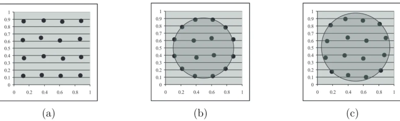
2.3 The result of the modified centroidal Voronoi tessellation algorithm for the unit square 27 2.4 Simplification of a circle to 100 of its points . . . 28
2.5 Reduction of the number of template points for chamfer matching . . . 29
2.6 Dilations of the head template to create the search regions . . . 29
2.7 The comparison of the equidistant and region-influenced centroidal Voronoi tessel- lation (RCVT) based reduction of one-pixel wide objects for chamfer matching for different distance maps . . . 30
2.8 The change of the distance map error at different levels of reduction of the points of the head template . . . 30
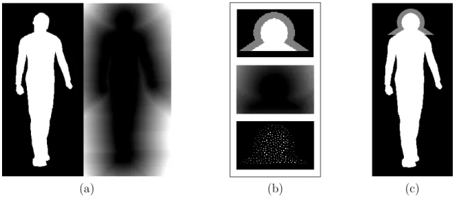
2.9 Thermal images captured under varying temperature conditions . . . 30
2.10 Best fitting positions of templates for a human silhouette represented by a snake . . 32
2.11 Point-wise goodness of fit distance profile for matching the head template against the body contour at different levels of template simplification and snake density . . 32
2.12 ROC curves for matching the head template against the body contour at different levels of template simplification and snake density . . . 33
2.13 Comparing the performance of simplified head templates with considering the sum of the distances between their best matching positions . . . 34
2.14 ROC curves to measure the performance of the simplification methods applied to the head template on an experimental dataset of target head objects . . . 34
2.15 Computation time of object matching vs. percentage of retained points of the original head template . . . 35
2.16 The result of the RCVT algorithm in 2D . . . 35
2.17 Chamfer matching of object parts . . . 36
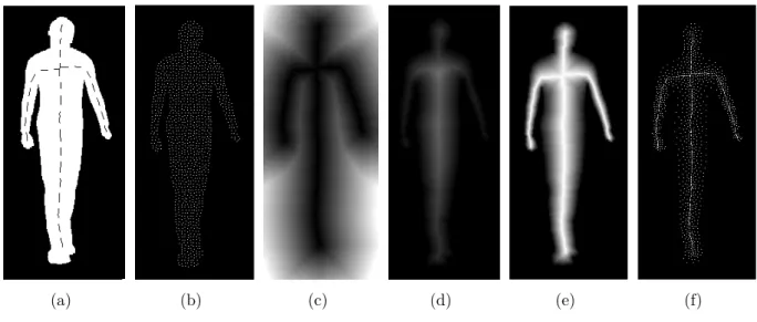
2.18 Simplification result of an object according to a weight function concentrating on its skeleton . . . 37
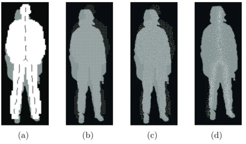
2.19 The result of fuzzy segmentation . . . 37
2.20 Best matching position for standing template . . . 38
2.21 Best matching position for walking template . . . 39
2.22 Components of a learning-based vessel segmentation algorithm . . . 40
2.23 Result of sampling strategies shown for a part of the vascular system . . . 41
2.24 Segmentation performance of sampling strategies regarding the level of simplification 42 2.25 Average segmentation performance of different sampling strategies . . . 43
2.26 Segmentation times regarding the level of simplification . . . 43
3 Piecewise linear digital curve representation and compression 3.1 Locating vertices and extracting the edges for the abstract curve graph. . . 48
3.2 The simplified abstract curve graph of the curve shown in Figure 3.1(a) . . . 48
3.3 Optimal curve tracing through a junction with finding the most straight direction and connecting corresponding edge end points . . . 50
3.4 Tracing the whole curve by choosing optimal directions at junctions . . . 50
3.5 An example alphabet of line segments of length at most six pixels . . . 51
3.6 Test curves of different types . . . 52
3.7 The partitioning of the test curves into line segments . . . 53
3.8 Closed curves for a comparative study with the MPEG-4 technique . . . 54
3.9 Weighting the abstract curve graph . . . 55
3.10 An example for the Chinese Postman Problem (CPP) algorithm . . . 57
3.11 Tracing a non-Eulerian curve using the CPP algorithm . . . 57
3.12 Distortion of an intersection during skeletonization . . . 59
3.13 Binary vessel map of the retina . . . 59
3.14 The result of the splitting step . . . 60
3.15 The reconnection step to fill in the gaps between the skeletons of the thick and thin vessels . . . 61
4 Combining algorithms for automatic detection of optic disc (OD) and macula in fundus images 4.1 Flowchart showing the steps of the proposed technique. . . 65
4.2 Refining the output of the optic disc (OD) and macula detectors by shifting. . . 66
4.3 False positive reported in the majority voting during OD detection. . . 67
4.4 Selecting final OD and macula candidates based on mutual spatial information. . . 68
4.5 A retinal image with a manually selected OD region . . . 71
4.6 Confidence maps of individual OD detector algorithms . . . 73
4.7 The confidence map of an OD detector with candidates meeting the OD-geometry constraint . . . 74
4.8 The increase of the Positive Predictive Value of the OD detector algorithms regard- ing the number of their candidates . . . 75
4.9 The clique meeting the geometric constraint and having maximal total weight . . . 76
4.10 Maps of member algorithms showing their confidence on whether an image pixel corresponds to the OD center or not . . . 81
4.11 The probability maps (density functions) of the member OD detectors . . . 82
4.12 Results of combination of the probability maps . . . 83
4.13 Locating the OD as the strongest peak covered by the OD template . . . 84
4.14 Performance of possible OD detector ensembles regarding their cardinality . . . 86
4.15 Results of the segmentation of the OD region based on aggregated probability maps using the Hidden Na¨ıve Bayes (HNB) model . . . 87
4.16 Results of the segmentation of the OD region by HNB model in images, where signs of other eye diseases can be found . . . 87
5 Generalizing the majority voting scheme to spatially constrained voting 5.1 The OD in a retinal image and the OD center candidates of individual detector
algorithms . . . 90 5.2 The graph of the probability termpn,k for classic majority voting . . . 92 5.3 The graph ofpn,k =k/n . . . 93 5.4 The graph ofpn,k for number of algorithmsn =9 and individual accuraciesp=0.9
with the geometric constraint to fall within a disc . . . 101 5.5 The geometric constraint applied to the candidates of the algorithms . . . 102 5.6 The graph ofpn,k forn =6 in our OD detector ensemble . . . 104 6 Creating ensembles for the automatic detection of microaneurysms (MAs)
6.1 The proposed framework for ensemble selection . . . 119 6.2 Results of microaneurysm (MA) detection by an ensemble . . . 123 6.3 Free-response receiver operating characteristic (F ROC) curve of the MA detector
ensemble on the Retinopathy Online Challenge . . . 126 6.4 Receiver operating characteristic (ROC) curve of the MA detector ensemble on the
Messidor database . . . 127 6.5 The effect of different preprocessing methods, where MAs are hard to detect . . . . 128 6.6 F ROC curve of the MA detector ensemble on the DiaretDB1 database . . . 129 6.7 F ROC curve of the MA detector ensemble on the Moorfields database . . . 129 6.8 Examples for MAs in different visual categories in a fundus image from the ROC
dataset . . . 130 6.9 MA categories based on visibility . . . 130 6.10 Examples for MAs in different spatial categories in a fundus image from the ROC
dataset . . . 131 6.11 MA categories based on spatial location . . . 132 7 An ensemble-based system for the automatic screening of diabetic retinopathy
(DR)
7.1 Flow chart of the proposed decision support framework . . . 139 7.2 Some representative visual features to be extracted from the images . . . 141 7.3 Representative images having grades R0, R1, R2, R3 from the Messidor database . 143 7.4 The difference between the actual and the detected OD and macula centers . . . 144 7.5 ROCcurves of automatic DR screening systems evaluated on the Messidor database
for the scenario R0 vs R1 . . . 150 7.6 ROCcurves of automatic DR screening systems evaluated on the Messidor database
for the scenario No DR/DR . . . 150
List of Tables
2 Optimal approach for fast object-template matching
2.1 Goodness of fit of simplified templates given in terms of the percentage of matched
template points . . . 38
3 Piecewise linear digital curve representation and compression 3.1 Comparative quantitative results with the JBEAM approach . . . 52
3.2 Comparative quantitative results with MPEG-4 coding . . . 54
3.3 Experimental results of separate (curve part) edge compression . . . 56
3.4 Improvement of the proposed method against classic skeletonization for vessel in- tersections . . . 62
4 Combining algorithms for automatic detection of optic disc (OD) and macula in fundus images 4.1 Candidates falling inside the manually selected OD patch . . . 71
4.2 Average Euclidean error of the OD candidates . . . 71
4.3 Candidates falling in the macula region . . . 72
4.4 Average Euclidean error of the macula candidates . . . 72
4.5 OD/macula candidates falling in OD/macula regions using mutual spatial information 72 4.6 Pearson’s correlation coefficients of the member algorithms . . . 83
4.7 Summary of the test datasets for OD detection using probability models . . . 84
4.8 OD detection results of member algorithms and ensembles on the databases Di- aretDB0, DiaretDB1 and DRIVE . . . 85
4.9 OD detection performance regarding healthy and unhealthy cases . . . 88
5 Generalizing the majority voting scheme to spatially constrained voting 5.1 Ensemble accuracy for classic majority voting . . . 92
5.2 Ensemble accuracy under the geometric constraint . . . 102
5.3 Change of the ensemble accuracy, when the sixth member is added to the ensemble of five algorithms . . . 105
5.4 The interval for the OD detector ensemble accuracy, if a new independent algorithm is added to a dependent system . . . 105
5.5 The interval for the OD detector ensemble accuracy, if a new independent algorithm is added to an independent system . . . 105
5.6 The interval for the minimal and maximal OD detector ensemble accuracy, if a new dependent algorithm is added to a dependent system . . . 106
5.7 The interval for the minimal and maximal OD detector ensemble accuracy, if a new dependent algorithm is added to a system with no dependency constraints . . . 106
5.8 Change of the ensemble accuracy for strict majority, when the sixth and seventh
members are added to the ensemble of five algorithms . . . 107
5.9 Overall system accuracies for the first set of classifier accuracies . . . 110
5.10 Overall system accuracies for the second set of classifier accuracies . . . 111
5.11 Overall system accuracies for the third set of classifier accuracies . . . 111
5.12 Selecting ensembles by using a weighted linear combination of generalized diversity measures . . . 114
6 Creating ensembles for the automatic detection of microaneurysms (MAs) 6.1 Comparison of the energy functions on the ROC database regarding the first case . 120 6.2 Comparison of the energy functions on the DiaretDB1 database regarding the first case . . . 120
6.3 Comparison of the energy functions on the Moorfields database regarding the first case . . . 121
6.4 Comparison of the energy functions on all three databases regarding the first case . 121 6.5 Comparison of the energy functions on the ROC database regarding the second case 121 6.6 Performance of the ensembles on the ROC database regarding the first case . . . 122
6.7 Performance of the ensembles on the DiaretDB1 database regarding the first case . 122 6.8 Performance of the ensembles on the Moorfields database regarding the first case . . 122
6.9 Performance of the ensembles on all three databases regarding the first case . . . 123
6.10 Performance of the ensembles regarding the second case . . . 123
6.11 Number of hpreprocessing method, candidate extractori (hPP, CEi) pairs selected for the ensembles in different test runs . . . 124
6.12 hPP, CEi pairs selected as members of the microaneurysm (MA) detector ensembles 126 6.13 Quantitative results of the Retinopathy Online Challenge for MA detection . . . 126
6.14 Results of diabetic retinopathy (DR) grading on the Messidor database based on the MA detection results . . . 127
6.15 The number of all MAs, images, and the MAs belonging to each category for the training and the test databases, respectively . . . 134
6.16 The number of true and false detections and the number of correctly recognized cases for each MA category in the training database . . . 135
6.17 The number of true and false detections and the number of correctly recognized cases for each MA category in the test database . . . 135
6.18 Comparison of the search- and the weighting-based MA detector ensembles . . . 136
6.19 Detailed quantitative results of the Retinopathy Online Challenge (including the weighted ensemble) . . . 136
7 An ensemble-based system for the automatic screening of diabetic retinopathy (DR) 7.1 Comparison of components of the automatic screening system . . . 138
7.2 Features for DR grading . . . 144
7.3 DR grading results for scenario R0 vs R1 on the Messidor database with forward search method using different fusion strategies and energy functions . . . 145
7.4 DR grading results for scenario R0 vs R1 on the Messidor database with backward search method using different fusion strategies and energy functions . . . 146
7.5 DR grading results for scenario No DR/DR on the Messidor database with forward search method using different fusion strategies and energy functions . . . 146
7.6 DR grading results for scenario No DR/DR on the Messidor database with backward
search method using different fusion strategies and energy functions . . . 147
7.7 DR grading results on the Messidor database with all of the classifiers included in the ensemble . . . 147
7.8 Comparison of the energy functions for the scenario R0 vs R1 . . . 148
7.9 Comparison of the energy functions for the scenario No DR/DR . . . 148
7.10 Comparison of the search methods for the scenario R0 vs R1 . . . 148
7.11 Comparison of the search methods for the scenario No DR/DR . . . 148
7.12 Comparison of classifier output fusion strategies for the scenario R0 vs R1 . . . 149
7.13 Comparison of classifier output fusion strategies for the scenario No DR/DR . . . . 149
7.14 Comparison of automatic DR screening systems . . . 149
7.15 Comparison of automatic DR screening systems evaluated on the Messidor database for the scenario R0 vs R1 . . . 150
7.16 Comparison of automatic DR screening systems evaluated on the Messidor database for the scenario No DR/DR . . . 150
Introduction
This dissertation presents some novel results concerning object detection tasks in digital images.
All the investigations were motivated by practical problems with the corresponding applied tech- niques belonging to the fields of discrete mathematics/geometry and information fusion. However, besides providing efficient practical tools, our aim was to discover also the theoretical background of the problems to be able to characterize and validate our solutions in an objective way. The work is built upon results [1–55] published after September 2003 – the time, when the author obtained his PhD degree. Among these publications [1–16] were published in journals covered by the Science Citation Index (SCI), while [17–55] are other indexed works. The structure of the dissertation basically follows a chronological order, which also reflects the logic how the results are built upon each other. This form of the presentation of the content also helps the reader understand why and how specific activities initiated each other. In this introduction, we shortly describe the practical problems that motivated the presented research and development activities.
For each chapter, we give some details about the challenges of the specific task investigated there with a more comprehensive introduction is enclosed at the beginning of each chapter. Similarly, we highlight here the importance of our related results with including the complete presentation in the corresponding chapters. In general, our new results presented in the dissertation proved to be competitive in the corresponding fields, in several cases outperforming the previously used or other state-of-the-art approaches/algorithms/techniques. It is supported also by the fact that these results were published in leading journals, e.g. of the IEEE1. All fields we investigate are of high relevance with important applications attracting the attention of many experts, having a vast literature. The description of the related works are outlined in the corresponding chapters, as well.
The practical problems addressed in the dissertation primarily come from two fields. Our first application relates to the project SHARE2, which aimed to develop an efficient rescue system for firefighters. Our task in the project was to extract interesting objects (e.g. humans) from videos acquired with thermal cameras by a rescue team. Thermal video analysis has great importance in such scenarios, since the infrared light has better penetration performance through smoke than the visible one. Thus, the firefighters can gather such information (e.g. door frames for exit, injured people on the floor, rescue team members) that they could not see by themselves or by using a camera working in the visible light domain. As a specific task, we focus on the recognition of human silhouettes in such videos.
Our second application relates to the project DRSCREEN3supervised by the author addressing the development of an automatic screening system for diabetic retinopathy (DR) based on the processing of retinal images. As for the clinical background of this field, more than 382 million people are suffering from diabetes in 2015 worldwide and the number of the diagnosed cases has been growing rapidly (e.g. in 2012 this figure was 360 million). Long-term diabetes also affects
1Institute of Electrical and Electronics Engineers.
2EU FP6 Information Society Technologies, FP6-004218, SHARE: Mobile Support for Rescue Forces, Integrating Multiple Modes of Interaction.
3TECH08-2 grant of the Hungarian National Office for Research and Technology (NKTH), DRSCREEN - De- veloping a computer-based image processing system for diabetic retinopathy screening.
the eye, resulting in a disease called diabetic retinopathy (DR). Automatic screening systems for DR are of great importance and our presented results also demonstrate our efforts to develop such a system. The corresponding tasks include the detection of the anatomic parts of the retina as well as the DR specific lesions. Using our object detection findings, we have developed a screening system to support everyday clinical routine.
As for the structure of the dissertation, some basic concepts and notations are introduced first in chapter 1. Besides the general formalism regarding digital images, we recall the necessary elements of discrete geometry and information fusion. From the tools of discrete geometry, we recall some graph theoretical concepts that are needed for the analysis of general digital curves representing e.g. object boundaries. In some of our fusion-based approaches, we also compose graphs having the candidates of object detectors as its vertices and apply weighted graph theoretical methods to extract the final suggested locations of desired objects. Since the dissertation focuses on fusion-based techniques in a large extent, we also recall the necessary concepts of this field.
Namely, we recall decision rules, whose extension to the spatial domain are considered for data aggregation in our models, and error measures to evaluate the performance of our corresponding ensemble-based systems. We also describe briefly the clinical field of retinal image analysis with highlighting those anatomical parts and lesions, whose detection will be primarily addressed. As certain general methodological components that will be considered in several chapters later on, we give some details on databases used for training and evaluation, and also on preprocessing and object detection algorithms that will be considered to build up our ensemble-based systems.
However, those algorithms and databases that are cited only in one chapter are going to be introduced there.
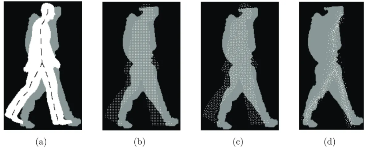
The novel contributions are started to be discussed from chapter 2 with the presentation of a method for object simplification [1, 17]. This technique can be considered as an optimal sampling providing an output, which can represent the original object with smallest error in a Hausdorff distance-based object matching scenario. As for methodology, we extend the sampling methods using the centroidal Voronoi tessellation (CVT) framework in a theoretical basis to reach our aim. Our sampling methods are suitable for the simplification of both contour- and region- like objects. It has been tested on human silhouettes extracted from thermal videos captured during rescue actions, to recognize firefighters and injured people in different poses. We have also demonstrated how the sampling can be directed by including a weight function in the model [18].
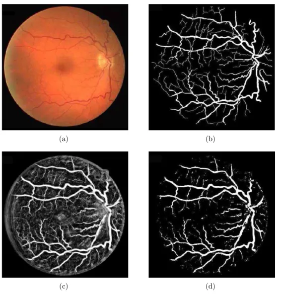
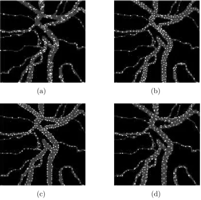
In this way, we can focus more on the main shape characteristics with suppressing the focus on the more various peripheral behavior of similar objects. As for their exploitations, our methods have been integrated in the project SHARE. The importance of our contributions lies in the fact that we can save computation time during the matching process, since it linearly drops with the level of simplification. Fast, online methods are highly welcome in rescue scenarios, where time is a key factor. Besides speeding up computations, our approach helps in composing such template databases that need smaller storage space to be carried more easily by devices having limited resources during a fire/rescue scene. Besides the chamfer matching-based approach, we investigate how the sampling strategies can be considered to speed up computations in learning- based segmentation [19]. Namely, we check whether an appropriate sampling strategy may help in giving a better representation of vessel points to separate them better from the background to segment the vascular system of the retina within our project DRSCREEN.
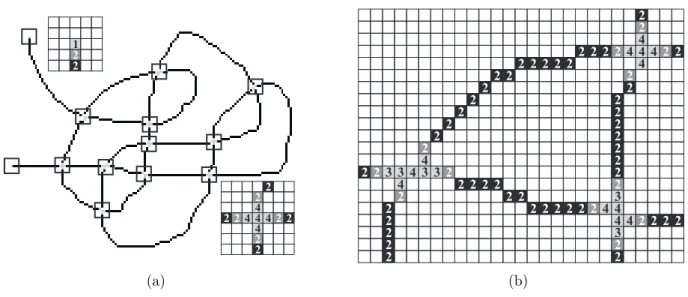
Still based on discrete geometric approaches, chapter 3 describes a method for digital curve compression [2, 20]. The motivation of this study originally was to compress the human body contours described in chapter 2 to support compact portability. We provide a graph theoretical approach to trace curves having arbitrary topology with assigning a graph to the digital curve and using letters from a Bezier-alphabet of linear line segments to approximate it. Because of the tracing step, the proposed method has better compression performance than the current state-of-
the-art approaches. This approach have also been incorporated in the SHARE system mentioned above to efficiently store human body contours. As a corresponding field, we also present some results regarding the possible improvement of the skeletonization of vessel intersections [21] with exploiting this result in our system DRSCREEN. Namely, when we extract a curve as the skeleton of a thicker object, the thinning process usually distorts the intersections, which can be improved by our proposed local method.
Though we also demonstrate the applicability of the above approaches there, from chapter 4 on we present our results focusing on the specific clinical challenge of developing an automatic screening system (DRSCREEN) for diabetic retinopathy (DR). To be competitive in this highly investigated field, we have considered approaches based on information fusion by combining the output of different image processing algorithms dedicated to specific object detection tasks. In this way, we generate ensemble-based systems, which are generally known to be more accurate and balanced. Moreover, this approach leads to very flexible systems, since any future individual algorithms can be integrated to improve the performance of the ensembles further. From both theoretical/practical points of view a serious challenge is to measure up/assure the diversity (inde- pendency) of the member algorithms, since a more diverse system is naturally expected to perform better, even if the individual accuracies are lower. We start the discussion with presenting our re- sults [3, 22] in chapter 4 for the detection of the optic disc (OD) and macula which are two normal anatomical parts of the retina. The detection of the OD is important to avoid its mis-recognition as a bright lesion (like exudate), while the macula has a significant role as being the center of the sharp vision. We propose approaches to automatically combine different OD and macula detec- tors, to benefit from their strengths while overcoming their weaknesses. In this study, we apply simple majority voting rule for the detection of the desired objects with selecting the region, where the largest number of the single candidates of the member algorithms fall. The dependency of the members is also addressed by assigning weights to their candidates considering appropriate pairwise statistics. To improve the detection accuracy, we include a strict geometric constraint on the mutual location of the OD and macula in our framework. Besides the simple majority voting-based rule, we apply weighted graph theoretical algorithms to perform the fusion of the individual detector outputs [23], when they are allowed to have multiple candidates. Moreover, we explain how to take advantage of all the available information from the output of the member al- gorithms supplied in terms of confidence maps [4]. We apply axiomatic and Bayesian approaches, as in the case of aggregation of judgments of experts in decision and risk analysis, to combine these confidence values. With the machine learning-based Bayesian models, we can also make a great effort to discover the dependencies among the member algorithms. Exhaustive experimental tests on publicly available datasets prove the competitiveness of all of these approaches against the state-of-the-art individual algorithms. Our experimental results verify the natural expectation that involving more information in the final decision raises accuracy and reliability, as well. These methods have also been considered in the automatic screening system DRSCREEN.
After reaching improvement in object detection using fusion-based approaches, we performed a thorough theoretical investigation to describe more precisely the behavior of voting models in the spatial domain. Accordingly, in chapter 5, we present our results on the generalization of the classic voting models to the spatial domain [5, 24], where the voters are detector algorithms with giving their votes in terms of single pixels for the location of the desired object. Beyond the generalization of the simple majority voting model, we explain how to assign weights to the member algorithms [25] based on their individual accuracies and the shape of the object to improve system performance. Concentrating only on independent components/variables is also a bottleneck in the theoretical investigations in this field. Since this approach can hardly be accepted in practice, we put effort to give an interval, where the accuracy of the system falls considering dependencies among the members in our models. Moreover, to address this issue better, we extend some
measures dedicated to discover the diversity of the members [26] to the spatial domain. This approach helps in composing efficient ensembles not purely based on the individual accuracies of the members, but also with taking the diversity issue also in account. The suitability of our models have been demonstrated in the OD detection task with empirical tests in publicly available databases showing improvement against the classic models.
Besides the detection of the normal anatomical components, the efficient detection of specific lesions is also a crucial task in an automatic screening system. In the case of DR, the most impor- tant lesions are the microaneurysms (MAs) and exudates. Regarding their detection with image processing techniques, the main difference is that MAs can be represented by single pixels, while exudates with regions. From the theory of information fusion, it is known that the combined system is more efficient in general, if the members are diverse. In chapter 6, we present our related results considering the fusion of lesion candidate extractors in a voting-based environment.
The larger diversity among the members is reached by applying different preprocessing algorithms before the lesion candidate extractor algorithms. This idea can be easily reasoned by the ob- servation that different preprocessors enhance image content differently. However, this approach is primarily meaningful in an ensemble-based scenario, where the possible deteriorations of the members can be compensated with increasing divergence between them. In this way, we create hpreprocessor, candidate extractori(shortlyhPP, CEi) pairs and compose combined systems from them [6, 27–29]. Then, we demonstrate how to create very competitive detectors in this way to recognize MAs [7, 30, 31]. This result of ours is internationally also admitted, as from 2012 till the submission of this dissertation our method is ranked as first in the Retinopathy Online Challenge4, which is the primary benchmark forum of MA detector algorithms. We also show that the detec- tion of MAs can be improved further by adding some contextual information regarding them [8, 32]
in the extracting process. Namely, the detection of MAs highly depends on the characteristics of the imaging device and other image properties (e.g. type of compression). As a result, some MAs can be easily spotted on the background of the retina, while the recognition of others are more difficult. Besides image characteristics, the spatial location also has influence on the detection of MAs (e.g. proximity of vessel parts, etc.).
The main purpose behind our activities dedicated to the processing of retinal images was to develop a decision support system for the automatic screening of diabetic retinopathy. The motivation for creating such reliable systems is to reduce the manual effort of mass screening, which also raises a financial issue. While several studies focus on the recognition of patients having DR and considering the specificity of the screening as a matter of efficiency, inchapter 7, we show how both sensitivity and specificity can be kept at high level by combining novel screening features and a decision-making process. Regarding decision making, automatic DR screening systems either partially follow clinical protocols or use a machine learning classifier. A common way to improve reliability in machine learning-based applications is to use ensemble-based approaches. For medical decision support, ensemble methods have been successfully applied to several fields. The proposed system [9, 33, 34] is ensemble-based at more levels: we consider ensemble systems both in image processing tasks and decision making. In chapter 7, we present how the characteristic features extracted by lesion detection and anatomical part recognition algorithms described in the subsequent chapters and others [35, 36] are integrated in a screening system. As for lesions, besides investigating fusion-based techniques, we have developed an individual MA detector [10, 37] based on intensity profile analysis, as well. This method – as a member algorithm – has also been incorporated in our detector ensemble considered for MA detection. Using similar principles as in our MA detector presented in chapter 6, we compose a fusion-based system also for the detection of exudates [38], which is a competitive approach for this task based on our experimental
4Retinopathy Online Challenge (ROC), http://webeye.ophth.uiowa.edu/ROC/
studies. All these features are then classified by using an ensemble of classifiers to reach the final decision. Besides the flow of decision making, we also present how to prefilter severely diseased cases or low quality images. The system has already been evaluated on publicly available databases providing high performance compared with other state-of-the-art techniques. These experimental tests suggest that our system is capable of performing automatic screening beyond simple decision support. Especially, our results are very close to meet the recommendations of the British Diabetic Association with its sensitivity 80% and specificity 95%.
Partially due to space reasons and to keep the content of the dissertation more coherent, some results are presented in a short form inchapter 8. These works are either loosely related or have just finished recently showing how the results of the dissertation are going to be improved or integrated in future activities. Regarding discrete geometry, we mention that in some of our works [11–14, 39] we investigate efficient ways of digital distance measurement. We also apply digital geometric approaches to improve the performance of active contour (snake) models in human body extraction tasks [40–42]. As for our clinical application, we have shown that the intensity profile analysis considered for MA detection can be successfully applied in retinal vessel segmentation, as well [43]. As a more theoretical approach, we investigate kernel functions leading to translation invariance in intensity [15] with demonstrating our findings in MA detection. Besides ensemble- based approaches, we have also developed a competitive methods for exudate detection in [16, 44–
47]. We determine clusters of retinal images coming from different sources to optimize MA [48] and exudate [49] detectors accordingly. As a natural step forward, we determine the optimal parameter settings in our ensemble-based MA [50] and exudate [51] detectors and give suggestions to make the stohastic search procedure computationally more efficient. XML-based metadata schemes have been created for the content description in both of our systems SHARE [52] and DRSCREEN [53].
To increase the performance of our system in DR screening, in [54, 55] we have shown that the inclusion of proteomic data gathered from tear as a secondary modality is a promising approach.
New scientific results are enclosed in terms of thesis points in theSummary of new scientific results. To preserve the readability of the dissertation, some proofs of technical nature have been moved to theAppendix. The author’s own papers cited in the dissertation are enclosed in chapter Author’s publications, while others’ works in theBibliography.
Chapter 1
Basic concepts and notations
1.1 General formalism . . . 8 1.2 Graph theory . . . 8 1.3 Information fusion . . . 8 1.3.1 Error measurement . . . 10 1.4 Clinical concepts . . . 12 1.5 Databases . . . 12 1.5.1 Retinopathy Online Challenge (ROC) database . . . 13 1.5.2 DiaretDB0 database . . . 13 1.5.3 DiaretDB1 database . . . 13 1.5.4 Database provided by the Moorfields Eye Hospital, London . . . 13 1.5.5 Messidor database . . . 13 1.5.6 DRIVE database . . . 14 1.6 Algorithms to create ensembles . . . 14 1.6.1 Preprocessing methods . . . 14 1.6.2 Single object (optic disc) candidate extractors . . . 15 1.6.3 Single object (macula) candidate extractors . . . 16 1.6.4 Multiple objects candidate extractors . . . 17
I
n this chapter, we recall some basic concepts, notations and results that will be needed later on. After the introduction of some general formalism, we collect the necessary elements of graph theory and information fusion according to the content of the dis- sertation. We also give a brief description of clinical concepts, databases and detector algorithms regarding the specific application fields.1.1 General formalism
Let the sets of natural, integer and real numbers be denoted byN=Z≥0, Z, andR, respectively.
To refer to their positive (resp. non-negative) subsets, we will use the notationsZ>0, R>0 (resp.
Z≥0, R≥0). With an m ∈ N, the m-dimensional (mD for short) number sets will be denoted by Zm, Rm, respectively. From 2D on (m ≥ 2), the elements of Zm, Rm will be written using the vector notation x = (x(1),x(2),. . .,x(m)). For scalars, we omit the vector format (e.g.k ∈ Z,p ∈ R). The spatial subsets will be denoted by capital letters as A,B,C ∈ Zm or Rm; the cardinality of a setAis written as|A|. To measure the distance of x,y ∈Zm orRm, usually their Euclidean distance d(x,y) will be considered.
This dissertation focuses on 2D digital images, where an intensity image I of l-levels (l ∈ N) over A ⊂ Z2 is defined as I : A → {0,. . .,l−1}. For a point x ∈ A, the pair (x,I(x)) is called a pixel. As most general instances, we will consider 8-bit (l = 256) intensity images of resolution r×c (r,c ∈ N) I : {1,. . .,c} × {1,. . .,r} → {0,. . ., 255}. Notice that this definition will allow us to refer to image point coordinates using the horizontal–vertical order. 24-bit RGB color images will be represented by a triplet (IR,IG,IB) of 8-bit intensity images of the same resolution dedicated to the red, green and blue color channels, respectively. As another family, we will also consider binary images withl =2.
1.2 Graph theory
In this dissertation, graphs will be considered in digital images, so we introduce the corresponding notations accordingly. A multigraph G is defined as a pair (V,E), where V ⊂ Z2 is a set of vertices, and E ⊆ V ×V = {(u,v) : u,v ∈ V} is a multiset of edges between the vertices. We focus on undirected graphs, so ∀u,v ∈ V : (u,v) ∈ E implies (v,u) ∈ E, and thus, we will write {u,v} for the edges from now on. In our graphs, we allow loops (edges of type {u,u}) and multiple edges (more edges between two vertices), which also reasons why we consider the more general formalism of multigraphs. If there are no multiple edges, we will use the classic graph concept, where E is supposed to be a set instead of a multiset. The degree of a vertex is the number of edges containing the vertex. A walk is a list of vertices {u1,u2,. . .,un} with {u1,u2},{u2,u3},. . .,{un−1,un} ∈ E, with u1 =un in the case of a cycle (closed walk). G is connected, if any two of its vertices have a walk connecting them. A walk which includes every edge ofGexactly once is called an Eulerian walk (or an Eulerian cycle, if the start and end vertices coincide). Notice that any Eulerian cycle is also an Eulerian walk. G is an Eulerian graph, if it has an Eulerian walk containing all of its edges. An Eulerian decomposition of G has the form G = Sn
i=1
Gi such that all theGi’s are edge-disjoint Eulerian graphs i.e. they do not contain common edges.
1.3 Information fusion
The results in the dissertation relating to information fusion are based on the consideration that merging the output of member components may lead to applications of higher accuracy. In our case, the member components are primarily image processing algorithms, whose outputs are merged for object detection purposes. Thus, besides introducing the general corresponding formalism, we also specialize our notation to this scenario.
As classic formulation [56], let D be a set (ensemble) of classifiers (voters) D1,D2,. . .,Dn, Di : Λ⊆Rm → RM≥0 (i = 1,. . .,n), and Ω = {ω1,ω2,. . .,ωM} is a set of finite class labels.
The classifier Di assigns the support values Di(χ) = (di,1(χ),. . .,di,M(χ)) to a feature vector χ∈Λ describing the opinion of the classifier on what degree χ should be labeled by ω1,. . .,ωM, respectively. Then, in a fusion-based scenario, the final class label is determined forχby applying some rule to the individual labels supported by the classifiers D1,. . .,Dn. Namely, as a general formulation, for each j (j =1,. . .,M) a discriminator function gj(χ) is calculated as
gj(χ) = F(d1,j(χ),. . .,dn,j(χ)), (1.1) where F is a combination function. According to the selection of the support values di,j(χ) and the combination function F, we can set up several decision rules and derive different ensemble classifiers like the following algebraic ones:
Davg(χ) =ωk ⇐⇒ gk(χ) = maxM
j=1 gj(χ) = 1 n
n X i=1
di,j(χ)
!
, (1.2)
Dpro(χ) =ωk ⇐⇒ gk(χ) = maxM
j=1 gj(χ) =
n Y i=1
di,j(χ)
!
, (1.3)
Dmin(χ) =ωk ⇐⇒ gk(χ) = maxM
j=1
gj(χ) =minn
i=1 (di,j(χ))
, (1.4)
Dmax(χ) =ωk ⇐⇒ gk(χ) = maxM
j=1
gj(χ) =maxn
i=1 (di,j(χ))
. (1.5)
Notice that these properties give constraints on which class labelωk should be selected forχ. Here, we apply the intuitive notation D(χ) =ωk instead of D(χ) = (0,. . ., 0
| {z } k−1
, 1, 0,. . ., 0
| {z } M−k
) with the only non-zero support 1 is put on the k-th label, since the overall aim for any ensemble classifier is to select a single class label as a final decision.
The simple majority voting based classic ensemble classifier can be derived by restricting the support of the individual classifiers with d1,j(χ) = 1, if the classifier Di labels χ in the class ωj and d1,j(χ) = 0, otherwise. The final labeling of the ensemble is based on determining the class received the largest support in terms of the number of votes:
Dmaj(χ) = ωk ⇐⇒ gk(χ) =maxM
j=1 gj(χ) =
n X i=1
di,j(χ)
!
. (1.6)
From the simple majority voting model we can easily derive a weighted one with assigning weightswi∈R≥0 to the classifiers Di implying the following final decision rule:
Dwmaj(χ) = ωk ⇐⇒ gk(χ) =maxM
j=1 gj(χ) =
n X i=1
widi,j(χ)
!
. (1.7)
In contrast to classic majority voting, here we consider each classifier output equipped with different weightswi(0≤wi≤1,i=1,. . .,n). It seems natural to give the classifiers with higher accuracies greater importance in making the final decision. Notice that the classic majority voting scheme can be considered as a special case of the weighted one, since in the majority rule the weight of each vote given by a classifier is constrained to 1 i.e.wi =1 for all i=1,. . .,n.
Several results of the dissertation correspond to ensembles A = {A1,A2,. . .,An} of object detector algorithms Ai (i =1,. . .,n). Based on the nature of the problem, we separate the cases when a single or multiple objects are to be detected in an image. Moreover, we focus on such objects that can be represented by single pixels with e.g. their centers. Thus, the object detector algorithms give their votes in terms of pixels as candidates for the location of the object. According to these considerations, we introduce the related notations as follows.
Let I be a digital image of size r×c. A candidate extractor algorithm for a single object detection scenario is defined as A˙i : I → P({1,. . .,c} × {1,. . .,r}) with i ∈ {1,. . .,n} and A˙i(I) = {c˙Ii,1,c˙Ii,2,. . .,c˙Ii,k}, where k ∈ N and P(A) denotes the power set of a set A. Notice that this definition allows a candidate extractor algorithm to give more candidates within an image for the possible location of a single object. However, we will investigate also such cases, when each ensemble member can have only one candidate for the location of the object. Thus, when with k =1 algorithm A˙i has a single candidate, we will write c˙Ii instead of {c˙Ii,1}for its candidate set.
If multiple objects may appear in the image, the definition of a candidate extractor algorithm is modified accordingly as A¨i : I → P({1,. . .,c} × {1,. . .,r}) with A¨i(I) ={c¨Ii,1,c¨Ii,2,. . .,c¨Ii,k}, where k ∈ N. Note the difference between the notation of the candidates corresponding to the single and multiple objects detection scenarios. Namely, c˙Ii,j for some j ≤ k is the j-th guess of A˙i for a single object, while c¨Ii,j (j ≤ k) predicts the appearance of a desired object in the corresponding location. Since the chapters of the dissertation separate the single and multiple object detection scenarios, we will use the simple notations Ai and cIi,. for both cases if it does not hurt clarity. As a further simplification of notation, we will omit the symbolI from the upper index of candidates, when only one image is concerned and write ci,. for short.
As from the candidate extractor algorithmsA1,A2,. . .,An ensemble candidates are composed via the fusion of their candidate sets A(I) = ∪ni=1Ai(I), we define a confidence measure to describe the rate of agreement of the members on the specific candidates. To do so, first we introduce a proximity relation =∼ to decide whether two candidates indicate the same object or not. With c1,c2 ∈ {1,. . .,c} × {1,. . .,r}, we say that c1 ∼= c2 if d(c1,c2) < Td with some distance threshold Td ∈ R≥0. As in our applications the objects to be detected are circular, Td can be selected as the diameter of the desired object. Now, the confidence of the ensemble on any of its candidatesc ∈ A(I) is defined as
confA(c) = |{Ai ∈ A : ∃c0∈ Ai(I) such that c∼=c0}| /|A|. (1.8) Notice thatconfA(c) ∈ {k/|A| : k=1,. . .,|A|}. We also classify the ensemble candidates based on the degree of confidence. Namely, the α–level candidates of A are defined as
A(I)α ={c∈ A(I) : confA(c) ≥α}, where 1/|A| ≤α≤1. (1.9) As specific cases, 1–level, α–level with α >1/2, and 1/|A|–level candidates are selected by each of, the majority of, and at least one of the members of the ensemble, respectively. For the latter case it should be noted that A(I) = A(I)1/|A|. Single algorithms are formally represented by ensembles consisting of one member providing |A| = 1 and α = 1–level confidence for all the candidates.
1.3.1 Error measurement
We also must set up a framework to measure the accuracy of ensemble-based approaches discussed in the dissertation for object detection in practical problems. We start with making the decision whether the candidates found by a specific member algorithm are true or false ones regarding some ground truth usually provided in terms of manual annotations.
LetGT (I)⊆ {1, . . ., c} × {1, . . ., r}be a so called ground truth set of candidates for an image I. For the classification of each candidatec∈ A(I) of an ensembleAin the same image regarding GT(I) and confidence level 1/|A| ≤ α≤1 we apply the following:
• c is an α–true positive (T Pα), ifc∈ A(I)α and ∃cgt ∈ GT(I) such that c ∼=cgt;
• c is an α–false positive (F Pα), if c∈ A(I)α and @cgt ∈ GT(I) such that c ∼=cgt.
Regarding a candidate cgt ∈ GT(I), we apply:
• cgt is an α–false negative (F Nα), if @c∈ A(I)α such thatc ∼=cgt. Finally,
• each point in {1,. . ., c} × {1, . . ., r}\(GT(I)∪ A(I)α) is an α–true negative (T Nα).
The set of all true positives, false positives, false negatives, and true negatives for a given image I will be denoted by T P(I)α,F P(I)α,F N(I)α, and T N(I)α, respectively. Notice that GT(I) is usually a manually annotated set created by experts of the application field. Moreover, since performance evaluation is usually expected to be given at database level, digital images are often organized into an image database DB. Now, to calculate the performance of an ensemble A regarding some ground truth, we introduce the following classic measures at both image and image database level [57, 58]:
• Sensitivity
SEN(I)α = |T P(I)α|
|T P(I)α|+|F P(I)α|, SEN(DB)α = X
I∈DB
SEN(I)α
|DB| , (1.10)
• Specificity
SP E(I)α = |T N(I)α|
|F N(I)α|+|T N(I)α|, SP E(DB)α = X
I∈DB
SP E(I)α
|DB| , (1.11)
• False Positive Rate
F P R(I)α =1−SP E(I)α, F P R(DB) = X
I∈DB
F P R(I)α
|DB| , (1.12)
• Positive Predictive Value
P P V(I)α = |T P(I)α|
|T P(I)α|+|F N(I)α|, P P V(DB)α = X
I∈DB
P P V(I)α
|DB| , (1.13)
• F-Score
F-Score(I)α = 2SEN(I)P P V(I)
SEN(I) +P P V(I), F-Score(DB)α = X
I∈DB
F-Score(I)α
|DB| , (1.14)
• Accuracy
ACC(I)α = |T P(I)α|+|T N(I)α|
|T P(I)α|+|F N(I)α|+|T N(I)α|+|F P(I)α|, (1.15) ACC(DB)α = X
I∈DB
ACC(I)α
|DB| , (1.16)
• Average False Positives per Image
F P I(DB)α= X
I∈DB
F P(I)α
|DB| , (1.17)
• Competition Performance Metrics CP M(DB) = X
g∈G
{SEN(DB)α: F P I(DB)α=g}
|G| , whereG=
1 8,1
4,1
2, 1, 2, 4, 8
. (1.18) As a specific case, we will omit the index α in the footnote of the above performance measures, if |A| = 1 and simply write e.g. SEN(I) instead of SEN(I)1/|A|, when our aim is to evaluate a single algorithm only.
1.4 Clinical concepts
In this section, we briefly describe those concepts of a clinical field that will be regularly referred in some chapters of the dissertation. Namely, some of our single and multiple objects detection results relate to some anatomical parts and diabetes specific lesions appearing in the human retina (fundus). In Figure 1.1(a), we can see the anatomic location of the retina as a layer in the eye dedicated to percept the light in the human vision system. The retinal anatomical parts and lesions are shown in Figure 1.1(b), whose detections are addressed in the dissertation. Namely, as single object detection problems, we will give results according to the localization of the optic disc (OD) and the macula. The localization of such lesions as microaneurysms (MAs) and exudates belong to the family of multiple objects detection problems, since usually several such lesions appear in a diseased case. More specific introductions regarding the detections of these objects will be given in the corresponding chapters. The region of interest (ROI) concept in the case of a retinal image generally corresponds to the useful content, that is, the image without its black background.
(a) (b)
Figure 1.1: Basic concepts of retinal image analysis; (a) the structure of the human eye and the location of the retina, (b) anatomical parts and diabetes related lesions of the retina.
1.5 Databases
In this section, we list those databases and give the corresponding characteristics of images be- longing to them which will be considered for training and evaluation in several chapters of the dissertation. Some other datasets that are used only in one chapter are going to be introduced there. The databases listed here relate to the field of retinal image analysis.