HEVENY VESTIBULARIS SZINDRÓMA KÉPÉBEN JELENTKEZÔ KÉSÔI MENINGITIS CARCINOMATOSA – KLINIKOPATOLÓGIAI ESETISMERTETÉS
Jarabin JA, MD, PhD; Klivényi P, MD, PhD; Tiszlavicz L MD;
Molnár FA; Gion K, MD; Földesi I, MD; Kiss JG, MD; Rovó L, MD; Bella Zs, MD
Ideggyogy Sz 2020;73(9–10):354–360.
Célkitûzés– Bár a szédülés a leggyakrabban elôforduló panaszok egyike, a vestibularis perifériák hirtelen kialakult tónusaszimmetriája hátterében mégis ritkán találunk peri - fériás eredetû betegséget utánzó malignus koponyaûri tumorokat. Dolgozatunk egy heveny vestibularis szindró- ma klinikai képében jelentkezô, késôi, temporalis csontot is beszûrô, disszeminált, generalizált mikrometa sztá zi sok - kal járó meningitis carcinomatosa esetet mutat be, ami egy primer pecsétgyûrûsejtes gyomorcarcinoma fel ébredé - sét követôen jelent meg.
Kérdésfelvetés– Célul tûztük ki, hogy azonosítjuk azon patofiziológiai folyamatokat, melyek magyarázatul szol- gálhatnak a daganat felébredésére, disszeminációjára.
A vestibularis tónusaszimmetria lehetséges okait szintén vizsgáltuk.
A vizsgálat alanya és módszerei– Ötvenhat éves férfi betegünk interdiszciplináris orvosi adatait retrospektíven elemeztük. Összegyûjtöttük és részletesen újraértékeltük az eredeti klinikai és patológiai vizsgálatok leleteit, majd új szövettani festésekkel és immunhisztokémiai módszerekkel egészítettük ki a diagnosztikus eljárásokat.
Background– Although vertigo is one of the most com- mon complaints, intracranial malignant tumors rarely cause sudden asymmetry between the tone of the vestibu- lar peripheries masquerading as a peripheral-like disor- der. Here we report a case of simultaneous temporal bone infiltrating macro-metastasis and disseminated multi- organ micro-metastases presenting as acute unilateral vestibular syndrome, due to the reawakening of a primary gastric signet ring cell carcinoma.
Purpose– Our objective was to identify those pathophysi- ological steps that may explain the complex process of tumor reawakening, dissemination. The possible causes of vestibular asymmetry were also traced.
Methods– A 56-year-old male patient’s interdisciplinary medical data had been retrospectively analyzed. Original clinical and pathological results have been collected and thoroughly reevaluated, then new histological staining and immunohistochemistry methods have been added to the diagnostic pool.
Results– During the autopsy the cerebrum and cerebel- lum was edematous. The apex of the left petrous bone
Correspondent: János András JARABIN MD, PhD, Department of Otorhinolaryngology and Head-Neck Surgery, University of Szeged; H-6725 Szeged, Tisza Lajos krt. 111. Telefon: +36(62) 545-310, fax: +36(62) 545-848.
E-mail: jarabin.janos.andras@med.u-szeged.hu; jarabinj@gmail.com https://orcid.org/0000-0003-3499-6371
Érkezett: 2020. május 9. Elfogadva: 2020. augusztus 11.
LATE SIMULTANEOUS CARCINOMATOUS MENINGITIS, TEMPORAL BONE INFILTRATING MACRO-METASTASIS AND DISSEMINATED MULTI-ORGAN MICRO-METASTASES PRESENTING WITH MONO-SYMPTOMATIC VERTIGO
– A CLINICO-PATHOLOGICAL CASE REPORT
János András JARABIN1, Péter KLIVÉNYI3, László TISZLAVICZ3, Fiona Anna MOLNÁR1, Katalin GION4, Imre FÖLDESI5, József Géza KISS1, László ROVÓ1, Zsolt BELLA1
1Department of Otorhinolaryngology and Head-Neck Surgery, University of Szeged, Szeged
2Department of Neurology, University of Szeged, Szeged, Szeged
3Department of Pathology, University of Szeged, Szeged
4Department of Radiology, University of Szeged, Szeged
5Department of Laboratory Medicine, University of Szeged; Szeged
| English| https://doi.org/10.18071/isz.73.0354 | www.elitmed.hu
B
ased on the estimation of global cancer statis- tics (GLOBOCAN), gastric cancer (GC) was responsible for over 1,000,000 new cancer cases in 2018 and the cause of an estimated 783,000 deaths.Incidence rates are significantly higher in Eastern Asia. Mortality is varying between the two sexes from 6.5 to 9.5% for females and males respective- ly1. The prognosis of GC is highly dependent on its stage at verification. Gastric carcinogenesis and the metastatic cascade is a multistep and multifactorial process. GC commonly causes metastasis to the peritoneal cavity, lymph nodes, liver and lung. The intracranium is a very rare site for metastasis. From a retrospective cohort of 8080 subjects with advanced GC, only 13 patients developed intracra- nial metastases (~1.6‰). Out of these 13 patients, 5 were shown to have carcinomatous meningitis
(CM) while the remaining 8 presented with paren - chymal metastasis2.
Dizziness is one of the most common complaints in medicine, affecting 15–35% of the general popu-
Eredmények– Kórboncolás során a nagyagy és a kisagy oedamás volt. A bal piramiscsont csúcsát egy 2 × 2 cm nagyságú daganatmassza szûrte be. A gyomorreszekátum eredeti szövettani metszeteinek újraértékelése submucosus daganatinfiltrációt igazolt vascularis invázió jeleivel.
Immunhisztokémiai vizsgálatokkal dominálóan
magányosan infiltráló daganatsejteket láttunk cytokeratin 7- és vimentinpozitivitással, valamint részleges E-kadherin szövettani festésvesztéssel. A kórboncolás során nyert szövetminták ezt követô hisztológiai vizsgálatai igazolták a disszeminált, többszervi mikroszkopikus daganatinváziót.
Megbeszélés– Az újabb eredmények igazolták, hogy a vimentin kifejezôdése, valamint az E-kadherin elvesztése szignifikáns (p < 0,05) kapcsolatot mutat az elôrehaladott stádiummal, a nyirokcsomóáttétek jelenlétével, a vascu- laris és neuralis invázióval, valamint a nem differenciált szöveti típussal. Betegünk középkorú volt és nem volt immunhiányos állapotban, így a gyomorcarcinoma kilenc éven át tartó alvó állapotot követô felébredését nem tudtuk megmagyarázni. A daganat szervspecifikus tropiz- musa, melyet a „seed and soil” teóriával magyaráznánk, kifejezetten váratlan volt, mivel a gyomorrákok ritkán képeznek áttétet az agyburkokon, hiszen a daganatsejtek elenyészô számban jutnak át a vér-agy gáton.
Következtetések– Az elôzményben szereplô malignus folyamat, valamint egy új neurológiai tünet megjelenése fel kell, hogy keltse a klinikus figyelmét a központi ideg - rendszer daganatos érintettségére, melyet adekvát, célzott diagnosztikus és terápiás stratégia megtervezése kell, hogy kövessen. Ehhez célzott szövettani festési eljárások, speci- fikus antitestek alkalmazása szükséges. A közelmúlt ered- ményei sejtkultúrákon igazolták a metformin epithelialis- mesenchymalis transitiót erôsen gátló hatását gyomor rák esetében. Így további kutatást kell végezni azon esetek- ben, amelyekben az epithelialis-mesenchymalis transitióra pozitív eredményeket kapunk.
Kulcsszavak: carcinomatosus meningitis, pecsétgyûrûsejtes gyomorcarcinoma, epithelialis-mesenchymalis transitio, heveny vestibularis szindróma was infiltrated and destructed by a tumor mass of 2x2 cm
in size. Histological reexamination of the original gastric resection specimen slides revealed focal submucosal tumorous infiltration with a vascular invasion. By immuno- histochemistry mainly single infiltrating tumor cells were observed with Cytokeratin 7 and Vimentin positivity and partial loss of E-cadherin staining. The subsequent histo- logical examination of necropsy tissue specimens con- firmed the disseminated, multi-organ microscopic tumor- ous invasion.
Discussion– It has been recently reported that the expres- sion of Vimentin and the loss of E-cadherin is significantly associated with advanced stage, lymph node metastasis, vascular and neural invasion and undifferentiated type with p<0.05 significance. As our patient was middle aged and had no immune-deficiency, the promoting factor of the reawakening of the primary GC malignant disease after a 9-year-long period of dormancy remained undis- covered. The organ-specific tropism explained by the
“seed and soil” theory was unexpected, due to rare occur- rence of gastric cancer to metastasize in the meninges given that only a minority of these cells would be capable of crossing the blood brain barrier.
Conclusion– Patients with past malignancies and new onset of neurological symptoms should alert the physician to central nervous system involvement, and the appropri- ate, targeted diagnostic and therapeutic work-up should be established immediately. Targeted staining with specific antibodies is recommended. Recent studies on cell lines indicate that metformin strongly inhibits epithelial-mes- enchymal transition of gastric cancer cells. Therefore, fur- ther studies need to be performed on cases positive for epithelial-mesenchymal transition.
Keywords: carcinomatous meningitis, signet ring cell carcinoma,
epithelial mesenchymal transition, acute vestibular syndrome
ABBREVIATIONS ALP: alkaline phosphatase
AVS: acute vestibular syndrome CM: carcinomatous meningitis CSF: cerebrospinal fluid CT: computer tomography
EMT: epithelial-mesenchymal transition GC: gastric cancer
IHC: immunohistochemistry MRI: magnetic resonance imaging SRCC: signet ring cell gastric carcinoma
lation at some point in their lives3. In a multi-na - tional, non-interventional observational study-co - hort of 3,676 patients with vertigo, only 133 (3.6%) had cranial tumors, neoplasms4. Malignant tumors even more rarely cause acute vestibular syndrome (AVS), characterized by a sudden asymmetry bet - ween the tone of the vestibular peripheries. In pa - tients with AVS the differentiation between ves ti - bular neuritis and cerebellar stroke is the key diffe - rential diagnostic question5–7.
In this report we present a patient who complai - ned of imbalance and showed the clinical signs of AVS. Ten years earlier, this patient had gastrec - tomy due to primary gastric signet ring cell carci - noma (SRCC). In the background of the fulminant neurological symptoms, the post-mortem examina- tions revealed the unrecognized CM and the con- comitant multi-organ micrometastases.
Case report
BRIEF PAST HISTORY
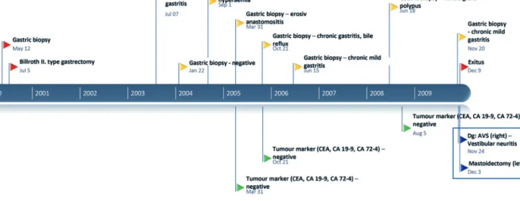
In July 2000 partial gastrectomy was executed ac - cording to Billroth II. (Dg: SRCC, diffuse mucosa type early carcinoma, on the basis of peptic ulcer;
Dukes 2c type, pT1Nx). The patient received no ad - juvant postoperative chemo- or radiotherapy. Re - peated gastric biopsy until July 2003 and June 2006 showed only mild chronic gastritis of the anastomo- sis. In 2008 fundic gland polypus was found. Signs of dysplasia, malignancy, Helicobacter pylori infec- tion were not seen at any point of the follow-up.
Repeated tumor marker analysis for CEA, CA 19-9
and CA 72-4 form March 2005 to August 2008 were also unremarkable. For milestones of patient past history see timeline (Figure 1).
PRESENTING COMPLAINTS AND INVESTIGATION RESULTS
In 2009, the 56-year-old male patient admitted to the Department of Otolaryngology, Head-Neck Sur gery, with 2.5 weeks long leading complaints of intense nausea and vomiting. He was also mild- ly imbalanced with fluctuating, left temporal and retro-auricular headache. On physical examination the eardrums were normal. Pure tone audiometry showed symmetric, mild-moderate sensorineural hea ring impairment. On routine vestibular tests, left-beating fast component horizontal jerk nystag- mus was seen at left gaze position. By clinical head-impulse test overt saccades were seen on both sides. Furthermore, the stance and gait deviated to the right side. Signs were regarded as AVS, sup- porting that the right labyrinth was less active.
Although the laboratory examinations at the point of the initial hospitalization showed only elevated alkaline phosphatase (ALP) level (212 U/l; normal value: <129), and otherwise were within or around normal ran ges, the patient was hospitalized with vestibular neuritis, due to a suspected gastrointesti- nal infection (complaints of intense nausea and vomiting) as a primary cause. As the patient’s blood pressure was elevated following the admin- istration of intravenous pentoxiphyllin (200 mg diluted in 500 ml NaCl), and the oral betahistine was not prescribed due to the past history of gastric surgery, only vestibular training was advised to perform regularly.
Figure 1. Timeline of past history
After the patient had been discharged with the complaints of mild imbalance and headache, 5 days later he was re-admitted with the symptoms of gen- eral malaise, nausea and vomiting. While observing at the intensive care unit the patient developed ge - ne ralized seizure. Neurological examination re vea - led the following symptoms:i)exophthalmos of the left eye, ii) left-beating fast component horizontal jerk nystagmus at left and center gaze position with iii)truncal ataxia, iv)uncoordinated gait and v) la - tent right sided hemiparesis.
The diagnostic lumbar puncture showed 9 lym- phocytes, 7 monocytes and 1 macrophage in 1 µL.
The total protein level was 1.52 g/L, while the glu- cose level was 0.6 mM/L (serum: 5.34 mM/L). Cy - to logical examination did not confirm cerebro spi nal fluid involvement with SRCC features.
Abdominal ultrasound identified mild hepa to - sple nomegaly without pathological lymph nodes.
On repeated ear examination infiltrated (i.e. thick) tympanic membrane was observed on the left side, therefore myringotomy was performed, and only a few drops of serous discharge was drained.
Horizontal plane skull computer tomography (CT) with intravenous contrast material showed the bone destruction with ragged edges of the left petrous bone apex(Figure 2).
Comparing the native and gadolinium enhanced T1-weighted magnetic resonance image (MRI) scans, contrast enhancement is showed at the apex of the left petrous bone, while the meninges were evaluated as normal (Figure 3). The most frequent signs of CM (i.e. subarachnoid and parenchymal enhancing nodules, diffuse or focal pial enhance- ment, nerve root enhancement and brain parenchy- mal metastases) were not identified.
In order to exclude an unrecognized middle ear infection, the patient finally underwent mastoidec- tomy, that revealed air filled mastoid cells without any sign of infection of the left middle ear. After surgery, the patient was observed at the intensive care unit with continuous leaking hemorrhage from the surgical area. Subsequently the patient’s state of consciousness began to fluctuate. Finally, deep coma developed, and the patient passed away. The cause of death was brain herniation due to increased intracranial pressure.
POSTMORTEM EXAMINATION AND REEVALUATION OF THE ORIGINAL GASTRIC RESECTION MATERIAL
During the autopsy the skull bones were intact. The dura mater was smooth and greyish-white in color.
The venous sinuses and the arachnoid mater were intact. The cerebrum and cerebellum was edema-
tous (1620gr). The sulci were shallow; the gyri were flattened. The cerebellar tonsils’ surface was excavated. The apex of the left petrous bone was infiltrated and destructed by a tumor mass of 2x2 cm in size (Figure 4). Other, remarkable macro- scopic discrepancy was not revealed. The subse- quent histological examination of necropsy con- firmed the microscopic invasion of the meninx, the trigeminal ganglion and nerve trunks. Hema to ge - nous micro-metastases were seen in the mediastinal hilar lymph nodes, in the lung and in the liver.
Figure 2.Horizontal plane skull CT with intravenous contrast material showed the bone destruction with ragged edges of the left petrous bone apex (black arrow)
Figure 3.Comparing the native(A)and gadolinium-enhanced (B)T1-weighted magnetic resonance image (MRI) scans, con- trast enhancement is showed at the apex of the left petrous bone (black arrows)
A B
Histological reexamination of the original gastric resection specimen slides revealed focal submucos- al tumorous infiltration with a vascular invasion, but lymph node metastasis was not noted within the perigastric adipose tissue. By immunohistochem- istry (IHC) mainly single infiltrating tumor cells were observed with Cytokeratin 7 and Vimentin positivity. Furthermore, gastric tumor cells showed partial loss of E-cadherin staining. These alterations indicate the epithelial-mesenchymal transition (EMT) phenomenon which can be a sign of a
metastatic phenotype (Figure 5.A–C). By IHC par- tial loss of E-cadherin membrane positivity was observed in the metastatic gastric carcinoma, with some perineural Vimentin positive carcinoma cells (Figure 6.A–C).
This case report was authorized by the Uni ver - sity of Szeged and National Scientific Research Ethi cal Review Board (No.4620; 21. Oct. 2019).
All therapeutic and diagnostic departments in vol - ved, consented to this retrospective study, with data security kept in mind.
Discussion
The initial neurootological tests suggested that the right peripheral vestibular organ was hypo-func- tioning, but the pathological examination revealed the left middle ear infiltrated by the metastasis, thus we retrospectively hypothesized that the symptoms of the AVS were attributable to an excitatory pa - thology of the left inner ear. Although both the weakness of the right labyrinth and the excitation of the left labyrinth were equally possible explanation.
Please note, that at the terminal stage of the disease neurootological examination couldn’t not be per- formed. The left temporal and retro-auricular head - ache was attributable to the involvement of the tri - geminal nerve trunk and ganglion (Figure 6.A), while further neurological and psychomotor signs then the central origin of the disease.
During the course of the metastatic cascade, malignant cells must adapt to new environments very different from the tissue of origin. This process includes local invasion, intra- and extravasation, and colonization, while becoming an apparent, de - tec table metastatic disease. This period may last for years as the dynamic equilibrium between tumor promoting and suppressing activities are in balance, termed as tumor dormancy and reawakening8. As our patient was middle aged and had no immune- deficiency, the promoting factor of the reawakening of the primary GC malignant disease after a 9-year- long period of dormancy remained uncovered.
The tropism of tumor cells to form metastases in preferred tissues gave the illustrative analogy of
“seed and soil” theory9. However, in our case pres- entation this tropism was a rare occurrence of GC to metastasize in the meninges given that only a minority of these cells would be capable of crossing the blood brain barrier. The explanation may be due to a cancer cell derived C3. This C3 activates the C3a receptor in the choroid plexus epithelium which leads to a disruption of the blood-cere- brospinal fluid (CSF) barrier. This would allow Figure 4.Destructive tumor mass at the apex of the left
petrous bone (black frame, the tumor mass is held by a forceps)
Figure 5.Surgical specimen histology. Top image: Intra mu co - sal diffuse sigillocellular infiltration (HE, 20x).A:Staining con- firmed the intracellular mucus vacuole (PAS-AB at pH 2.5, 10x).
B:Vascular invasion and nerve trunks (HE, 10x).C:Peri vas - cular Cytokeratin 7 positive single tumor cells (IHC, 10x) HE: hematoxylin and eosin, PAS-AB: periodic acid-Schiff-AB, IHC: immunohistochemistry, CK-7: cytokeratin 7
plasma components, including amphiregulin and other mitogens to enter the CSF and promote can- cer cell growth, probably also in the meninges10.
Recent studies emphasize the influence of tumor microenvironment on the characteristics of malig- nant cells, where pathological gastric EMT activa- tion is considered to be significant11. This switch between these two histological modalities of the GC cells in which epithelial cells exhibit reduced intercellular adhesion and acquire migratory fibrob- lastoid properties, increase initiation and invasion, metastasis formation and chemo-resistance11–14. It has been recently reported that the expression of Vimentin and the loss of E-cadherin are significant- ly associated with advanced stage, lymph node metastasis, vascular and neural invasion and undif- ferentiated type with p<0.0515. From the mechanis- tic aspect, the leptomeningeal dissemination proba- bly occurred via the Batson’s vertebral venous plexus that might give the explanation for paradox- ical, intracranial tumor spread16–19. CSF abnormali- ties were nonspecific in this case. The sensitivity could be increased with obtaining large sampling volumes or a repeated CSF analysis20, 21. The most sensitive technique of radiology is MRI with gadolinium enhancement22, 23. The prognosis of CM is poor; the treatment is almost always palliative24. In a single-center retrospective study with 155 con- secutively analyzed patients with CM the overall median survival time was 4.8 months25.
Conclusion
Patients with past malignancies and new neurologi- cal symptoms should alert the physician to central nervous system involvement. Consequently, the appropriate, targeted diagnostic and therapeutic work-up should be established immediately. On the other hand, EMT is a significant predictor of aggres-
sive metastatic behavior. Therefore, targeted stain- ing with specific antibodies is recommended.
Recent studies on cell cultures indicated that met- formin strongly inhibited EMT of gastric cancer.
Furthermore, wound-healing and invasion assays showed a significant decrease in cell migration and invasion after metformin treatment26. Therefore, fur- ther clinical studies need to be performed on cases positive for epithelial-mesenchymal tran sition.
CONFLICT OF INTEREST
The authors declare that they have no conflict of interest.
REFERENCES
1.Bray F, Ferlay J, Soerjomataram I, Siegel RL, Torre LA, Jemal A.Global cancer statistics 2018: GLOBOCAN esti- mates of incidence and mortality worldwide for 36 cancers in 185 countries. CA Cancer J Clin 2018;68:394-424.
https://doi.org/10.3322/caac.21492
2.Kim M. Intracranial involvement by metastatic advanced gastric carcinoma. J Neurooncol 1999;43:59-62.
3.Neuhauser HK. The epidemiology of dizziness and vertigo [Internet]. 1st ed. Handb. Clin Neurol Elsevier B.V.; 2016.
4.Agus S, Benecke H, Thum C, Strupp M. Clinical and de - mographic features of vertigo: Findings from the REVERT registry. Front Neurol 2013;4:1-8.
https://doi.org/10.3389/fneur.2013.00048
5.Kattah JC, Talkad AV, Wang DZ, Hsieh YH, Newman-Toker DE. H.I.N.T.S. to diagnose stroke in the acute vestibular syndrome – three-step bedside oculomotor exam more sen- sitive than early MRI DWI. Stroke 2009;40:3504-10.
https://doi.org/10.1161/strokeaha.109.551234
6.Halmagyi GM, Curthoys IS. A Clinical Sign of Canal Pa - resis. Arch Neurol 1988;45:737-9.
https://doi.org/10.1001/archneur.1988.00520310043015 7.Halmagyi GM, Chen L, MacDougall HG, Weber KP, Mc Gar -
vie LA, Curthoys IS. The video head impulse test. Front Neu - rol 2017;8. https://doi.org/10.3389/fneur.2017.00258 8.Boire A, Coffelt SB, Quezada SA, Vander Heiden MG,
Weeraratna AT.Tumour Dormancy and Reawakening: Op - Figure 6.Necropsy histology. Top image: Meningeal carcino- matous infiltration from gastric carcinoma (HE, 10x).A: Tumo - rous infiltration around of trigeminal nerve trunks (HE, 20x).
B:some Vimentin positive carcinoma cells around a nerve (IHC, 20x).C: Partial loss of E-cadherin membrane positivity in metastatic gastric carcinoma (IHC, 20x)
HE: hematoxylin and eosin, IHC: immunohistochemistry
portunities and Challenges. Trends in Cancer [Internet].
Elsevier 2019;5:762-5.
https://doi.org/10.1016/j.trecan.2019.10.010
9.Liu Q, Zhang H, Jiang X, Qian C, Liu Z, Luo D. Factors involved in cancer metastasis: A better understanding to
“seed and soil” hypothesis. Mol Cancer. Molecular Cancer 2017;16:1-19. https://doi.org/10.1186/s12943-017-0742-4 10.Boire A, Zou Y, Shieh J, Macalinao DG, Pentsova E MJ.
Complement component 3 adapts the cerebrospinal fluid for leptomeningeal metastasis. Cell 2017;168:1101-13.
https://doi.org/10.1016/j.cell.2017.02.025
11.Lei Huang, Ruo-Lin Wu A-MX. Epithelial-mesenchymal tran- sition in gastric cancer. Am J Transl Res 2015;7:2141-58.
12.Xue Y, Zhang L, Zhu Y, Ke X, Wang Q, Min H. Regulation of proliferation and epithelial-to-mesenchymal transition (EMT) of gastric cancer by ZEB1 via modulating Wnt5a and related mechanisms. Med Sci Monit 2019;25:1663-70.
https://doi.org/10.12659/msm.912338
13.Zhang W, Sun J, Chen J, Xu C, Zhang L. Downregulation of miR-95 in gastric cancer promotes EMT via regulation of Slug, thereby promoting migration and invasion. Oncol Rep 2019;41:1395-403. https://doi.org/10.3892/or.2018.6911 14.Xia P, Xu XY. Epithelial-mesenchymal transition and gast-
ric cancer stem cell. Tumor Biol 2017;39:1-5.
15.Ryu HS, Park DJ, Kim HH, Kim WH, Lee HS.Combination of epithelial-mesenchymal transition and cancer stem cell- like phenotypes has independent prognostic value in gast- ric cancer. Hum Pathol [Internet]. Elsevier Inc 2012;43:
520-8. https://doi.org/10.1016/j.humpath.2011.07.003 16.Batson OV. The function of the vertebral veins and their
role in the spread of metastases. 1940. Clin Orthop Relat Res 1995;4-9.
17.Brook R, Tung K, Oeppen R.Batson’s plexus and retrogra- de venous spread of malignancy – a pictorial review. Can - cer Imaging 2014;14:P40.
https://doi.org/10.1186/1470-7330-14-s1-p40
18.Gottlieb MD, Roland JT. Paradoxical spread of renal cell carcinoma to the head and neck. Laryngoscope 1998;108:
1301-5.
https://doi.org/10.1097/00005537-199809000-00007 19.Nathoo N, Caris EC, Wiener JA, Mendel E.History of the
vertebral venous plexus and the significant contributions of breschet and batson. Neurosurgery 2011;69:1007-14.
https://doi.org/10.1227/neu.0b013e3182274865
20.Chamberlain MC, Glantz M, Groves MD WW.Diagnostic Tools for Neoplastic Meningitis: Detecting Disease, Iden ti - fying Patient Risk, and Determining Benefit of Treatment.
Semin Oncol [Internet]. Elsevier Inc 2009;36:S35-45.
https://doi.org/10.1053/j.seminoncol.2009.05.005
21.Chamberlain MC.Leptomeningeal metastasis. Curr Opin Oncol 2010;22:627-35.
22.Le Rhun E, Taillibert S, Chamberlain M. Carcinomatous me - ningitis: Leptomeningeal metastases in solid tumors. Surg Neurol Int [Internet] 2013;4:S265-88.
https://doi.org/10.4103/2152-7806.111304
23.Assi R, Hamieh L, Mukherji D, Haydar A, Temraz S, El-Dika I, et al. Leptomeningeal metastasis as initial manifestation of signet ring colorectal adenocarcinoma: A case report with review of literature. J Gastrointest Oncol 2015;6:E89-101.
24.Gábor C, Tibor V, Norbert T, Zoltán G, Tamás V, Péter S, et al.Meningitis carcinomatosa. Lege Artis Med 2007;17:
688-93.
25.Herrlinger U, Förschler H, Küker W, Meyermann R, Bamberg M, Dichgans J, et al.Leptomeningeal metastasis:
Survival and prognostic factors in 155 patients. J Neurol Sci 2004;223:167-78.
https://doi.org/10.1016/j.jns.2004.05.008
26.Valaee S, Yaghoobi MM, Shamsara M. Metformin inhibits gastric cancer cells metastatic traits through suppression of epithelial-mesenchymal transition in a glucose-indepen- dent manner. PLoS One 2017;12:1-12.
https://doi.org/10.1371/journal.pone.0174486