INTRODUCTION
Even the most modern intravenous radiographic contrast media (ICM) can induce allergic reactions, including urticaria, hypotension and respiratory failure. Acute drug- induced immune thrombocytopenia is a very rare event following ICM. Only a few cases have been reported so far (1-6). Thrombocytopenia occurs usually within a few hours, without bleeding or evidence of disseminated intravascular coagulation and microangiopathic thrombocytopenia.
An immunoallergic mechanism is postulated to be in the background of such an idiosyncratic reaction.
Thrombocytopenia may develop after the very first administration of ICM.
Acute Severe Thrombocytopenia Following Non-Ionic Low-Osmolarity Intravenous Contrast Medium Injection
Pal Bata, MD
1, Adam Domonkos Tarnoki, MD
1, David Laszlo Tarnoki, MD
1, Evelin Horvath, MD
2, Viktor Berczi, MD, PhD
1, Ferenc Szalay, MD, PhD, DSc
2Departments of 1Radiology and Oncotherapy and 2Internal Medicine, Semmelweis University, Budapest 1083, Hungary
Intravenous contrast medium (ICM) rarely induces anaphylactic reactions, including urticaria, hypotension and respiratory failure. Even the most modern ICM may cause such adverse events. Thrombocytopenia has been reported as an extreme rare consequence of ICM. Here we report on a case of a 72-year-old male patient with a self-limiting severe acute thrombocytopenia following administration of intravenous non-ionic low-osmolarity contrast medium. No such low platelet count has ever been reported. We also present a review of the literature.
Index terms: Intravenous contrast material; Thrombocytopenia; Immunoallergic mechanism
Received July 1, 2011; accepted after revision October 13, 2011.
Corresponding author: Ferenc Szalay, MD, PhD, DSc, Department of Internal Medicine, Semmelweis University, 2/A Koranyi Sandor street, Budapest 1083, Hungary.
• Tel: (361) 4591500 • Fax: (361) 4591500/53171
• E-mail: szalay@bel1.sote.hu
This is an Open Access article distributed under the terms of the Creative Commons Attribution Non-Commercial License (http://creativecommons.org/licenses/by-nc/3.0) which permits unrestricted non-commercial use, distribution, and reproduction in any medium, provided the original work is properly cited.
Korean J Radiol 2012;13(4):505-509
Here we report on a case of a 72-year-old male patient with a self-limiting severe acute drug-induced immune thrombocytopenia following exposure to non-ionic low osmolarity intravenous radiographic contrast medium. No such low platelet count has ever been reported before.
CASE REPORT
A 72-year-old extremely obese (Body Mass Index: 42.3 kg/m2) man was admitted to our radiology department for an abdominal CT scan. The patient had a history of a solitary papillary carcinoma (TCC, T1G3) in the urinary bladder which had been treated by transurethral resection one year earlier. No recurrence was detected by regular cystoscopy and cytology controls. A contrast enhanced CT scan was performed to clarify the cause of pyelectasia and mixed echodensity in the parenchyma in both kidneys detected by US at the latest urological check up four weeks earlier.
The patient had metabolic syndrome with obesity, a history of hypertension for 10 years, diabetes mellitus type II. for 6 months without the necessity of medical treatment.
A small benign colonic polyp was removed endoscopically four years earlier. His daily medication consisted of
pISSN 1229-6929 · eISSN 2005-8330
tamsulosin 0.4 mg, carvedilol 12.5 mg, enalapril 20 mg, enalapril-maleate 20 mg, hydroclorothiazide 12.5 mg and doxazosine 4 mg. He was on a sugar-free and a low carbohydrate diet.
An abdominal, single dose, non-ionic low osmolarity contrast agent (120 mL, Optiray 350©, Covidien, 25 February, 2009, 1st day, 4 pm) (Fig. 1) enhanced CT scan detected a left renal cyst and an enlarged prostate with an inhomogeneous structure. No sign of malignancy was detected. No immediate allergic reaction was observed.
Forty minutes after the examination, he exhibited sudden respiratory distress, creeps started, and elevated blood pressure (190/100 mm Hg). No chest pain, swelter, urticaria, itches, rash, sore throat or cough were noticed.
The patient vomited once, and enuresis and encopresis developed. No black-outs or seizures were noticed. Oxygen therapy and non-invasive ventilation was applied. The patient was then admitted to the intensive care unit.
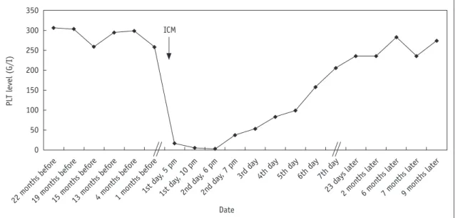
Myocardial infarction and pulmonary embolism were excluded. A transient fever of 38.0°C was seen. Blood pressure returned to normal and the presenting symptoms spontaneously ceased. After some hours, petechiae appeared on the extremities. Severe thrombocytopenia with a lowest value of 2 x 10⁹ Lit-1 was the only laboratory abnormality. The patient received methylprednisolone (125 mg) intravenously and a platelet suspension (8 units).
The platelet count gradually returned to the normal range by day 5 and remained normal during 20 months follow up (Fig. 1). A fecal occult blood test was positive, but no
macroscopic bleeding was observed. Moderate and transient elevations of white blood cell (WBC) counts (15.4 G/Lit) and C-reactive protein (CRP) levels (up to 43 IU) were observed on day 3. These parameters returned to the normal range by day 6. No eosinophilia was observed. There was no significant change in hemoglobin, hematocrit, mean corpuscular volume (MCV), mean cell hemoglobin (MCH), content or mean cell hemoglobin concentration (MCHC) values. The transient and minor increase in mean platelet volume (MPV) is considered as the natural consequence of the compensatory accelerated platelet production (Table 1). Hematological or immunological diseases and actual malignancies were excluded.
The chest X-ray showed a light transparency decrease on the right side at day 1, which was not present at day 3.
On day 5, a chest CT was performed which found a small fibrotic linear shadow and three small nodules (2-3 mm) on the right side. No sign of pneumonia, or indirect signs of emboli or malignancy were detected. The patient was discharged on the 7th day.
Follow Up
Ten months later, a cranial CT examination was performed using no ICM due to neurological symptoms of transient sensory disturbances of the right hand. Leucoarainosis around the ventricles and two small (6-7 mm) hypodensity chronic ischemic lesions were detected. No signs of bleeding or malignancy were detected.
Platelet counts were in the normal range during the 14
Fig. 1. Platelet (PLT) level before and after development of acute severe thrombocytopenia. It should be noted that time scale is non- proportional; it refers to Dates studied.
Date ICM
PLT level (G/I)
350 300 250 200 150 100 50 0
19 months before 15 months before
13 months before 4 months before
1 months before 1st day, 5 pm
1st day, 10 pm 2nd day, 6 pm
2nd day, 7 pm 3rd day
4th day 5th day
6th day 7th day
23 days later 2 months later
6 months later 7 months later
9 months later 22 months before
month follow-up period. No recurrence of the colonic polyp or urinary bladder malignancy and no hematological disease were detected. The patient successfully reduced his body weight using a strong diet. Blood sugar, blood pressure and all laboratory findings moved into the normal range. The patient was without any complaint.
In Vitro Test with ICM on the Blood of the Patient To investigate the incidental direct effect of the ICM (Optiray 350©, Covidien) on the patient’s platelets we performed an in vitro test. ICM solution was added to both EDTA and citrate anticoagulated blood samples of the patient in concentrations corresponding to the estimated in vivo concentration at the time of contrast enhanced CT.
Platelet counts were measured every 10 minutes throughout the 3 hours and then every 20 minutes up to 5 hours. There was no significant change in the platelet counts during the observed time period. The values varied between 246 and 266 G/L.
The experiments were repeated by adding surplus plasma of the patient to the tubes to exclude the lack of a sufficient amount of complement and immunoglobulins in the system. No change in the platelet count was observed following the administration of ICM in this setting.
These tests were performed six months after recovery.
DISCUSSION
The history of development of intravenous contrast materials clearly shows a significant reduction of adverse events. However, even the most modern non-ionic low molecular contrast materials may induce adverse events including acute severe thrombocytopenia as a rare complication in some patients who usually have a genetically determined idiosyncrasy or most frequently an immunologically mediated hypersensitivity. The curiosity of our present case was the acute very severe thrombocytopenia with an extremely low platelet count and rapid recovery.
In a recent retrospective review from the Mayo clinic, adverse events were reported in 0.11% of more than four hundred thousand cases with low-osmolar iodinated and gadolinium contrast agent investigations; 0.15% for low osmolar iodinated and 0.04% for gadolinium contrast doses (7). The most common adverse effects were hives and nausea. Only 16 cases of adverse effects necessitated transfer for further observation or treatment in other unit than the radiology department. Only one death in the study period occurred after administration of low-osmolar iodinated contrast material. The patient had no symptoms during the contrast administration or imaging but died Table 1. Laboratory Test Characteristics before and after Development of Acute Severe Thrombocytopenia
1 Months
Before 1st Day 2nd Day
6 am 2nd Day
7 pm 3rd Day
7 am 4th Day 5th Day 6th Day 7th Day
Plt (G/L) 257 4 2 37 52 82 99 157 205
MPV (fL) 8.5 - - 9.1 - 11.7 10.4 10.9 9.9
Hgb (G/L) 116 - 104 105 105 110 108 110 109
Ht (L/L) 0.35 - 0.34 0.34 0.34 0.36 0.35 0.36 0.35
MCV (fL) 72 - 74.2 75 75 75.7 74.7 74.9 75.5
MCH (pg) 23.9 - 22.6 23 23.1 23 23 23.1 23.3
MCHC (G/L) 332 - 304 307 308 304 307 308 309
WBC (G/L) 9.3 - 15.4 11.4 11.5 11.9 6.7 8.2 9.8
L-Neu (%) 62.7 - 88.3 93 84.3 80.8 64.7 69.1 71.2
Eo (%) 6.1 - 0.1 0.1 0 0.8 0.2 0.6 2.6
Basophil (%) 1 - 0.4 0.1 0.1 0.8 0.8 0.6 0.6
Mo (%) 6.6 - 5.5 1 6.4 7 7.7 7.3 7.3
Ly (%) 21.2 - 5.8 5.8 9.2 10.6 26.6 22.4 18.3
CRP (mg/L) 2.5 - 40.7 - 43.8 - - 6.1 -
INR - - 1.24 - - 1.17 - 1.1 -
Note.— Basophil = basophil leucocyte, CRP = C-reactive protein, Eo = eosinophil leucocyte, Hgb = haemoglobin, Ht = hematocrit, INR = international normalized ratio, Ly = lymphocytes, L-Neu = neutrophil leucocyte, MCH = mean cell haemoglobin content, MCHC = mean cell haemoglobin concentration, MCV = mean cell volume, Mo = monocytes, MPV = mean platelet volume, Plt = platelet, WBC = white blood cell
suddenly within 30 minutes of receiving the dose. No data on the platelet count of this patient was reported. In our case the symptoms started 40 minutes after receiving the intravenous contrast material.
Authors of papers from the nineteen eighties reported acute thrombocytopenia as a complication of iodinated contrast medium (2-5). Severe thrombocytopenia and recurrent thrombocytopenia are considered rare
complications of ICM (1, 8). In our case, the lowest platelet count was 2 x 10⁹ L-1 at 20 hours after the administration of the contrast medium. No such low platelet count has ever been reported before. Most of the studies reported platelet counts between 4 and 30 G/L in ICM induced thrombocytopenia.
The mechanism of ICM induced thrombocytopenia remains unclear. Most commonly, an immunoallergic mechanism is postulated. In vitro studies of the effects of iodinated contrast media on platelets have yielded conflicting results.
Schulze et al. (9) have shown that fibrinogen consumption and platelet aggregation with resultant thrombocytopenia can be induced in whole blood that is preincubated with high concentrations of iodinated contrast media and then exposed to platelet-rich plasma. Chang et al. (4) could not detect intravenous contrast materia (Renografin-76) dependent platelet aggregating factor, and concluded that ICM-induced thrombocytopenia was not caused by in vivo aggregation of platelets.
In our case, the in vitro test did not prove the decrease of platelet count when intravenous contrast material was added to the blood sample of the patient. It should be mentioned that the test was performed, as it usually is in other reported cases, at the time when the platelet count of the patient returned to the normal range. However, the experiment confirmed that the ICM has no direct toxicity on the platelets of the patient.
We also postulate an immunoallergic mechanism, although all the immunological tests were negative. The history, the hematological data and the follow up did not prove coincidence with immune thrombocytopenia. Based on the patient’s history of a malignant disease-urinary bladder carcinoma-the possibility of a paraneoplastic phenomenon was taken into consideration. However, there was no sign of recurrence of the tumor at the time of investigation nor during the 14 months follow-up.
The transient elevation of CRP on days 2 and 3 indicates an acute inflammatory reaction most probably in connection with the rapid drop in platelet count. No sign of an
intercurrent viral infection or other infection was detected.
Since the CRP value was perfectly normal on day 6, it also indicates an acute phase reaction.
Since the patient was treated for hypertension with different drugs, theoretically, an interaction between ICM and a drug can be supposed. However, there is no evidence supporting this possibility in this case or in the literature.
As suggested in the literature, we also administered corticosteroids intravenously and administered a platelet suspension to prevent fatal bleeding complications. The time needed for recovery of platelet counts to normal levels was several days, as reported by others (4, 8). The effect of intravenous human globulin or plasmapheresis has not been proven in similar cases (10).
Even the most modern non-ionic low osmolarity intravenous radiographic contrast media can induce acute severe thrombocytopenia by an idiosyncratic mechanism in hypersensitive patients. The mechanism is still unknown.
Mostly, an immunoallergic mechanism is supposed. No such low platelet count, as in our case of a 72 year old man, has ever been reported before.
REFERENCES
1. Wiemer M, Kreuzpaintner G, Lauer B, Kiefel V, Schultheiss HP, Horstkotte D, et al. [Recurrent immune thrombocytopenia: a rare complication after contrast medium injection]. Dtsch Med Wochenschr 1995;120:1236-1240
2. Lacy J, Bober-Sorcinelli KE, Farber LR, Glickman MG. Acute thrombocytopenia induced by parenteral radiographic contrast medium. AJR Am J Roentgenol 1986;146:1298-1299
3. Shojania AM. Immune-mediated thrombocytopenia due to an iodinated contrast medium (diatrizoate). Can Med Assoc J 1985;133:123
4. Chang JC, Lee D, Gross HM. Acute thrombocytopenia after i.v.
administration of a radiographic contrast medium. AJR Am J Roentgenol 1989;152:947-949
5. Wein P, Handler M, Chadda KD. Severe thrombocytopenia as a result of contrast left ventricular angiography. Cathet Cardiovasc Diagn 1982;8:495-499
6. Ural AU, Beyan C, Yalçin A. Thrombocytopenia following intravenous iopamidol. Eur J Clin Pharmacol 1998;54:575-576 7. Hunt CH, Hartman RP, Hesley GK. Frequency and severity
of adverse effects of iodinated and gadolinium contrast materials: retrospective review of 456,930 doses. AJR Am J Roentgenol 2009;193:1124-1127
8. Bonnard P, Sarraj A, de Cagny B, Lassoued K, Slama M.
Recurrent severe thrombocytopenia and non cardiogenic pulmonary edema following intravenous contrast medium. Eur J Radiol Extra 2004;50:27-29
9. Schulze B, Riester P, Blanke D, Fees K, Lehnard G. [Effect of
iodinated roentgen contrast media on coagulation, fibrinolysis and platelets]. Arzneimittelforschung 1977;27:2128-2133
10. Aster RH, Bougie DW. Drug-induced immune thrombocytopenia. N Engl J Med 2007;357:580-587