ABSTRACT
A variety of therapeutic modalities can be used for the endodontic treatment of a traumatized tooth with internal root resorption (IRR). The authors present a case report of the successful restoration of a traumatized upper central incisor that was weakened due to severe IRR and subsequent periapical lesion formation. A 20-year-old female patient was referred to our clinic with severe internal resorption and subsequent periapical pathosis destroying the buccal bone wall. Root canal treatment had been initiated previously at another dental practice, but at that time, the patient's condition could not be managed even with several treatments. After cone-beam computed tomography imaging and proper chemomechanical cleaning, the tooth was managed with a mineral trioxide aggregate plug followed by root canal filling using short fiber-reinforced composite, known as the Bioblock technique. This report is the first documentation of the use of the Bioblock technique in the restoration of a traumatized tooth. The Bioblock technique appears to be ideal for restoring wide irregular root canals, as in cases of severe internal resorption, because it can uniquely fill out the hollow irregularities of the canal. However, further long-term clinical investigations are required to provide additional information about this new technique.
Keywords: Short fiber-reinforced composite; Bioblock technique; Reinforcement;
Internal resorption; Mineral trioxide aggregate plug; Cone-beam computed tomography
INTRODUCTION
Internal root resorption (IRR) has been described as a resorptive defect of the internal aspect of the tooth root caused by odontoclastic activity that is mainly associated with chronic pulpal inflammation and trauma [1]. IRR is an inflammatory condition that results in the progressive destruction of intraradicular dentin along the middle and apical thirds of the root canal [2]. Early diagnosis and treatment are important because the lesion progresses over time, leading to potential perforation and/or thinning of the root canal walls. Due to the potential difficulty of accessing, cleaning, and perfectly sealing the perforation site, the presence of perforation in cases of severe IRR further complicates and may even affect the prognosis of the endodontic treatment [3-6]. As stated by Yildirim and Elbay [7], the presence of perforation is one of the most important factors affecting the prognosis of IRR treatment. As advanced IRR progressively destroys and consequently weakens the tooth and
Case Report
Received: Apr 2, 2020 Revised: May 27, 2020 Accepted: May 27, 2020
Fráter M, Sáry T, Garoushi S
*Correspondence to Márk Fráter, DMD, PhD, MSc
Assistant Professor, Department of Operative and Esthetic Dentistry, University of Szeged Faculty of Dentistry, Tisza Lajos Krt. 64-66., Szeged H-6720, Hungary.
E-mail: meddentist.fm@gmail.com Copyright © 2020. The Korean Academy of Conservative Dentistry
This is an Open Access article distributed under the terms of the Creative Commons Attribution Non-Commercial License (https://
creativecommons.org/licenses/by-nc/4.0/) which permits unrestricted non-commercial use, distribution, and reproduction in any medium, provided the original work is properly cited.
Conflict of Interest
No potential conflict of interest relevant to this article was reported.
Author Contributions
Conceptualization: Fráter M; Investigation:
Sáry T; Project administration: Sáry T;
Supervision: Garoushi S; Writing - original draft: Fráter M; Writing - review & editing:
Garoushi S.
ORCID iDs Márk Fráter
https://orcid.org/0000-0002-0365-1613 Tekla Sáry
https://orcid.org/0000-0002-3889-9045 Sufyan Garoushi
https://orcid.org/0000-0001-9457-2314
Márk Fráter ,1* Tekla Sáry ,1 Sufyan Garoushi 2
1Department of Operative and Esthetic Dentistry, University of Szeged Faculty of Dentistry, Szeged, Hungary
2 Department of Biomaterials Science and Turku Clinical Biomaterials Center, Institute of Dentistry, University of Turku, Turku, Finland
Bioblock technique to treat severe internal resorption with subsequent periapical pathology: a case report
Provisional
Provisional
space preparation should be conducted so as to avoid the removal of radicular dentin [10].
This may leave the practitioner with a mismatch between the FRC post and the root canal diameter, which, depending on the actual cross-section, may leave considerable space for the inferior luting material. In cases of IRR, given the increased surface irregularity of the canal wall, this problem is even more pronounced.
Several methods have been proposed for the construction of individual FRC posts, either via a multi-post technique [11] or by relining an FRC post with composite [12]. However, these techniques cannot be applied in most IRR cases, as the coronal structure is usually well- preserved, making insertion of a well-fitting individualized post impossible. Some authors have recommended the use of short-fiber reinforced composite (SFRC) in the root canal, with this material serving a double purpose of filling and reinforcement [13,14]. In 2019, an improved method called the Bioblock technique was reported. This technique yielded good results in terms of leakage between the fiber-reinforced material and the canal walls, curing inside the canal, and fracture resistance [15].
This case report describes the application of this new technique in a central incisor with severe IRR.
CASE REPORT
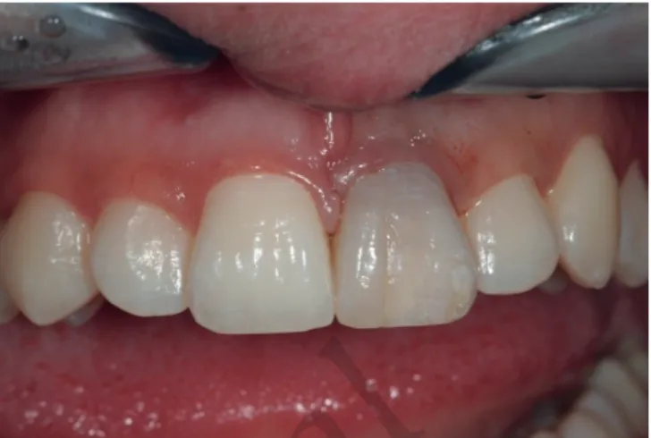
A 20-year-old woman with a history of root canal treatment of the upper maxillary (left) central incisor was referred to our department from a private clinic. The root canal treatment had been initiated 4 months prior but could not be finished. A clinical examination revealed a temporary filling on the palatal surface. The coronal part of the tooth was discolored, and multiple visible cracks were present on the buccal surface (Figure 1). The tooth was asymptomatic; the mobility was normal, and the probing depth at all sites was ≤ 3 mm. A periapical radiograph revealed a lesion around the apex and an enlarged and clearly irregular root canal (Figure 2A). When specifically asked, the patient denied knowledge of any previous dental trauma. Upon checking the electronic documentation of the faculty of the institution, we found that the patient had visited the department 2 years prior for other reasons, and X-rays were taken at that appointment. From a comparison of the periapical X-rays, it was clear that something must have happened since her last visit (Figure 2B). After gaining her confidence, the patient and her mother admitted that the patient had suffered from domestic abuse at the hands of her boyfriend, but that had been taken care of by the police. At this point, a small-field-of-view high-resolution cone-beam computed tomography (CBCT) scan was taken to investigate the extent and irregularity of the lesion. The scan confirmed the presence of a periapical lesion, which had destroyed most of the buccal bony wall around
Provisional
Provisional
the root. The scan also showed massive internal resorption inside the root canal (Figure 2C).
The patient was informed of the findings and the poor prognosis of the tooth, but since she desperately wanted to keep her tooth, she provided consent for endodontic treatment, which was initialized at a later appointment. The patient was also informed that due to the irregular nature and more pronounced buccolingual extent of the resorptive lesion and the massive destruction of the buccal bony plate, which cannot be seen or monitored on periapical X-rays, it would be beneficial to control the steps of the treatment and perform the monitoring via CBCT scans. The patient was informed of the extra radiation exposure resulting from the CBCT scans, but she insisted on undergoing scans instead of periapical X-rays when deemed beneficial by the clinicians.
The working length was established with an electronic apex locator (Root ZX, J. Morita Corp., Tokyo, Japan) and confirmed via radiography. The canal was subjected to instrumentation only with NiTi files (K-File Nitiflex, Dentsply-Maillefer, Ballaigues, Switzerland), stainless steel hand files (K-File, Dentsply-Maillefer), and Hedstrom files (ReadySteel, Dentsply- Maillefer). The last instrument used for apical preparation was a size 120 file, and at that point a definite apical stop could be established. The site was irrigated with copious amounts of 5% sodium hypochlorite. At the time of drying, the electronic apex locator, the hand files, and the paper points indicated no sign of perforation. At the most apical part of the Figure 1. Photograph of the patient upon presentation.
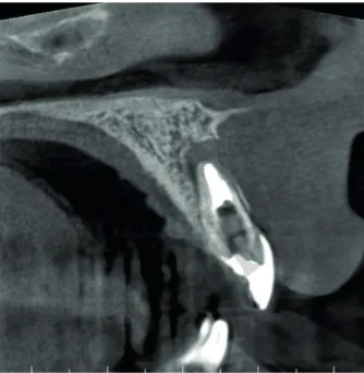
A B C
Figure 2. Images of the patient's tooth upon presentation and 2 years prior. (A) X-ray image upon presentation.
(B) X-ray of the same tooth from 2 years prior. (C) Cone-beam computed tomography image of the tooth upon presentation.
Provisional
Provisional
resorption defect, the root canal appeared extremely thin; thus, the use of warm gutta-percha for obturation was deemed risky and difficult to control. An approximately 4- to 5-mm-thick mineral trioxide aggregate (MTA) plug (MTA+, CERKAMED Medical Company, Stalowa Wola, Poland) was made to establish a proper apical barrier without error in the affected apical part (Figure 3). The MTA was covered with a moist cotton pellet, and the tooth was temporized with Cavit W filling material (3M ESPE, Seefeld, Germany) for 48 hours. At the next appointment, the root canal was adhesively treated with a dual-cure self-etch adhesive system (Gradia Core Self-Etching Bond, GC Europe, Leuven, Belgium) and was filled with SFRC (EverX Posterior, GC Europe) according to the Bioblock technique [15] to the point of the root canal orifice. During this procedure, an approximately 4-mm-thick increment of SFRC material was placed in the root canal and applied to the most apical part to make contact with the MTA plug. This can be accomplished by pressing alternately with any small- headed microbrush and a periodontal probe or plugger. After the first layer appeared to be in position, a light-transmitting FRC post (1.4 mm GC Fiber Post, GC Europe) was inserted into the canal to facilitate the transmission of the light to the apically-positioned layers. The light-transmitting post was withdrawn to 0.5–1 mm from the surface of the uncured SFRC layer so as not to directly contact it. The first layer of SFRC was light-cured through the post for 80 seconds using a light source with an average power density of 900 mW/cm2. This was continued to the level of the cemento-enamel junction. After the root canal was filled to that level, the coronal portion of the tooth was internally bleached several times with sodium perborate (made by the pharmacy of the University of Szeged) mixed with distilled water at a ratio of 2:1 (g/mL) and covered with glass ionomer filling (Equia Fore, GC Europe). After the desired color was reached, the coronal cavity and the SFRC material in the orifice were refreshed with a diamond bur, adhesively treated (G-premio Bond, GC Europe), and restored with SFRC and a 2-mm-thick composite covering on the palatal aspect (G-aenial Anterior, GC Europe) (Figure 4).
Figure 3. Cone-beam computed tomography image after the establishment of the apical mineral trioxide aggregate plug.
Provisional
Provisional
A follow-up CBCT scan was taken 1 year after the final root canal treatment and revealed continued healing of the periapical radiolucency and reformation of the buccal bony wall around the apex (Figure 5). The tooth has remained asymptomatic ever since, and the patient was satisfied with the result.
DISCUSSION
IRR is a relatively rare entity, but when present, it can pose a challenge from both the endodontic and restorative points of view. As seen in the present case, IRR may go unnoticed until the lesion has advanced significantly, resulting in perforation or symptoms of acute or chronic apical periodontitis after the entire pulp has become necrotic [2].
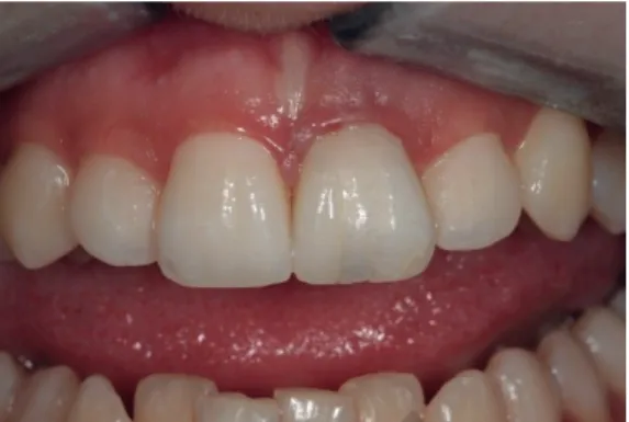
It should be emphasized that the quality and longevity of restorations in root canal-treated teeth play an important role in the outcomes, and restoration must be considered a critical final step for successful endodontic therapy [16]. This is extremely important in cases Figure 4. Final outcome after intracoronal bleaching and permanent composite restoration.
Figure 5. Follow-up cone-beam computed tomography scan taken 1 year after the finalization of root canal treatment, showing the homogeneity of the short-fiber reinforced composite inside the canal.
Provisional
Provisional
optimal for such applications, given that it is applied centrally, far from the interface.
As pointed out by Juloski et al. [18], to form a reinforced, mechanically homogeneous unit, a restorative material with mechanical properties similar to those of dentin should ideally be directly bonded to the dentin of the root canal. Conventional composite materials resemble dentin in most of their mechanical properties, but their fracture toughness is significantly lower. A material with high fracture toughness has the ability to better resist crack initiation and propagation. Consequently, fracture toughness is of key importance in the longevity of dental materials [19]. Bijelic-Donova et al. [20] showed that SFRC had a statistically significantly higher fracture toughness (2.4 MPa·m1/2) and fatigue limit than conventional composite resins (range, 0.9–1.1 MPa·m1/2). As a result, even if a fracture develops, the pattern is mostly reparable. This was demonstrated by Fráter and colleagues [15,21] in the context of SFRC use in both shallow and deep cavities, as well as inside the root canal [22].
In the Bioblock technique, SFRC is directly and closely adapted to the root canal wall, avoiding the drawbacks of the use of luting cement or the biomechanically incorrect positioning of the FRC post. This has the potential to eliminate the damaging tensile stress produced when a restoration is loaded [15] and enables the clinician to fill and restore any root canal with an irregular cross-section deemed not ideal for FRC post insertion. With the Bioblock technique, the number of fibers can also be maximized in the critical cervical area of the tooth, which appears to be important regarding future stress accumulation [23]. It should be highlighted that the Bioblock technique minimizes the number of interfaces within the restoration-tooth complex, which has been shown to be beneficial, as interfaces increase the amount of stress within this unit [24]. SFRC contains randomly-oriented short glass fiber fillers on the millimeter scale, and these fibers can conduct and scatter light. SFRC has been determined to be suitable for bulk use in 4-mm increments in coronal cavities [25,26]. In this specific technique, the SFRC inside the root canal is light-cured through a light-transmitting FRC post, which is only used for light transmission and therefore is removed after this step.
The light-transmitting potential of FRC posts has been demonstrated by multiple studies [27-30] and may compensate for the reduced energy of the light coming from a longer distance due to the depth of the restored cavity. Still, the length of the tooth must be taken into consideration in every case. Since the amount of light energy transferred decreases with the length of the tooth, the length of the post should be set at the minimum at which the operator can simply remove it from the canal. This enables the light source to be placed as close as possible to, and in direct contact with, the tooth.
In the present case, the working length was 20 mm, of which the MTA plug took up 5 mm.
The FRC post was cut back to the lower margin of the cavity orifice. The operator also had to calculate the thickness of the shallowest layer of SFRC in the canal (4 mm), which left us
Provisional
Provisional
with an approximately 6-mm-long FRC post for light transfer. As light transfer to the apically placed SFRC is a clinically important part of the Bioblock technique, it has been verified using the microhardness test in previous in vitro studies, which have clearly shown that SFRC can be appropriately cured in the canal with extended light-curing time (80 seconds) [15,31]. The authors would like to emphasize that in our studies and in this case, the SFRC was placed not more than 6–7 mm deep to the canal orifice, which seems to be a depth at which the setting and curing of SFRC can be controlled. The placement of SFRC has not been attempted in deeper areas and cannot be currently recommended.
In this severe case of IRR, after the MTA plug was set, the root canal was restored with SFRC directly placed on top of the MTA without any traditional root canal obturation material in between, a technique recommended by many to reinforce immature teeth [32]. The authors would like to emphasize that, based on their experience, the Bioblock technique can be easier and faster than FRC post insertion with core build-up, assisting with the everyday work of clinicians.
CONCLUSIONS
This case indicates that the Bioblock technique could be an effective and relatively easy way to restore and likely to reinforce even advanced cases of IRR in the anterior dentition. The case also emphasizes the importance of using CBCT in cases of resorption when periapical X-ray imaging does not provide sufficient information about the extent of the lesion.
REFERENCES
1. Kaval ME, Güneri P, Çalışkan MK. Regenerative endodontic treatment of perforated internal root resorption: a case report. Int Endod J 2018;51:128-137.
PUBMED | CROSSREF
2. Patel S, Ricucci D, Durak C, Tay F. Internal root resorption: a review. J Endod 2010;36:1107-1121.
PUBMED | CROSSREF
3. Aktemur Türker S, Uzunoğlu E, Deniz Sungur D, Tek V. Fracture resistance of teeth with simulated perforating internal resorption cavities repaired with different calcium silicate-based cements and backfilling materials. J Endod 2018;44:860-863.
PUBMED | CROSSREF
4. Abuabara A, Costa RG, Morais EC, Furuse AY, Gonzaga CC, Filho FB. Prosthetic rehabilitation and management of an MTA-treated maxillary central incisor with root perforation and severe internal resorption. J Prosthodont 2013;22:413-418.
PUBMED | CROSSREF
5. Bendyk-Szeffer M, Łagocka R, Trusewicz M, Lipski M, Buczkowska-Radlińska J. Perforating internal root resorption repaired with mineral trioxide aggregate caused complete resolution of odontogenic sinus mucositis: a case report. J Endod 2015;41:274-278.
PUBMED | CROSSREF
6. Küçükkaya Eren S, Aksel H, Askerbeyli Örs S, Serper A, Koçak Y, Ocak M, Çelik HH. Obturation quality of calcium silicate-based cements placed with different techniques in teeth with perforating internal root resorption: a micro-computed tomographic study. Clin Oral Investig 2019;23:805-811.
PUBMED | CROSSREF
7. Yıldırım S, Elbay M. Multidisciplinary treatment approach for perforated internal root resorption: three- year follow-up. Case Rep Dent 2019;2019:5848272.
PUBMED | CROSSREF
8. Jacobovitz M, de Lima RK. Treatment of inflammatory internal root resorption with mineral trioxide aggregate: a case report. Int Endod J 2008;41:905-912.
PUBMED | CROSSREF
Provisional
Provisional
PUBMED
13. Bijelic J, Garoushi S, Vallittu PK, Lassila LV. Short fiber reinforced composite in restoring severely damaged incisors. Acta Odontol Scand 2013;71:1221-1231.
PUBMED | CROSSREF
14. Forster A, Sáry T, Braunitzer G, Fráter M. In vitro fracture resistance of endodontically treated premolar teeth restored with a direct layered fiber-reinforced composite post and core. J Adhes Sci Technol 2017;31:1454-1466.
CROSSREF
15. Fráter M, Lassila L, Braunitzer G, Vallittu PK, Garoushi S. Fracture resistance and marginal gap formation of post-core restorations: influence of different fiber-reinforced composites. Clin Oral Investig 2020;24:265-276.
PUBMED | CROSSREF
16. Akman S, Akman M, Eskitascioglu G, Belli S. Influence of several fibre-reinforced composite restoration techniques on cusp movement and fracture strength of molar teeth. Int Endod J 2011;44:407-415.
PUBMED | CROSSREF
17. Turkaslan S, Bagis B, Akan E, Mutluay MM, Vallittu PK. Fracture strengths of chair-side-generated veneers cemented with glass fibers. Niger J Clin Pract 2015;18:240-246.
PUBMED | CROSSREF
18. Juloski J, Radovic I, Goracci C, Vulicevic ZR, Ferrari M. Ferrule effect: a literature review. J Endod 2012;38:11-19.
PUBMED | CROSSREF
19. Lassila L, Säilynoja E, Prinssi R, Vallittu P, Garoushi S. Characterization of a new fiber-reinforced flowable composite. Odontology 2019;107:342-352.
PUBMED | CROSSREF
20. Bijelic-Donova J, Garoushi S, Vallittu PK, Lassila LV. Mechanical properties, fracture resistance, and fatigue limits of short fiber reinforced dental composite resin. J Prosthet Dent 2016;115:95-102.
PUBMED | CROSSREF
21. Fráter M, Forster A, Keresztúri M, Braunitzer G, Nagy K. In vitro fracture resistance of molar teeth restored with a short fibre-reinforced composite material. J Dent 2014;42:1143-1150.
PUBMED | CROSSREF
22. Sáry T, Garoushi S, Braunitzer G, Alleman D, Volom A, Fráter M. Fracture behaviour of MOD restorations reinforced by various fibre-reinforced techniques - An in vitro study. J Mech Behav Biomed Mater 2019;98:348-356.
PUBMED | CROSSREF
23. Vallittu PK. Are we misusing fiber posts? Guest editorial. Dent Mater 2016;32:125-126.
PUBMED | CROSSREF
24. Belli S, Eraslan O, Eskitascioglu G, Karbhari V. Monoblocks in root canals: a finite elemental stress analysis study. Int Endod J 2011;44:817-826.
PUBMED | CROSSREF
25. Garoushi S, Gargoum A, Vallittu PK, Lassila L. Short fiber-reinforced composite restorations: a review of the current literature. J Investig Clin Dent 2018;9:e12330.
PUBMED | CROSSREF
26. Lempel E, Őri Z, Szalma J, Lovász BV, Kiss A, Tóth Á, Kunsági-Máté S. Effect of exposure time and pre- heating on the conversion degree of conventional, bulk-fill, fiber reinforced and polyacid-modified resin composites. Dent Mater 2019;35:217-228.
PUBMED | CROSSREF
Provisional
Provisional
27. Seyam RS, Mobarak EH. Reinforcement of teeth with simulated coronal fracture and immature weakened roots using resin composite cured by a modified layering technique. Oper Dent 2014;39:E128-E136.
PUBMED | CROSSREF
28. Galhano GA, de Melo RM, Barbosa SH, Zamboni SC, Bottino MA, Scotti R. Evaluation of light transmission through translucent and opaque posts. Oper Dent 2008;33:321-324.
PUBMED | CROSSREF
29. Aksornmuang J, Nakajima M, Panyayong W, Tagami J. Effects of photocuring strategy on bonding of dual- cure one-step self-etch adhesive to root canal dentin. Dent Mater J 2009;28:133-141.
PUBMED | CROSSREF
30. Bell-Rönnlöf AL, Jaatinen J, Lassila L, Närhi T, Vallittu P. Transmission of light through fiber-reinforced composite posts. Dent Mater J 2019;38:928-933.
PUBMED | CROSSREF
31. Fráter M, Sáry T, Néma V, Braunitzer G, Vallittu P, Lassila L, Garoushi S. Fatigue failure load of immature anterior teeth: influence of different fiber post-core systems. Odontology 2020 May 2. doi: 10.1007/
s10266-020-00522-y. [Epub ahead of print]
PUBMED | CROSSREF
32. Linsuwanont P, Kulvitit S, Santiwong B. Reinforcement of simulated immature permanent teeth after mineral trioxide aggregate apexification. J Endod 2018;44:163-167.
PUBMED | CROSSREF