II./1.: Outer Ear Diseases
Magdolna Szőnyi
II./1.1.: Developmental Abnormalities
Often associated with middle or inner ear developmental abnormalities, or with heart developmental abnormalities.
Figure 1.: The helix is decreased in size, deformed, behind the helix a screw of the hearing aid (bone anchores hearing aid, BAHA) is visible.
We can talk about only cosmetic disorder: e.g. sticking out ear. Recurring inflammatory diseases can be caused by fistules, and cyst around the ear.
The fistules could have a dead end, or can spread deeply even into the pharynx.
Figure 2.:Preauricular Fistule Opening
It can be a serious deformity that affects the hearing, like the absence of the helix, or outer ear canal atresia.
The injury of the mucosal surface of the ear canal could, in rare cases, cause closure of the ear canal.
Therapy:
What to do in case of helix or inner ear canal injuries?
With plastic surgery most of the mild cases can be treated well. In case of total absence of the helix, the epitasis can have a better result. In case of ear canal-, middle-, and inner ear disorders imaging modalities like CT, MRI is important before surgery. With the help of these modalities we can visualize if the middle ear ossicles, nerves, the cochlea, and the tympanic membrane is involved. The imaging modality helps to localize anatomical variations that can cause surgical difficulties e.g. it helps to track the facial nerve.
The cysts and fistules have to be completely removed. If part of their
mucosal surface remains in the deep, they begin to recidivate, and because of the scar tissue their complete elimination is more difficult on the previously operated area.
II./1.2.: Injuries
Sharp injuries: cut, torn, bit injuries of the helix
Blunt injuries occur due to sheer power, othaematoma could be the result.
Figure 3.: Othaematoma
Frostbite, or congelatio, burn
In case acid, base, petrol gets into the ear canal it can cause severe skin injury or even perforation of the tympanic membrane.
After trauma, in case of bleeding from the ear, the fracture of the skull base has to be excluded, e.g. the fracture of the pyramids and the liquorrhea. The most suitable for this is the CT. The tympanic membrane and the ossicular chain have to be examined.
In the ear canal most of the injuries are caused by the cotton bud. The foreign bodies in the ear canal in case of children are mostly small pieces of toys, pearls, in case of adults they are cotton wool, sometimes insects.
Therapy:
Thorough disinfection of the injury of the helix (3% hydrogen peroxide, betadine), antibiotics, and tetanus injection.
Connection with Hearing Diagnosis chapter
The naked cartilage pieces are to be removed, and the edges of the wound are to be sewn. If a bigger cartilage piece remains naked, then free edge skin transplantationor cover it with rotated skin.
The torn helix part or the whole helix possibly has to be transported on ice in sterile sheet with physiological saline. Keeping it alive effectively has a chance only if the surgery occurs within one or two hours. Othaematoma caused by blunt injury is a haemmorhage between the cartilage and the perichondrium. A smaller othaematoma can be treated with paracentesis, bigger ones with incision, then pressure bandage has to be applied. It is also recommended to give per os antibiotics to avoid infection. Regular control is needed, the bandage has to be changed to notice the reoccurrences of the haematoma. In relapsing cases x shaped skin surgery is possible with cutting the opposing corners.
How to remove the foreign body that got into the ear canal?
In case of frostbite and burning keeping the surface sterile is important, besides, restoring the circulation anticoagulant therapy can be considered.
In case of ear canal injuries, when there is no sign of tympanic membrane or ossicular chain injury, a sterile Burow stripe with antibiotics can be inserted into the ear canal.
Recognising the injury of the tympanic membrane and ossicular chain is of utmost importance. Tympanic membrane can be examined with otoscopy, microscope, hearing examination (threshold audiometry, tympanometry).
The circular foreign bodies must not be removed with forceps from the ear canal but with foreign body hook. The foreign body can fall out of the forceps and get stuck in the ear canal at the border of the bony and the cartilaginous parts, which is very narrow, and if it moves onward it may cause tympanic membrane injury.
The annoying movements of living insects can be blocked with filling up the ear canal with water or ethyl alcohol. Similarly to the cerumen, they can be removed with water injection, suction, maybe forceps, or with a hook.
II./1.3.: Inflammatory Diseases
Helix
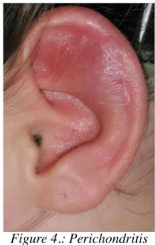
In case of perichondritis the inflammation spreads between the cartilage and the perichondrium of the helix. Pus gathers under the perichondrium
blocking the blood supply of the cartilage from the perichondrium. In case of untreated cases damage of the cartilage, shrinking, and permanent shape disorder may form.
Figure 4.: Perichondritis
In case of erysipelas the inflammation of the skin develops in the helix and its surroundings. It is mainly caused by Streptococcus.
Etiology:
Injury, surgery, furuncule Symptoms:
The skin of the helix is hyperaemic, oedematous, thickened, painful when moving
Pus may pass through fistule Therapy:
Is it necessary to clean the ear canal regularly?
Local and systematic antibiotics, surgery if necessary. In mild cases bandage with antibiotics, in case of spreading into the ear canal a Burow strip should be inserted. Per os antibiotics (amoxicillin, cephalosporin, macrolides).
In case of an extended case, diabetes, or immunosuppressed state surgery of the pus under the perichondrium, parenteral antibiotics.
Outer Ear
The inflammatory origin of the outer ear canal can be categorised according to its severity from the mild inflammatory form to the potentially life threatening diseases mostly occurring in elderly patients (necrotising malignant otitis externa).
Which symthoms suggest the
inflammation of the ear canal?
Forms:
acute diffuse inflammation
circumscribed inflammation (furunculosis)
chronic ear canal inflammations (dermatological diseases: e.g.
underlying eczema, atopic dermatitis, psoriasis) Etiology:
Risk factors:
slackening of the skin caused by humidity, water polluted with bacteria and fungi local trauma (ear cleaning stick) absence of cerumen
ear canal narrower than usual wearing hearing aid
According to the self-cleaning mechanism of the ear canal the skin layer that covers the outer ear canal and the outer surface of the tympanic
membrane continuously wanders outwards thus forcing the cerumen and the polluting materials out to the external opening of the canal.
Connection to the hearing diagnostics chapter
Preventing the inflammation: earplugs when swimming (oily cotton wool, silicon wax, PVC), dripping acid solutions (acetic acid, boric acid) and drying out the ear canal with low temperature hair dryer.
The use of cotton buds must be avoided.
Symptoms: itching
pain (intensifying when chewing, and moving the mandible, and when there is pressure on the tragus)
a feeling of blockage, hearing loss
congestion and swelling of the outer ear canal complete block of the ear canal
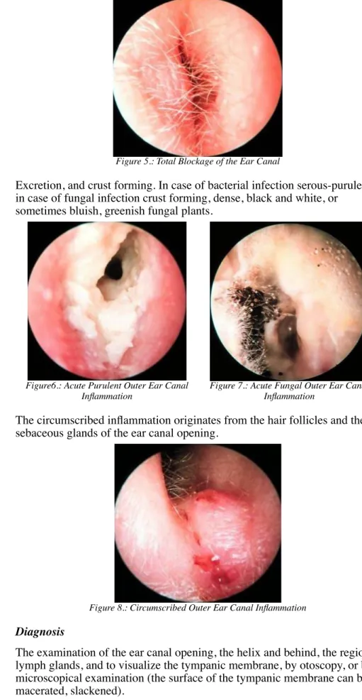
Figure 5.: Total Blockage of the Ear Canal
Excretion, and crust forming. In case of bacterial infection serous-purulent, in case of fungal infection crust forming, dense, black and white, or
sometimes bluish, greenish fungal plants.
Figure6.: Acute Purulent Outer Ear Canal Inflammation
Figure 7.: Acute Fungal Outer Ear Canal Inflammation
The circumscribed inflammation originates from the hair follicles and the sebaceous glands of the ear canal opening.
Figure 8.: Circumscribed Outer Ear Canal Inflammation
Diagnosis
The examination of the ear canal opening, the helix and behind, the regional lymph glands, and to visualize the tympanic membrane, by otoscopy, or by microscopical examination (the surface of the tympanic membrane can be macerated, slackened).
Differential diagnosis: Acute and chronic otitis media persist the same time, and the excretion through the perforated tympanic membrane causes the otitis externa. Audiometry, Tympanometry, plain X-ray (sinuses, and
Schüller), and CT scan to exclude further complications. Oedema around the ear, and lymph gland swelling could develop, and in case of elderly or immunosuppressed patients spread of the inflammation to the parotis, or to the temporo-mandibular joint (malignant necrotizing otitis externa). In these very rare cases, the pain is extraordinarily strong, the excretion is feculent, granulation tissue appears, inflammation can spread to the mastoid and to the skull base, and cranial nerve damage could also occur. Severe
chondritis, osteomyelitis, and ear canal stenosis can happen.
Pathogens
The most typical bacteria are Pseudomonas aeruginosa, Staphylococcus aureus and some anaerobic bacteria. Fungal infections: Aspergillus niger and Candida species.
Therapy
Careful and gentle cleaning of the ear canal (wipe and suction). Local disinfection, anti-inflammatory drugs, antibiotics, and anti-fungal
medications. In mild cases ear drops, in severe stenosis strips with acidic- disinfectant solutions (Burow, or boric acid solution) should be applied into the ear canal. Painkillers. Avoid water, humidity, and slackening of the skin.
Bacterial culture (recurrent, or persisting infection). Oral antimicrobial treatment (in case of immunosuppressed patients, and if the inflammation spreads to the regional tissue). In case of necrosis incision, surgery, and parenteral antibiotics should be applied. In chronic cases, the underlying dermatological conditions should be treated, e.g. local steroids.
Diseases caused by Herpes virus
Apart from the simptoms on the skin what other deformations can herpes viruses cause?
Herpes zoster is the viral infection of the sensory ganglia, which causes severe pain in its dermatome, and later shingles forming. The eruption is common at the cavum conchae, at the ear canal opening, and on the dorsal ear canal wall. The typical shingles usually dry out soon, and transform into yellowish slough, then heal. The viral infection can affect the
vestibulocochlear nerve, so it can cause vestibular symptoms, and the motor fibers of the facial nerve can be affected, too, causing total or partial palsy of the facial nerve. (Ramsay-Hunt syndrome herpes zoster oticus). If the vagal nerve is affected the inflammation, or in some rare cases the
haemorraghic bullae of the ear canal’s inner third, and the upper-dorsal part of the tympanic membrane could happen, and palsy, and ulcers on the same side of the larynx. Since in most of the palsy cases there are symptoms persist after the infection, the highest possible dose antiviral treatment should be started as soon as possible, even in parenteral way (e.g.
Acyclovir), huge dose B12, non-steroid anti-inflammatory drugs, and antiglobulin therapy can be considered.
II./1.4.: Neoplasms
Benign lesions
The most common benign tumours are: papillomas, angiomas, fibromas, and adenomas. Their therapy is surgical remove. The osteoma of the outer ear canal can appear as a solitary or multiplex neoplasm that spreads into the lumen, or as a hyperostosis that narrows the lumen from the frontal or from the inferior wall. If the lumen is blocked and the self-clearing
mechanism of the ear canal is inhibited, surgical removal is recommended.
Can chronic excresion from the ear canal be the sign of malignant
neoplasm?
Atheromas are formed from sebaceous glands with clogged ducts. The sebum accumulated inside them could get infected so the process gets very painful. In the phase of acute inflammation it needs to be drained and then later, a total removal is needed.
In case of surgical solutions of keloids the incision line is recommended to follow the lesion’s border, local steroid infiltration and post-surgical irradiation decreases the risk of recurrence.
Chondrodermatitis is a pressure-sensitive, itchy, node that ulcerates in its centre.
Precancerous states are senile keratosis and the cornu cutaneum.
Malignant Neoplasms
Basalioma (basal cell carcinoma)
Figure 9.: Basalioma of the Helix
In most instances the basalioma starts on the sun-exposed parts of the helix as an itchy and later ulcerating node covered with slough. The tumour grows slowly but continuously, its complete surgical removal essential.
Squamous Cell Carcinoma
It starts as an ulcerating process on the helix. In the ear canal purulent- sanguineous excretion is visible, a tumour that mimics granulation tissue or polyp. Its identification is challenging, histology is recommended from these lesions.
Melanoma malignum
It is a fast-growing, painless dark coloured tumour. Total surgical removal is needed, if necessary, irradiation and chemotherapy is also required.
Rare malignant tumours: adenocarcinomas and sarcomas.
Basaliomas rarely, but squamous cell carcinomas and melanomas frequently have metastasis lymph nodes located under the ear, on the neck, in the parotis, and on the skull base.
Therefore, in case of metastasis suspect lymph node the neck ultra sound, fine needle aspiration biopsy, and CT scan is needed during the patient’s examination. In case of metastasis surgical removal is necessary. When it is not possible, chemotherapy and irradiation is needed.