Inhibitors of Protein Synthesis
C. T . Caskey
I. Introduction 131 I I . Protein Biosynthesis 132 I I I . Inhibitors of Peptide Chain Initiation 139
A. Aurintricarboxylic Acid 139
B. Pactamycin 141 C. Kasugamycin 142 D . Sodium Fluoride ( N a F ) 143
IV. Inhibitors of Peptide Chain Elongation 144
A. A m i n o a c y l - t R N A Binding 144
B. Translocation 150 C. Peptide Bond Formation 155
V. Inhibitors of Peptide Chain Termination 163
A. Antibodies to R F 1 and R F 2 163 B. Antibiotics I
63
V I . Premature Peptide Chain Termination 165
A. Puromycin 165 B. Aminooligonucleosides 166
V I I . Other Inhibitors 167 A. Colicin E3 167 B. G D P C P 168 V I I I . Summary 169
References.. 170
I. INTRODUCTION
The process of protein biosynthesis has been investigated in bacterial, mammalian, and plant cells (1). These studies indicate that cells of
all types have a similar mechanism for protein biosynthesis, although the protein factors and ribosomal structure of these cells differ consider
ably (1). The structural differences in these proteins are partly responsi
ble for the cellular differences in sensitivity to inhibitors of protein bio- 131
synthesis (2). Furthermore, the differences in permeability of cells to certain inhibitors contribute to their differences in inhibitor sensitivity.
These mechanisms of differential cellular sensitivity to protein biosynthe
sis inhibitors are the basis of the therapeutic usefulness of many anti
biotics. These cellular differences occur in spite of overwhelming informa
tion indicating that both the genetic code and the mechanisms for its translation are universal among all cell types (3). It is not clear why cells have evolved with these differences, but it appears likely that bio
synthesis of antibiotics may have contributed to the genetic pressure for the divergence of the prokaryotic and eukaryotic cells. While the reasons for these differences are not clear, it is well known that a given inhibitor may exert its effect on only particular cells and/or only under in vitro conditions. Care must therefore be taken in extrapolating infor
mation obtained from one cell type to that of another or from in vitro experiments to in vivo effects. Recently, several investigators (4, 5) have indicated that data obtained from study of antibiotic effects on in vitro partial reactions of protein biosynthesis do not accurately predict their major sites of action either in whole cells or in polysome protein biosynthesis. The reason for these apparent discrepancies may relate to the interdigitation of intermediate reactions occurring in protein bio
synthesis mechanisms. In such cases a given inhibitor may predominately affect a single intermediate event and indirectly inhibit other events occurring at an adjacent ribosomal site. Furthermore, while antibiotic effects are observed in partial reactions, the addition of other components essential for complete protein biosynthesis may modify the antibiotic binding and thus its primary action. Because of these complexities, this review will deal primarily with the in vitro effects of protein inhibitors and, where possible, will indicate their in vivo effectiveness and site of action. The reader may consult other reviews (2, 6), which attempt to correlate in vivo and in vitro inhibitor actions.
II. PROTEIN BIOSYNTHESIS
The linear linkage of amino acids through amide bonds into protein molecules is a highly ordered process directed by mRNA. The special codons utilized for initiation (AUG), termination (UAA, UAG, or UGA), and chain elongation of protein synthesis are identical in all cells thus far studied (7). The translational process occurs on the ribosome and involves an ordered interaction of mRNA, soluble protein factors, amino-
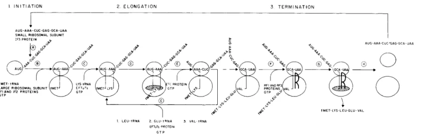
acyl-tRNA, GTP, and ribosomal subunits. An outline of this process is shown in Fig. 1. While Fig. 1 applies specifically to bacterial protein biosynthesis, many similar intermediate steps have been identified in mammalian and yeast extracts and will be referred to in the text.
The initial step (A, Fig. 1) of protein biosynthesis is attachment of mRNA to the smaller ribosomal subunit. This attachment is stimulated by a soluble protein factor, IF3, and positions mRNA for the process of translation. Several forms of IF3 isolated from bacterial cells have differences in activity for chain initiation with specific mRNA types
(i.e., T4 bacteriophage and RNA bacteriophage mRNA) (8, 9). The multiple forms of IF3 are well characterized from bacterial cells and are known to be involved in the dissociation of the ribosomal 70 S complex into its 30 and 50 S subunits (10). The precise manner and timing of this ribosomal-dissociating activity are not unanimously agreed upon. While the mechanism of ribosomal exchange is not fully understood it is clear that both prokaryotic and eukaryotic ribosomal subunits undergo cyclic exchange (11, 12) during protein biosynthesis. The mechanism of this exchange is only partially clarified at this time. On the one hand, 70 S ribosomal couples are said to be separated by IF3 following release from the mRNA at chain termination (13). Alternately, it has been suggested that 70 S ribosomal couples are prevented in their forma
tion by IF3 (14)- Comparable studies in mammalian extracts of an IF3-like activity are not available at this time. A factor washed from avian muscle ribosomes has been identified which is required for transla
tion of avian myosin mRNA in a rabbit reticulocyte cell-free extract (15). This factor is similar in some respects to bacterial IF3, which suggests that mammalian and bacterial cells initiate translational events in a similar manner.
Step B results in the formation of active ribosomal complex (70 S) consisting of two nonidentical ribosomal subunits (30 and 50 S), mRNA, fMet-tRNA, and transiently GTP, and two soluble protein factors. The factor IF1 has a molecular weight of 9000, binds to the smaller ribosomal subunit, and dissociates from the ribosomal subunit upon formation of the ribosomal duplex (70 S complex) (16). The factor IF2 exists in two forms (80,000 and 100,000 daltons), binds to the ribosomal complex
(70 S) with GTP, and dissociates upon hydrolysis of its y-phosphate residue (16). There are no known functional differences in the two forms of IF2, and recent structure studies of the two forms indicate that they have many common tryptic peptides (17). It is not known if the smaller molecule represents a posttranslational modified molecule or if it is the product of a partially reduplicated gene. The end result of such interac-
A U G - A A A - C U C - G A G - G C A - U A A
I
SMALL RIBOSOMAL SUBUNIT
1. L E U - tRNA 2. GLU- t R N A 3. V A L - t R N A EFTuTs PROTEIN
FIG. 1. Intermediate steps in protein biosynthesis.
tions is formation of the ribosomal duplex containing mRNA with initial codon, AUG, and fMet-tRNA. This series of events is not entirely pro
grammed by mRNA and the ribosome, as indicated by the recent dis
covery of a complex consisting of IF2, fMet-tRNA, and GTP that forms in the absence of mRNA and the ribosome {18). The role of IF1 in the formation and regulation of these complexes is not clear at this time.
These complexes form and are subsequently programmed for ribosomal binding by the mRNA initiator codon, resulting in the delivery of fMet- tRNA and GTP to the proper ribosomal site. To date, only one of two cellular species of Met-tRNA is known to initiate protein synthesis. Formyl- Met-tRNA initiates protein synthesis in bacterial cells (19) while Met- tRNA initiates mammalian (20) and yeast protein biosynthesis (21).
These special initiator species of Met-tRNA have common characteris
tics. It is clear, however, that both yeast and mammalian cells lack the enzyme responsible for the formation of fMet-tRNA from Met-tRNA and tetrahydrofolic acid (THFA) (22). Only fMet-tRNA, not Met-tRNA, initiates bacterial proteins, while Met-tRNA is the initiator species in prokaryotic cells. Protein factors have been identified from a variety of prokaryotic sources which not only affect the binding of Met-tRNA to ribosomes but also interact with GTP (23-25). These factors bear a func
tional similarity to bacterial IF1 and IF2 factors.
In both bacterial and mammalian extracts, GTP hydrolysis is neces
sary for formation of the first peptide bond. The binding of fMet-tRNA to bacterial ribosomes occurs with either GTP or an analog, 5'-guanylyl- methylenediphosphonate (GDPCP), which cannot undergo y-phosphate hydrolysis (26). There is a distinctive difference, however, in the char
acter of ribosomal-bound fMet-tRNA programmed with GTP and GDPCP. In the first case, fMet-tRNA participates in formation of the first peptide bond, while complexes formed with GDPCP do not. Thus, GTP hydrolysis is a requisite event that allows the participation of ribosomal-bound fMet-tRNA in the first peptide bond. There is no ac
companying movement of mRNA along the ribosome concomitant with this GTP hydrolysis, and thus it does not appear to be a translocational event (27). These results have been interpreted in the light of the ribosomal two-site model to indicate that fMet-tRNA enters directly into the ribosomal holding site (P) but must be "accommodated" by GTP hydrolysis for activation (27). Alternately, it has been suggested that accommodation is merely a reflection of removal of IF2 and IF1 from the ribosome following GTP hydrolysis. These concepts will certainly be modified as more detailed information on ribosomal structure and function become available.
The presentation of the second aminoacyl-tRNA to the proper bac
terial ribosome site requires two soluble protein factors, EFTu and EFTs, and GTP (28, 29). A eukaryotic factor, EF1, has functions similar to those of the bacterial EFTuTs complex (30). As indicated in Fig. 1 ( C ) , a preribosomal complex consisting of Lys-tRNA-EFTu-GTP is formed independent of mRNA. The equivalent eukaryotic complex is Lys- tRNA-EFl-GTP. The complex is then programmed for ribosomal binding by the mRNA codon AAA. In response to codon recognition the aminoacyl-tRNA is transferred to a site on the ribosome, allowing interaction of the above with the ribosome and subsequent release of Pi and the binary complex EFTu-GDP (31). A second protein, EFTs, displaces GDP from EFTu-GDP and thus EFTu, permitting its repeated participation in identical ribosomal events. In eukaryotic cells, EF1 interacts with GTP and aminoacyl-tRNA prior to ribosomal binding of aminoacyl-tRNA to ribosomes (32). It is not clear whether EF1 disso
ciates from the ribosome and GDP in a fashion identical to that of EFTu. Several forms of EF1 are known, but no functional differences are attached to each at this time (33).
The result of these first few intermediate steps is the alignment of two aminoacyl-tRNA species on the ribosome in response to mRNA codons. Peptidyltransferase, a ribosomal activity, catalyzes peptide bond formation with these aligned aminoacyl-tRNA species. This is achieved by transfer of fMet from its ester linkage to the amino group of Lys- tRNA. The ribosome catalyzes this event independent of soluble factors or GTP (34). The ribosomal proteins responsible for this activity are not characterized. The enzyme activity is known to reside exclusively with the larger ribosomal subunit in the case of both eukaryotic (35) and prokaryotic ribosomes (36). Peptidyltransferase not only catalyzes peptide bond formation but has been shown to be capable of transesteri- fication (37, 38) and hydrolysis (30). The ability of peptidyltransferase to hydrolyze peptidyl-tRNA has implicated it in the peptidyl-tRNA hydrolysis event of peptide chain termination (39). Thus peptidyltrans
ferase is one of a number of enzymes with the capacity to catalyze nucleophilic attack of chemical bonds. (This point will be further dis
cussed below.)
Following peptide bond formation by peptidyltransferase, mRNA and the newly formed peptidyl-tRNA must be moved with respect to the ribosome for translation of the next codon and further extension of the peptide chain. This step (E) is translocation, and requires both a soluble protein factor and GTP (40). A bacterial factor, EFG, of M W 80,000 is required for translocation. A mammalian factor, EF2, is analogous
in function to EFG but is not interchangeable with EFG HI). The GTP hydrolysis occurring with translocation is a ribosomal event and requires formation of a EFG-GTP-ribosome intermediate. The soluble factor EFG has no GTP hydrolysis capacity alone. Recently, investiga
tors have found that removal of two ribosomal proteins (13,000 M W ) from the 50 S subunit suppresses the GTPase activity of both EFTu and EFG (42, 43). The activity is restored upon addition of the purified proteins back to the depleted ribosomal subunits (48). This and other studies which will be discussed in the sections concerning thiostrepton and fusidic acid suggest that the GTP hydrolysis occurring with amino
acyl-tRNA binding and peptidyl-tRNA translocation require common ribosomal proteins or binding sites. The result of translocation is move
ment of peptidyl-tRNA from the binding (A) to the holding (P) site.
Concomitant with this event, the deacylated tRNA is displaced from the ribosome (44)- The mRNA movement positions the next codon for translation in the binding site (A) and a new round of elongation can begin. The process of aminoacyl-tRNA binding ( C ) , peptide bond for
mation ( D ) , and translocation (E) then repeats in a cyclic manner for the number of mRNA codons available for translation. These cyclic events result in peptide chain elongation and are interrupted by peptide chain termination.
Peptide chain termination is the final ribosomal event in completion of a protein. The event is coded by one of three codons in both mam
malian and bacterial cells (UAA, UAG, or UGA) (45, 46)- While a single codon is adequate for peptide chain termination, recent studies of naturally occurring chain termination codons indicate that they can occur in pairs (UAA-UAG) (47, 48). Such tandem codon arrangements would give a measure of assurance for chain termination and would be particularly protective in cells that carry suppressor genes. It is unre
solved at this time if such tandem codons occur for the above reasons or if they perhaps signal additional events—ribosomal dissociation, mRNA degradation, mRNA displacement, etc. Several soluble protein factors (RF1, RF2, and RF3) (49, 50) that have been identified in bacterial cells participate in peptide chain termination in vitro. The RF1 and RF2 factors recognize termination codons, possess no nucleic acid, are approximately 45,000 M W (51), and recognize codons with specificity (RF1, UAA, or UAG; RF2, UAA, or UGA). They are required for ribosomal-mediated hydrolysis of peptidyl-tRNA, although RF1 and RF2 have no hydrolysis activity in the absence of ribosomes. In reactions containing 20% ethanol RF1 or RF2 can bind to ribosomes independent of codon recognition and promote peptidyl-tRNA hydrolysis (52). These
studies indicate that codon recognition is not required for peptidyl-tRNA hydrolysis. Thus, RF1 and RF2 are required for two separate interme
diate events in peptide chain termination. A third protein factor, RF3, has no codon specificity but stimulates the activity of RF1 and RF2 (50). There are conflicting reports over whether RF3 is equivalent to or differs from EFTu (50y 53). While the precise character of the protein is not clear, several activities of the RF3 are recognized. It binds to GDP and GTP.
Furthermore, RF3 stimulates the ribosomal binding of RF1 and RF2 and becomes part of the ribosomal complex consisting of R F 1 - or R F 2 - RF3-terminator codon-ribosome (54). This complex rapidly dissociates upon addition of GTP or GDP. Thus RF3 has several general properties of the fMet-tRNA and aminoacyl-tRNA binding factors IF2 and EFTu.
Thus far an absolute GTP requirement for in vitro bacterial peptide chain termination has not been demonstrated. Conversely, requirement for GTP can be easily demonstrated for in vitro peptide chain termina
tion with mammalian extracts (46). A protein fraction (RF reticulocyte), from rabbit reticulocytes has been purified some 800-fold. This RF has an apparent molecular weight of 255,000 by Sephadex chromatography and on SDS gel analysis has two bands of lower molecular weight (40,000- 50,000) (55). Reticulocyte RF participates in peptide chain termination with oligonucleotides containing any of the three terminator codons UAA, UAG, or UGA. Attempts to separate codon-specific RF's have not succeeded and indirect evidence suggests that a single component of reticulocyte RF recognizes all three codons (55). These studies with reticulocyte RF collectively suggest it is a protein complex with the activities of bacterial RF1, RF2, and RF3. Additional studies are required to define the precise function of these subunits. The hydrolysis of nascent pep
tidyl-tRNA occurring with RF requires GTP, is inhibited by GDPCP, and is not affected by GDP or GMP (55). An uncoupled hydrolysis of the y-phosphate of GTP occurs in reactions containing RF and ribosomes.
This GTP hydrolysis is stimulated by terminator oligonucleotides and occurs in the absence of peptidyl-tRNA hydrolysis. The binding of RF to ribosomes directly by terminator oligonucleotides has been shown to require GTP or GDPCP. Since hydrolysis of GTP is required for peptidyl-tRNA hydrolysis but not RF binding to ribosomes, GTP is assumed to be required for an essential ribosomal event following RF binding. There is no indication that GTP is actually required in the hydrolysis of peptidyl-tRNA with RF. Thus, these studies with RF are consistent with those demonstrating that IF2 and EFTu can participate in ribosomal binding of fMet-tRNA and aminoacyl-tRNA with GDPCP but that these ribosomal complexes do not initiate or extend peptide
chains. In all of these cases GTP hydrolysis is required for a ribosomal event.
The hydrolysis of peptidyl-tRNA occurring at peptide chain termina
tion apparently requires an interaction of RF molecules with ribosomal elements. In studies of both bacterial and mammalian peptide chain termination, RF1, RF2, or reticulocyte RF are required for hydrolysis although they cannot promote such a reaction independently. Peptidyl- tRNA hydrolysis of nascent peptidyl-tRNA can be mediated in vitro by the ribosomal enzyme peptidyltransferase in the absence of release factor. The inhibitors of mammalian and bacterial peptidyltransferase have been found to also inhibit RF-mediated peptide chain termination
{56). These studies suggest strongly that the completion of peptide chains requires the cooperative interaction of both RF and the peptidyltrans
ferase activity of the ribosome. The mechanism of mRNA, RF, tRNA, and ribosomal subunit dissociation (H) is not totally assessed at this time. Messenger RNA has been shown to separate from ribosomes at peptide chain termination directly by in vitro study and indirectly by genetic study of polarity {57, 58). The mechanism of this dissociation is not known.
Since the soluble factors required for protein biosynthesis were initially isolated and characterized in a number of laboratories, earlier papers devoted to their description did hot have a uniform nomenclature. Most investigators now agree on the number and character of these factors and have attempted to minimize the confusion by the acceptance of a uniform nomenclature. The new nomenclature has been used through
out this chapter {59). For the benefit of those referring to older papers for more detailed information, the old and new terminology of these soluble factors are given in Table I {28, 49, 53, 54, 60-68).
III. INHIBITORS OF PEPTIDE CHAIN INITIATION
A. Aurintricarboxylic Acid
Aurintricarboxylic acid (ATA) is one of several triphenylmethan dyes {69) found to be effective in blocking initiation of protein biosynthesis in both prokaryotic and eukaryotic extracts. Since ATA does not enter many cells, most studies have been performed using mammalian and bacterial cellular extracts. Aurintricarboxylic acid is by far the most extensively studied triphenylmethan dye.
T A B L E I
UNIFORM NOMENCLATURE FOR TRANSLATION FACTORS
Factors Abbreviation Nomenclature replaced
Prokaryotic factors
Initiation factor 1 IF1 F l (60), A (61), F I (62) Initiation factor 2 IF2 F2 (60), C (61), F i l l (62) Initiation factor 3 IF3 F3 (60), B (61), F I I (62)
Elongation factor T u E F T u T u (28), F IU (63), S3 (64)
Elongation factor Ts E F T s Ts (28), F IS (63), Si (64)
Elongation factor G E F G G (28), F I I (63), S2 (64), translocase (65)
Release factor 1 R F 1 R (66), R l US) Release factor 2 R F 2 R 2 (49) Release factor 3 R F 3 a (53), S (54)
Eukaryotic factors
Elongation factor 1 EF1 Transferase I (67), T F I (68) Elongation factor 2 E F 2 Transferase II (67), T F I I (68)
Addition of ATA (7.0 X 10~
3
M) to bacterial extracts prior to addition of bacteriophage mRNA totally inhibits phage protein biosynthesis. If ATA is added to extracts following the establishment of phage mRNA translation a slow cessation of protein synthesis occurs. While ATA in
hibits the overall rate of protein synthesis under these conditions, there is little apparent effect on the process of elongation and termination as determined in pulse chase experiments (70). Furthermore, in the pres
ence of ATA, polysomes slowly convert to monosomes at higher levels of ATA (2 X 10~
4
M) and chain elongation is known to be affected, suggesting the lack of specificity of the antibiotic at high concentrations.
Similar ATA effects have been observed using reticulocyte extracts synthe
sizing globin (70). These studies indicate that ATA has an inhibitory effect on peptide chain initiation with both prokaryotic and eukaryotic cells. This inhibition of initiation apparently exerted by preventing formation of mRNA-ribosome complexes as determined with bacterial and mammalian ribosomes (71); ATA has little capacity to promote breakdown of pre
formed complexes. Since formation of these complexes requires not only the ribosome but also soluble protein factors, ATA could produce this effect by interacting with either soluble or ribosomal components. The
dye ATA binds strongly to a variety of proteins. The recent finding that [
3
H ] A T A binds to the bacterial 30 and 50 S ribosomal subunits is highly suggestive that this effect is exerted against ribosomal rather than soluble proteins (2).
B. Pactamycin
Pactamycin is a potent inhibitor (10 _ 6
-10 -7
M) of initiation of protein synthesis in both prokaryotic and eukaryotic extracts and cells (72, 73). Because of its effectiveness with intact cells it has received more widespread usage than ATA recently in the study of mammalian viral protein synthesis (74). Its effects differ somewhat from those of ATA and it therefore probably affects separate ribosomal proteins or inter
mediate events of the initiation process.
Both ATA and pactamycin effectively totally inhibit protein biosyn
thesis if added to extracts prior to mRNA addition and produce a slow cessation of protein biosynthesis if added to extracts following establish
ment of mRNA translation. Pactamycin has little to no effect on peptide bond formation (75), chain elongation, or chain release (76). Further
more, polysomes are known to give rise to monosomes in the presence of pactamycin. At high concentrations (10~
5
M) pactamycin has been shown to inhibit chain elongation and polysome dissociation. Thus, its specificity, just as that of ATA, is concentration dependent (73).
The question of pactamycin binding to soluble or ribosomal protein function appears to be well resolved. Not only has [
3
H]pactamycin been found to bind to 80 and 40 S ribosomal subunits (77), but experiments with mixed ribosomal and soluble fractions from reticulocytes reincu-
bated with and without pactamycin indicated that ribosomes rather than soluble proteins are sensitive to the antibiotic. Studies with bacterial ribosomes indicate that [
3
H ] pactamycin binds to 30 but not 50 S ribo
somal subunits (78). The precise ribosomal protein responsible for pac
tamycin binding is not known, but the availability of resistant bacterial cells offers an approach to the resolution of this question. Pactamycin inhibition differs from that of ATA since it not only inhibits formation of initiation complexes consisting of peptidyl-tRNA-mRNA-ribosome but also promotes a dissociation of preformed complexes (78). Pacta
mycin resembles streptomycin in this latter respect. Streptomycin can also inhibit the formation and dissociation of initiation complexes (79).
These studies with pactamycin indicate several similarities to strepto
mycin and suggest that the pactamycin-binding protein may be at a
30 S ribosomal position near the streptomycin-binding protein. It is clear, however, that the "pactamycin protein" must differ from the "streptomy
cin protein" since pactamycin does not induce the coding ambiguity found with streptomycin and is an effective inhibitor of prokaryotic and eukaryotic ribosomes. These studies suggest that pactamycin inhibits initiation with prokaryotic and eukaryotic ribosomes by both decreasing stability and preventing formation of initiation complexes consisting of peptidyl-tRNA, mRNA, and ribosomes.
C. Kasugamycin
This aminoglycoside antibiotic differs from others such as streptomy
cin, kanamycin, neomycin, and gentamycin, since it does induce miscoding (80) and the kasugamycin resistance marker maps near the leucine marker and is distinct from streptomycin resistance (81). Kasugamycin exerts its inhibitory action on prokaryotic protein biosynthesis via the 30 S rather than the 50 S ribosomal subunit and has no effect on eukary
otic extracts.
Binding of fMet-tRNA to 30 S ribosomal subunits directed by poly (A,U,G) or bacteriophage f2 mRNA is inhibited by kasugamycin (2 X 10~
4
M) but not by streptomycin, kanamycin, or gentamycin (82).
Streptomycin is an inhibitor of this reaction with 70 S ribosomes when GTP, not GDPCP, is used. The latter effects may relate to the recent finding that streptomycin induces release of fMet-tRNA from 70 but not 30 S ribosomal initiation complexes (79) and further that IF2 can prevent ribosomal binding of streptomycin (83). Thus, the two anti
biotics apparently differ in their effects on the intermediate events of initiation. Kasugamycin inhibits initial binding of fMet-tRNA to the 30 S ribosomal subunit, while streptomycin apparently exerts its effect on initiation only after junction of the 50 S ribosomal subunit and GTP hydrolysis. Additional in vitro studies have identified other features of kasugamycin inhibition that distinguish it from that of other amino
glycosides. Although kasugamycin effectively inhibits formation of initia
tion complexes, it also inhibits less effectively Phe-tRNA binding to 70 S ribosomes with and without GTP and EFTuTs (25-40% inhibition, 5 X 10-
4
M) (82). Binding of fMet-tRNA to 70 S ribosomes directed by f2 RNA or AUG was inhibited 98 and 100% at the same kasugamycin concentrations. Thus, while kasugamycin preferentially affects formation of initiation complexes, its in vitro specificity of action would appear to be affected by antibiotic concentration. Kasugamycin does not sig-
nificantly inhibit peptidyltransferase or translocation reactions by 70 or 50 S bacterial ribosomes (84).
D. Sodium Fluoride (NaF)
Although not an antibiotic, NaF has been succssfully used as a re
versible inhibitor of protein synthesis in both rabbit reticulocytes and extracts of reticulocytes (85-87). There is little reported use of NaF in other cellular types, but apparently NaF is an effective reversible inhibitor of protein biosynthesis in cultured mammalian cells (88).
Initial studies indicated that only a portion of protein biosynthesis was sensitive to NaF. Peptides synthesized in reticulocyte extracts with
[ 1 4
C]valine in the presence of NaF were found to contain little N-termi- nal valine. Other studies indicated that NaF had little or no effect on the incorporation of other amino acids during completion of peptide chains (89). Thus, it appears that NaF preferentially inhibits initiation of new peptide chains rather than completion of those already committed to synthesis. These interpretations are compatible with the observed conversion of reticulocyte polysomes to monomeric ribosomes by NaF.
It was found that these monomeric ribosomes lacked nascent peptide chains but were capable of cell-free synthesis of hemoglobin peptides.
Comparison of cell-free protein synthesis with naturally occurring mRNA-directed hemoglobin synthesis indicated a striking inhibition of NaF-derived monosomes, but not regular ribosomes, by NaF. These NaF-derived monosomes incorporated N-terminal valine in an amount expected for uniform labeling of the globin and thus appeared to be highly dependent on initiation for protein synthesis. Regular ribosomes did not have such a rigid requirement for initiation of protein synthesis and therefore most of the valine incorporated occurred during chain completion (89). The differential in vitro effect of NaF on protein bio
synthesis with these two ribosomal types therefore supports the primary site of NaF action at chain initiation.
Although studies with naturally occurring mRNA have not been per
formed for delineation of NaF site of action, a number of studies per
formed with polyphenylalanine synthesis directed by poly U appear to be relevant to the issue. Synthesis of polyphenylalanine directed by poly U is very sensitive to inhibition of NaF with both regular and NaF- treated ribosomes (87). The site of inhibition is chain initiation. Studies with this artificial mRNA probably bear resemblance to the NaF effect on naturally occurring peptide chain initiation. The formation of Phe-
tRNA-poly U-ribosome complexes is inhibited by NaF (0.02 M) both with and without EF1 and GTP. This inhibition is reduced by preincuba
tion of all reaction components. The most effective combination of com
ponents preventing NaF inhibition was found to be Phe-tRNA, poly U, and ribosomes. Little to no protection by preincubation of individual or combinations of these components was observed. Later studies indicated that t R N A
P he
could be substituted for Phe-tRNA. Since these complexes are formed in the absence of initiation factors, NaF would appear to exert its effect on the initiation process by interacting with a site blocked by the formation of Phe-tRNA or t R N A
p h e
- p o l y U-ribosome complexes.
The precise molecular definition of the NaF inhibitory effect cannot be made at this time. It is clear, however, that NaF exerts its reversible inhibitory effect at initiation of protein biosynthesis.
IV. INHIBITORS OF PEPTIDE CHAIN ELONGATION
A. Aminoacyl-tRNA Binding
1. TETRACYCLINE
Tetracycline is a potent inhibitor of bacterial protein biosynthesis both in vitro (90) and in vivo (91) and has only weak effects on mam
malian extracts. Most studies attempting to elucidate the mechanism of tetracycline action have been performed with bacterial extracts. Both the 30 and 50 S bacterial ribosomal subunits bind radioactive tetracy
cline, although the 30 S subunit binds twice the amount of the 50 S subunit (92, 93). The precise quantity of tetracycline bound appears to vary with in vitro conditions and divalent cation conditions in par
ticular (94). A value as high as 300 molecules per ribosome subunit has been reported (92). No ribosomal mutants resistant to tetracycline have been identified to date.
Tetracycline exerts its major effect on protein biosynthesis by inhibit
ing the binding of codon recognition molecules to the ribosome (90, 95). It has little to no effect on translocation or transpeptidation (5, 96) and does not induce miscoding. Both bacterial and mammalian ribo
somes have at least two easily distinguishable ribosomal sites for amino
acyl-tRNA binding, A and P. An aminoacyl-tRNA is said to be located in the ribosomal P site if it can react with puromycin to yield an amino
acyl puromycin product. Conversely, it is located in the ribosomal A site when there is no puromycin compound formed. The formation of
the aminoacyl puromycin compound is catalyzed by the ribosomal pep
tidyltransferase. Tetracycline and chlortetracycline have been shown in a number of studies to inhibit aminoacyl-tRNA ribosomal binding to the A site.
At concentrations of KH-IO -5
M, tetracycline reduced Phe-tRNA ribo
somal binding to bacterial ribosomes by 50% (96). Analysis of the Phe- tRNA bound to ribosomes in the presence of the tetracycline indicated that 98% was located in the P site or reacted to yield Phe-puromycin.
The relative amounts of Phe-tRNA bound to A and P ribosomal sites and the efficiency of tetracycline inhibition, on this binding varies with M g
2+
concentration (97). For example, Phe-tRNA binding to ribosomes at 0.01 M occurs primarily at the P ribosomal site and is insensitive to tetracycline inhibition, while Phe-tRNA binding at 0.013 M occurs at both A and P ribosomal sites and is partially sensitive to low levels of tetracycline. Thus, the percentage of inhibition of tetracycline on ribo
somal binding of aminoacyl-tRNA is condition dependent and perhaps dependent on the particular aminoacyl-tRNA species chosen. For exam
ple, several reports indicate that polylysyl-tRNA ribosomal binding is less sensitive to tetracycline (98, 99). Conflicting reports on the sensi
tivity of Ac-Phe-tRNA ribosomal binding to tetracycline inhibition exist.
In no case has tetracycline or one of its derivatives been found to pro
mote dissociation of preformed aminoacyl-tRNA-mRNA-ribosome complexes.
Tetracycline and streptomycin inhibit binding to the same ribosomal site, although they bind to separate ribosomal proteins since streptomy
cin-resistant organisms continue to be tetracycline sensitive. The inhibi
tion of t R N A p he
on ribosomal binding of Phe-tRNA is mediated at a site that differs from the tetracycline and streptomycin site (100).
There are additional reports indicating that tetracycline inhibits ribo
somal binding not only of aminoacyl-tRNA species but of other codon recognition molecules such as the initiating tRNA species fMet-tRNA (96) and the termination factors RF1 and RF2 (52). In the case of fMet-tRNA, higher levels of tetracycline (10~
4 -10
-3
M) are required for inhibition of ribosomal binding. While these data were initially de
scribed as evidence for entry of the fMet-tRNA into the ribosomal A site, more recent studies indicate that fMet-tRNA binds first to the ribosomal P site. Thus, tetracycline inhibits A site binding preferentially but at higher concentrations can affect P site binding. Tetracycline is an effective inhibitor of peptide chain termination in bacterial extracts
(10~
4
M) (49) and works only poorly at 10 -3
M with mammalian extracts (46). This inhibition occurs by preventing the binding of RF1 or RF2
to the ribosomes. This inhibition also occurs with RF3 added to reactions (54). Tetracycline has no effect on peptidyl-tRNA hydrolysis (101, 102).
These studies indicate that tetracycline can inhibit the binding of fMet-tRNA, aminoacyl-tRNA, and RF1 and RF2 to the bacterial ribo
some. Its effect is greatest with aminoacyl-tRNA and RF1 and RF2 binding to the A site, although at high concentrations P site binding may be affected. The ribosomal proteins to which it binds are not known.
The antibiotic has minimal effects on the mammalian ribosome and these are less well studied.
2. STREPTOMYCIN
Streptomycin is the most extensively studied aminoglycoside. This may account in part for its reported pleiotropic effect on bacterial protein biosynthesis. It is an effective antibiotic inhibitor of bacterial protein biosynthesis both in vivo and in vitro (103, 104). It has no effect on eukaryotic protein biosynthesis. There is apparently little difference in the effect of several other aminoglycoside antibiotics, including neomycin and kanamycin (105, 106). Kasugamycin differs somewhat in its effects and has been previously described.
Radioactive dihydrostreptomycin binds only to the 30 S ribosomal subunit (107) and exerts its inhibitory effect on protein biosynthesis by this binding. Ribosomal subunits prepared from streptomycin-re
sistant strains of E. coli do not bind streptomycin and are streptomycin resistant on this basis. The ribosomal protein essential for this binding has been identified as a result of comparative study of the 30 S ribosomal subunit proteins from streptomycin-sensitive and -resistant strains (108).
Since the 30 S ribosomal subunit could be successfully fractionated into its separate proteins and RNA component and reconstituted to full activity, the substitution of ribosomal components from streptomycin- resistant and -sensitive ribosomes permitted a method of analysis for the resistant compound. These studies initially indicated that the resis
tance component was ribosomal protein, not RNA. Further study of the isolated proteins permitted the identification of the ribosomal P10 protein
(109) as a critical molecule for streptomycin binding. The resistant ribo
some was found to be altered in P10. This protein does not bind dihydro
streptomycin alone, but ribosomal subunits missing P10 are incapable of binding the radioactive dihydrostreptomycin. The precise molecular alteration of P10 leading to streptomycin resistance and the additional ribosome proteins that participate in streptomycin binding are not known.
This protein is also important in the sensitivity of E. coli ribosomes to kana
mycin (105) but not spectinomycin (110). In the case of spectinomycin, another aminoglycoside, resistance is conferred by alteration of another 30 S ribosomal subunit protein, P4 (110). While mutants resistant to aminoglycosides are easily obtained and the mutant protein responsible for resistance identified, our understanding of the primary action of streptomycin inhibition of protein synthesis is not clear. The antibiotic is known to produce a variety of effects on protein biosynthesis, which will now be reviewed.
Some aminoglycosides produce miscoding (106) of mRNA, which has been shown to occur not only with in vitro (111) but also with in vivo (112) mRNA translation. Clearly the most qualitatively dramatic alter
ation of translational miscoding occurs with neomycin (113). Neomycin has the capacity to alter translational specificity such that denatured DNA, ribosomal RNA, and transfer RNA, which ordinarily cannot direct in vitro protein biosynthesis, now behave as active mRNA templates
(113). Similar changes occur with kanamycin, less well with streptomy
cin, and not at all with kasugamycin. These antibiotic effects do not occur with ribosomes from streptomycin-resistant cells. In vitro studies that utilize synthetic polyribonucleotides as mRNA templates indicate that certain aminoglycosides can also alter the specificity of codon trans
lation. For example, polyuridylic acid directs the polymerization of phenylalanine but not of isoleucine, serine, and tyrosine (111). However, when identical reactions contain streptomycin at molar ratios of 1:1 with respect to ribosomes, polymerization of phenylalanine is inhibited and isoleucine, serine, and tyrosine incorporation is stimulated (HI).
Similar streptomycin-dependent miscoding was observed when strepto
mycin-sensitive ribosomes were used to study the codon recognition prop
erties of several aminoacyl-tRNA species for poly U. Poly U stimulates He-, Leu-, and Ser-tRNA binding in the presence, but not absence, of streptomycin (114). The streptomycin-induced miscoding does not occur with streptomycin-resistant ribosomes. The specificity of this base mis
conducting was examined more precisely using synthetic polyribonu
cleotides of defined sequence. Streptomycin was found to bring about misreading of pyrimidines more than purines and predominantly only one base of a given codon, usually internal and 5' base residues (115).
Not all aminoglycosides induce misreading. Streptomycin appears to be most effective since low drug:ribosome ratios (1:1) give maximal effects. Kanamycin, neomycin, and gentamycin all give increasing mis
reading up to ratios of 1000:1. Spectinomycin and kasugamycin produce no misreading at concentrations that effectively inhibit protein biosyn-
thesis. Thus, structural differences in the aminoglycosides correlate with misreading effects. All of the antibiotics with miscoding properties con
tain a deoxystreptamine or streptamine residue. Recently, deoxystrep- tamine and streptamine were discovered to induce miscoding without inhibition of protein biosynthesis (116).
Conversely, since a number of aminoglycosides do not induce miscod
ing, the killing effect of this class of antibiotics would appear to be related to another effect other than miscoding. All inhibit protein biosyn
thesis, and several recent studies appear to provide some additional information. Streptomycin is a potent inhibitor of natural mRNA trans
lation. Recently, a number of investigators have suggested that this in
hibition is due to an interruption of the normal ribosomal physiological cycle (117). Addition of streptomycin to sensitive E. coli rapidly inhibits protein synthesis (90% in 10 minutes) and alters the polysome pattern.
There is a gradual decrease of polysomes and free 30 and 50 S ribosomal subunits. As a result of streptomycin, 70 S monomers accumulate at a slow rate (30 minutes). These monomers contain a stabilized mRNA.
These poststreptomycin 70 S ribsomes are inhibited in their ability to form initiation complexes with a new mRNA, unlike 70 S ribosomes from untreated cells. In more detailed studies of the streptomycin effect on initiation complexes, fMet-tRNA binding to 70 S ribosomal subunits was reduced by streptomycin (79). Streptomycin is without effect on complexes formed with 30 S ribosomal subunits or in the presence of GDPCP (79). These studies suggest that streptomycin not only alters aminoacyl-tRNA binding to the A site but creates instability of the aminoacyl-tRNA bound to the ribosomal P site. These observations are further substantiated by the report that IF3 inhibits streptomycin ribo
somal binding (83).
Streptomycin inhibits formation of R F 1 - or RF2-UAA-ribosome com
plexes and via this mechanism inhibits the overall process of polypeptide chain termination (118). The inhibition is not as effective as that seen with tetracycline. This action is presumably due to the inhibition of codon recognition molecules at the ribosomal A site.
In summary, aminoglycosides bind to 30 S ribosomal proteins. While the ribosomal protein P10 is associated with streptomycin resistance and P4 is associated with spectomycin resistance, more than one contact point on the ribosome may be involved in binding of these antibiotics.
All aminoglycosides inhibit protein biosynthesis but not miscoding. The killing action of streptomycin is related to its ability to bind to the bac
terial ribosome. Conversely, the deoxystreptamine and streptamine resi
dues alone produce miscoding but no inhibitory effect on protein biosyn-
thesis. Streptomycin inhibits aminoacyl-tRNA binding to the ribosomal A site and creates instability of aminoacyl-tRNA molecules bound to the P site. Streptomycin induces polysome loss and accumulation of 70 S monomers that contain mRNA and do not participate in initiation of protein synthesis. The precise effect of streptomycin and its related aminoglycosides on microbial cells is not defined. There is no effect on transpeptidation or translocation.
3. EDEINE
This basic polypeptide occurs in two forms, A and B {119, 120) and has an action on bacterial protein biosynthesis similar to that of tetracycline.
While it has a potent effect on bacterial protein biosynthesis (90%
inhibition at 10~
7
M), there is less effect (20% inhibition at 1 0 4
M) on mammalian protein biosynthesis {121). Studies utilizing radioactive edeine indicate that it binds with higher affinity to 30 S ribosomal subunits (2-3 moles/mole of ribosome) than to 50 S ribosomal units
(1-2 moles/mole of ribosome) (122).
The prebinding of edeine to bacterial ribosomes is inhibitory to subse
quent aminoacyl-tRNA and fMet-tRNA binding. There is little or no effect of edeine on preformed aminoacyl-tRNA or fMet-tRNA-mRNA- ribosome complexes (123). The edeine inhibition on this binding has greater but not exclusive effects on ribosomal P site binding. Furthermore, these differential effects on A and P sites are dependent on in vitro conditions. In studies that utilized synthetic polyribonucleotides, Phe- tRNA binding to 70 S ribosomes was preferentially inhibited for the P, not the A, site. Under different conditions (lower Mg
2 +
, 0.003 M), both A and P site binding were affected. In studies that utilized naturally occurring mRNA (bacteriophage MS2), the effect of edeine on the bind
ing of fMet-tRNA and Ala-tRNA was examined (first two amino acids of MS2 coat protein). Using ribosomal subunits pretreated with edeine, fMet-tRNA binding was more effectively inhibited (96%) with 30 S ribosomal subunits than with 50 S ribosomal subunits (60% inhibition).
There was a concomitant reduction in Ala-tRNA binding of equal mag
nitude under identical conditions. These and the previous studies suggest that edeine binds preferentially to the 30 S ribosomal subunit and exerts its most prominent effect on the binding of recognition molecules to the ribosomal P site (124). This interpretation is in agreement with earlier work indicating that the 30 S ribosomal subunit posseses two sites. Presumably the 30 S edeine-sensitive site is part of the P site that can be identified with the 70 S ribosomal unit. These in vitro studies
therefore suggest that edeine inhibits primarily at chain initiation rather than at chain elongation.
B. Translocation
1. FUSIDIC ACID
Fusidic acid, a steroidal antibiotic {125), is an inhibitor of transloca
tion in both bacterial (126) and mammalian (127) extracts. Numerous bacterial mutants (128, 129) resistant to fusidic acid have been isolated.
In each well-characterized case the resistance can be attributed to a mutant soluble protein factor, EFG, not to altered ribosomal proteins.
Binding of [ 3
H ] fusidic acid to resistant EFG has been examined by equilibrium dialysis and found to be significantly lower than that found for wild-type EFG. Maximal fusidic acid binding to wild-type EFG occurs at fusidic acid:EFG molar ratio of 1:1. Equivalent'studies with mammalian EF2, are not available.
Ribosomal-dependent GTPase is a characteristic of both bacterial EFG (28) and mammalian EF2 (67). This y-phosphate hydrolysis reac
tion occurs in the absence of ribosomal-bound aminoacyl-tRNA, tRNA, mRNA, or other soluble protein factors. Even in reactions where protein biosynthesis is proceeding, the stoichiometry of the ribosomal-dependent GTPase requiring EFG or EF2 indicates that excess GTP is cleaved with respect to translocation events. Thus, the EFG or EF2 ribosomal- dependent GTPase is said to be uncoupled. Fusidic acid significantly inhibits this hydrolysis reaction (mammalian EF2 95% inhibited,
1.6 X lO"
3
M) bacterial EFG 95% inhibited, 1.8 X 10~
3
M) (126, 127).
The inhibition of this intermediate step in protein biosynthesis parallels the inhibition of this antibiotic on peptide chain elongation and is pre
sumed to be the principal site of action.
The molecular basis for this inhibition is now partially understood.
Fusidic acid allows the ready isolation of a complex consisting of EFG, GDP, fusidic acid, and ribosome (130) by retention on Millipore filters, sedimentation in sucrose gradients, or equilibrium dialysis. Careful stoi
chiometric measurement indicates that they occur in a molar ratio of 1:1:1:1 (131). The use of EFG from bacterial cells resistant to fusidic acid reduces significantly (90%) the amount of complex isolated under identical conditions (132). These stable complexes are formed in the presence of GTP or GDP. In all cases, however, only GDP is found in the complex (133). Thus, while fusidic acid appears to be capable of stabilizing EFG and ribosomal complexes it does not totally inhibit
GTP hydrolysis. These fusidic acid effects appear to be best correlated in studies that examined both the formation of the above ribosomal complexes and the inhibition of GTP hydrolysis as they vary with the ratio of GTP and EFG. At limiting GTP, fusidic acid has no detectable inhibition on y-phosphate hydrolysis, while significant stimulation of ribosomal complex formation is evident. As the molar ratio of GTP/EFG is increased to values greater than 1, fusidic acid significantly inhibits the extent of GTP hydrolysis but not its initial rate. In separate studies, the amount of fusidic-acid-dependent ribosomal complexes isolated varied with the concentration of GTP or GDP. These studies have there
fore suggested that fusidic acid does not actually inhibit GTP hydrolysis with EFG but does effectively stabilize EFG on the ribosome in a com
plex containing fusidic acid and GDP and therefore limits its participa
tion in cyclic GTP hydrolysis events (134). Furthermore, since ribosomal complexes dependent on fusidic acid form readily with GDP, GTP hy
drolysis is not required for complex formation. Based on the above and other studies, a model has been suggested which takes into account these data and localizes the intermediate step affected by fusidic acid (134).
1. E F G + G T P + ribosome ^± E F G - G T P - r i b o s o m e 2. E F G - G T P - r i b o s o m e ^ E F G - G D P - r i b o s o m e + Pi 3a. E F G - G D P - r i b o s o m e ^ E F G + G D P + ribosome 3b. E F G - G D P - r i b o s o m e + F A ^ E F G - G D P - r i b o s o m e 3c. E F G + G D P + ribosome + F A ^ E F G - G D P - r i b o s o m e
In step 1 a complex consisting of EFG, GTP, and ribosome is formed.
Hydrolysis of GTP follows (step 2) and translocation occurs concomitant with this event. Fusidic acid (FA) is suggested to have its action at step 3. Fusidic acid either binds directly to EFG-GDP-ribosome com
plexes or facilitates formation of a complex containing fusidic acid, with the overall effect of reducing soluble EFG and converting ribosomes to a stable, presumably nonfunctional complex for protein synthesis.
Both or either effect would be expected to impair protein synthesis sig
nificantly since EFG-ribosome molar ratios are approximately 1 in vivo.
If such a model for fusidic acid action is correct, one translocation event might be expected to occur before fusidic acid exerts its effect. One report, however, suggests that fusidic acid actually inhibits translocation of peptidyl-tRNA from A to P site. Additional studies are required to examine this question in which EFG is in stoichiometric equivalence or excess to ribosomes. Fusidic acid has no inhibitory effect on the ribo
somal-dependent GTPase activity of bacterial IF2 (135) or EFTuTs (136). Several investigators (137, 138) have examined the effect of
EFG-GDP-fusidic acid-ribosome complexes on the ability of EFTu to conduct ribosomal binding of aminoacyl-tRNA and mediate ribosomal- dependent GTPase. In both cases, the above complex formation inhibited these activities of EFTu. These investigators suggest that EFTu and EFG share common ribosomal binding sites and perhaps similar GTPase catalytic centers on the ribosome. In another study, the GTPase activity of reticulocyte RF was stimulated by fusidic acid (56).
Fusidic acid is an inhibitor of chain elongation in bacterial and mam
malian extracts and mediates its action by promoting formation of a stable translocational intermediate consisting of EFG, GDP, fusidic acid, and ribosome. This complex can prevent the elongation of peptide chains by blocking the function of EFTu. It is unclear whether fusidic acid blocks one round of translocation prior to formation of the complex or if the complex forms in the absence of translocation and thereby effectively inhibits protein biosynthesis.
2. DIPHTHERIA TOXIN
Diphtheria toxin has a molecular weight of 62,000 daltons and is re
leased extracellularly by Cory neb act erium diphtheria strains lysogenic for A or B temperate phage (139). A peptide fragment of this molecule can be generated in vitro by reduction of a disulfide residue followed by enzymic peptide cleavage (lJfi). The resultant fragments, A ( 2 4 , 0 0 0
daltons) and B (38,000 daltons), are easily distinguishable physically and functionally (11+1). The precursor molecule and fragment B have no identifiable enzymic activity, do not inhibit in vitro protein biosynthe
sis, and are required for cellular toxicity. Toxin alone or fragment B together with fragment A are cellular toxic agents. Fragment A has no cellular toxicity alone, is an inhibitor of protein synthesis in vitro alone and in vivo in the presence of fragment B , and has demonstrable enzymic activity. The fragmentation of diphtheria toxin appears to be necessary for its inhibitory effect on protein biosynthesis (142, 143).
This cleavage has not been detected in blood or mouse L cells, but it occurs in HeLa cells. It is speculated that enzymic cleavage of diph
theria toxin occurs at the cell surface following absorption of the com
plete diphtheria toxin. While the details of this reaction and sources of enzyme (s) responsible for the cleavage have not been elucidated, a recently described diphtheria toxin mutant provides some insight. A stable mutant of phage B (45) can lysogenize with a sensitive C7 (—) strain of C. diphtheria and produce a nontoxic, immunologically positive fragment of 45,000 M W with enzymic activity. The toxin fragment is
synthesized under conditions of phage repression. Since the crm45 frag
ment gives rise to fragment A in vitro, but has no toxicity in vivo, investigators have suggested that the A fragment is NH2-terminal and thus the carboxyl-terminal fragment of the molecule is the B fragment
and is responsible for cellular absorption (143).
Diphtheria toxin fragment A can catalyze the following reaction (144) •
N A D + + E F 2 — > A D P - r i b o s y l - E F 2 + nicotinamide + H+
fragment
A stable bond is formed between ADP-ribose and EF2 that can be readily detected using radioactive N A D
+
labeled in the ribosyl residue.
Fragment A can catalyze this event with EF2 isolated from a number of eukaryotic sources such as yeast, wheat, lobster, frog, and mammalian cells (142). There is no reaction with bacterial soluble factor EFG,
which has functional characteristics of EF2. Thus, the catalytic transfer of ADP-ribose to EF2 by fragment A appears to be specific for eu
karyotic "translocase*." Rat liver EF2 has been purified to homogeneity and found to have a molecular weight of 87,500-105,000 and to be a single polypeptide chain. A specific breakdown to fragments of 64,000, 42,000, and 37,000 M W occurs at 50°C. Although the two smaller frag
ments are enzymically inactive, they accept ADP-ribosyl residues in the presence of diphtheria toxin fragment A (145). The toxin-catalyzed reaction shown above renders EF2 inactive for ribosomal-dependent
GTPase activity and for complementation of EF1 in protein biosynthesis.
A similar reaction occurs in vivo with mammalian cells exposed to diph
theria toxin and presumably explains the inhibited protein biosynthesis of such exposed cells (142). It is not certain, however, that this is the only activity of diphtheria toxin and fully accounts for its profound cellular toxicity.
3. EFG ANTIBODY
Rabbit immunoglobulin prepared against highly purified bacterial EFG is an effective inhibitor of in vitro translocation (lift, 147). There was no apparent immunological reactivity of the antibody with bacterial initiation, elongation factor EFTuTs, yeast, guinea pig liver, or rat liver extracts. Anti-EFG could effectively inhibit both the ribosomal-dependent GTPase of EFG and its complementarity of EFTuTs in protein biosyn
thesis. Furthermore, in experiments that utilized oligoribonucleotides of defined sequence (AUGUUU), anti-EFG prevented formation of fMet- Phe-puromycin but not fMet-Phe. Translocation of fMet-Phe-tRNA from the A to P site is essential for this puromycin reaction. In control
studies anti-EFG had no effect on the enzymic ribosomal binding of fMet-tRNA, Phe-tRNA, or peptide bond formation. Thus, anti-EFG is a highly specific inhibitor of translocation. No equivalent antibody has been prepared with mammalian EF2.
4. COMBINED INHIBITION OF AMiNOACYL-tRNA AND EFG RIBOSOMAL BINDING: SIOMYCIN
Siomycin, a new antibiotic, is chemically related to thiostrepton and thiopeptin (148). All are sulfur-containing peptides and were initially reported as inhibitors of translocation (149, 150). It is now apparent that these antibodies also inhibit aminoacyl-tRNA binding (151). They are effective against bacterial but not mammalian ribosomes (149).
Siomycin exerts its effect on the 50 S but not the 30 S ribosomal particle (152). The binding is of high affinity and possibly covalent, since exten
sive dialysis does not remove the inhibitory effect. Furthermore, the addition of untreated ribosomes to dialyzed, treated ribosomes results in no inhibition to the untreated ribosomes. Thus far the precise ribo
somal protein (s) to which siomycin and its related antibiotics bind have not been reported, but ribosomal binding of siomycin does not apparently inhibit chloramphenicol, lincomycin, or erythromycin binding (152).
Ribosomal 50 S particles devoid of the two helical proteins required for EFG GTPase activity maintain their capacity to bind thiostrepton and siomycin (43). Thus, these antibiotics may not bind to the GTPase catalytic site.
Siomycin (10~
6
M), thiostrepton (10"
6
M), and thiopeptin (5 X 10"
6
M) have all been reported to effectively inhibit the ribosomal-dependent GTPase activity associated with EFG (153-155). The same antibiotics have been found to also inhibit the ribosomal-dependent GTPase activity associated with EFTu (151, 153). They have no effect on the formation of EFTu-GTP-aminoacyl-tRNA. In other studies investigating the ribo
somal binding of EFTu-GTP-aminoacyl-tRNA complexes and EFG, siomycin, thiostrepton, and thiopeptin were inhibitory to each. Thus, these antibiotics prevent EFTu and EFG ribosomal binding and their respective ribosomal-dependent GTPase reactions. These data are sug
gestive but do not prove that GTP hydrolysis of at least two inter
mediate events in protein biosynthesis may involve a common ribosomal or catalytic site. The effect of these antibiotics on the ribosomal-de
pendent GTPase activities of IF and RF factors have not been reported.
There is apparent specificity of these sulfur-containing peptides for in
hibiting some steps in protein biosynthesis. They do not inhibit formation
of fMet-tRNA ribosomal complexes or peptidyltransferase activity (15, 152).
C. Peptide Bond Formation
1. SPARSOMYCIN
This sulfur-containing antibiotic (156) is a potent inhibitor of peptide bond formation in both mammalian (157) and bacterial cells (158).
It is effective both in vivo and in vitro. Mutants resistant to sparsomycin have been searched for in a variety of laboratories without success.
It is therefore presumed that this antibiotic affects a critical function in protein synthesis.
Sparsomycin inhibits peptide chain elongation (10~
6
M, 1 0 0 % inhibi
tion) and prevents polysome breakdown (157). Sparsomycin has no in
hibitory effects on fMet-tRNA, aminoacyl-tRNA, and RF (118) ribo
somal binding or translocation. Thus, the apparent remaining site for sparsomycin action is peptide bond formation and the ribosomal peptidyltransferase.
In vitro reactions containing fMet-tRNA, ribosomes, Mg 2 +
, K +
, 2 0 % ethanol, and puromycin catalyze formation of fMet-puromycin, the equivalent of a peptide bond (159). This simple reaction isolates the peptide-bond-forming reaction of other intermediate steps required for mRNA-directed protein biosynthesis. Sparsomycin is a potent inhibitor of this reaction. In two ways its site of action has been identified to be on the subribosomal bacterial 5 0 S and yeast 6 0 S particle. Sparso
mycin inhibits the above puromycin reaction when 5 0 or 6 0 S subribo
somal particles are substituted for intact ribosomes (160). This puro
mycin reaction was further modified to use 3'-terminal fragments of fMet-tRNA and several other iV-acylaminoacyl-tRNA's [CACCA (Ac- Leu) and CACCA (Ac-Phe) ] . These tRNA fragments can interact in an unstable manner with the larger ribosomal subunits in the presence of ethanol and allow reaction with puromycin (161-165). Sparsomycin also effectively inhibits this reaction, thus further limiting the involved contact sites with the ribosome and tRNA. Sparsomycin stabilizes the formation of CACCA (Ac-aminoacyl) ribosomal complexes in an unreac- tive form (163). Such complexes are readily identified with mammalian
(160) or bacterial ribosomes (163) in the presence of sparsomycin.
The precise mechanism of these two sparsomycin effects is not known.
No other inhibitors of peptidyltransferase appear to produce these effects on both eukaryotic and prokaryotic ribosomes. For these reasons it has
been suggested that sparsomycin actually binds to the peptidyltrans
ferase catalytic site. Sparsomycin is a rather specific inhibitor of pep
tidyltransferase of mammalian and bacterial ribosomes, promoting for
mation of stable nonreactive peptidyl-tRNA-ribosome complexes.
2. GOUGEROTIN, A M I C E T I N , AND B L A S T I C I D I N S
These three aminohexose pyrimidine nucleoside antibiotics are the most extensively studied in this group. Each is an inhibitor of protein biosynthesis in bacterial and mammalian extracts (166). Gougerotin and amicetin antibiotics are more effective with bacterial ribosomes, while blasticidin S is more effective with the mammalian ribosome (2).
Gougerotin and amicetin inhibit peptide chain elongation in extracts from bacteria, rabbit reticulocytes, and mouse liver without promoting polysome breakdown (167). This inhibition is exerted with naturally occurring mRNA and synthetic polyribonucleotides. While the synthesis of proteins by some polyribonucleotides appears to be more sensitive to gougerotin inhibition than others, all are sensitive (168). For exam
ple, proline incorporation directed by Poly C is inhibited more by gougerotin (10~
7
M) than is phenylalanine incorporation directed by poly U, which is in turn more sensitive than lysine incorporation directed by poly A. It should be noted that similar types of template sensitivity have been observed with other antibiotics and thus may not relate to the antibiotic action but rather to slightly different ribosomal interac
tions occurring with these various templates and their respective aminoacyl-tRNA.
Since antibiotics that prevent polysome breakdown and chain elonga
tion are commonly found to be inhibitors of peptidyltransferase, gougerotin and amicetin were examined for this site of action by a num
ber of investigators. Both antibiotics effectively inhibit formation of iV-acylaminoacyl puromycin compounds with bacterial ribosomes when either fMet-tRNA or Ac-Phe-tRNA are used as donors (169). Furthermore, they act in a manner similar to sparsomycin in reactions containing Mg
2 + , K
+
, 20% ethanol, 50 S ribosomal subunits, and 3'-terminal frag
ments of fMet-tRNA [CAACCA(fMet) ] : (a) fMet-Puromycin is in
hibited by both antibiotics (10~
4 -10-
5
M); (b) CAACCA(Ac-aminoacyl) binding to 50 S ribosomal subunits is stimulated by amicetin and gougerotin (170). The latter effect is not as great as that with sparso
mycin. Several reports (114, 171, 172) indicate that amicetin and gou
gerotin inhibit ribosomal binding of CACCA-Phe to ribosomes. Thus, these studies collectively indicate that amicetin and gougerotin are in-
hibitors of the peptidyltransferase. It is not resolved at this time if the inhibition of CACCA-Phe represents the critical partial reaction of peptidyltransferase inhibition. While it was indicated earlier that these antibiotics also affect the peptidyl transferase mammalian ribosomes, their sensitivity is lower than that of the bacterial ribosome.
Gougerotin and amicetin have no effect on formation of or ribosomal binding of fMet-tRNA, aminoacyl-tRNA, RF, or the ribosomal GTPase activities of their respective soluble protein binding factors. Similarly they exert no inhibition on translocation. There are no reported mutants resistant to these antibiotics. Their effects on peptide chain termination will be dealt with in a later section.
Blasticidin S is an antibiotic chemically related to gougerotin and amicetin but differing somewhat in its effects. Whereas bacterial ribo
somes are more sensitive to gougerotin and amicetin, mammalian ribo
somes are more sensitive to blasticidin. Blasticidin binds to bacterial ribosomes at a level of approximately one molecule per 5 0 S ribosomal subunit. This binding is inhibited by gougerotin but not chloramphenicol, lincomycin, erythromycin, or puromycin (173). It is an inhibitor of chain elongation directed by natural mRNA or synthetic polyribonucleotides (174) and of formation of peptidyl puromycin compounds. These proper
ties suggest that blasticidin is also an inhibitor of peptidyltransferase.
3. LINCOMYCIN
Lincomycin is an effective inhibitor of protein biosynthesis for gram- negative prokaryotic cells but not for all gram-positive prokaryotic cells
(175). It has no effect with eukaryotic cells (176). The resistance of gram-positive bacteria to lincomycin is due to differences in protein bio- synthetic components, not to permeability (177). One lincomycin- resistant mutant of Bacillus stearothermophilus has been described which acquired resistance by lack of lincomycin permeability rather than altera
tion of protein biosynthetic machinery. Other E. coli lincomycin-resistant mutants have been reported to have at least an altered protein of the 5 0 S ribosomal subunit (177). The precise molecular nature of this al
teration is not defined. Lincomycin is a product of Streptomyces lincol- nensis, differs in structure from other classes of antibiotics, and is antag
onistic to the action of erythromycin (178).
The antagonism of erythromycin and lincomycin has been described by a variety of experimental approaches. Radioactive lincomycin binds to 5 0 S but not 3 0 S subribosomal particles at a molecular ratio of 1:1 (179). Lincomycin binding to B cores of the 5 0 S subribosomal particle
is quite low. This binding is competively inhibited by addition of eryth
romycin. A variety of other antibiotics including chloramphenicol also inhibits this binding. In vivo antagonism between erythromycin and lin
comycin has been demonstrated using staphyloccocus strains resistant to erythromycin (178). These studies have been interpreted by Vazquez as suggesting that the topographic binding of lincomycin to the 50 S subribosomal particle is related to but not identical with those of eryth
romycin, chloramphenicol, and perhaps puromycin (179).
Lincomycin has been shown to be an inhibitor of peptidyltransferase activity (158). Polymerization of Phe-tRNA and Lys-tRNA directed by poly U and poly A, respectively, are inhibited (50%) at 5 X 10~
4
M lincomycin (177, 180). Studies that determined lincomycin sensitivity for polyphenylalanine synthesis with ribosomal subunits from sensitive and resistant bacterial stains indicated that lincomycin sensitivity was associated with the 50 S ribosomal subunit (177). In other studies linco
mycin promoted polyribosome breakdown, in marked contrast to the action of other peptidyltransferase inhibitors (181). Lincomycin has lit
tle or no effect on translocation (182).
Study of partial reactions of peptidyltransferase indicate that lincomy
cin inhibits this activity. Lincomycin inhibits formation of peptidyl puro
mycin compounds utilizing a variety of substrates (158) [poly Phe-tRNA, fMet-tRNA, ACACCA(fMet), and CACCA (Ac-Leu) ] , indi
cating its inhibition of peptidyltransferase activity. Recently, two addi
tional peptidyltransferase reactions, transesteriflcation and hydrolysis of fMet-tRNA, were found to be affected by lincomycin (37, 39). Linco
mycin effectively inhibits formation of fMet-ethyl ester catalyzed by peptidyltransferase from fMet-tRNA, ethanol, and directed by CCA.
Peptidyltransferase hydrolysis of fMet-tRNA, also directed by CCA in reactions containing acetone, is stimulated 150% by lincomycin (10~
4
M). Thus, while lincomycin inhibits peptidyltransferase reactions it ap
pears to be capable of differential inhibition of the three reactions now described for peptidyltransferase, which suggests that it is an inhibitor affecting the peptidyltransferase catalytic site. It is possible that the lincomycin-induced instability of polysomes is related to this differential effect on peptidyltransferase resulting in hydrolysis of peptidyl-tRNA.
Lincomycin is an inhibitor of E. coli peptide chain termination with both RF1 and RF2 (183). There is no inhibition of their ribosomal binding as determined by the formation of RF-[
3
H]UAA-ribosome com
plex. This inhibition is parallel to the effect on peptidyltransferase inhibi
tion and therefore is probably not a specific inhibition of chain termina
tion (184), as will be discussed in a later section.