Optimizing Callus Induction and Proliferation for Agrobacterium-mediated Transformation
of Brachypodium distachyon
A. Behpouri1,4*, A. perochon1,2,3, F.M. DoohAn1,2,3 and c.K.-Y. ng1,2,3
1School of Biology and Environmental Science, University College Dublin, Belfield, Dublin 4, Ireland
2UCD Centre for Plant Science, University College Dublin, Belfield, Dublin 4, Ireland
3UCD Earth Institute, University College Dublin, Belfield, Dublin 4, Ireland
4Department of Agroecology, Darab College of Agriculture and Natural Resources, Shiraz University, Darab, Iran
(Received 2 February 2017; Accepted 13 September 2017)
Brachypodium distachyon has emerged as the model species for important temperate grass crops such as wheat and barley and the genome of the B. distachyon community inbred line Bd21 has been sequenced. Methods for tissue culture and Agrobacterium-mediated transformation have been developed for this model grass as a resource for reverse genetics and functional genomic analyses. In order to obtain a high quantity and quality of compact embryogenic callus (CEC) in B. distachyon, it is important to examine and optimize the optimal concentration of the auxin 2,4-D (dichlorophenoxyacetic acid) to use in both callus induction and callus proliferation media. Here, we investigated the effects of different con- centrations of 2,4-D on callus induction and callus proliferation of B. distachyon Bd21. Our results showed that 2.5 mg l–1 2,4-D is an optimal concentration to use for both callus induc- tion and proliferation, although 5.0 mg l–1 may also be used for callus proliferation.
Additionally, the suitability of hygromycin or bialaphos as selectable markers was examined and results indicated that hygromycin is significantly more efficient than bialaphos when using the Agrobacterium-mediated transformation system.
Keywords: Brachypodium distachyon, immature embryos, callus induction and prolif- eration, Agrobacterium-mediated transformation, selectable marker
Introduction
In recent years, Brachypodium distachyon has become the new model species for temper- ate grass research and its genome was sequenced in 2010 (The International Brachypo- dium Initiative 2010). Although B. distachyon is not agriculturally important, it is a useful experimental plant model for understanding the genetics, cell and molecular biology, and physiological responses of temperate monocotyledonous plants. Its suitability as a tem- perate grass model for tissue culture was first investigated in the mid-1990s, testing dif- ferent media and conditions for their suitability in generating and sub-culturing of em- bryogenic callus and cell suspension cultures from various explants (Bablack et al. 1995).
*Corresponding author; Email: behpoori@shirazu.ac.ir
Importantly, Christensen et al. (2005) showed that there is a high level of genetic varia- tion among B. distachyon accessions, and Vogel et al. (2006b) evaluated 29 different ac- cessions of B. distachyon and conducted inbreeding experiments before identifying the diploid inbred line Bd21 as the community standard for genome sequencing due to its potential for both embryogenic callus production and plant regeneration (Vogel et al.
2006a). Since then, B. distachyon Bd21 has been used by several research groups to de- velop and improve methods for Agrobacterium tumefaciens-mediated transformation (Vogel et al. 2006b; Vogel and Hill 2008; Vain 2008; Alves et al. 2009).
The first attempts for determining suitable explants and medium for Brachypodium tissue culture showed that immature embryos and Linsmaier and Skoog (LS) agar plates supplemented with 2.5 mg l–1 synthetic auxin, 2,4-dichlorophenoxyacetic acid (2,4-D), were the best explants and media, respectively, for generating Brachypodium embryo- genic callus (Bablack et al. 1995). Draper et al. (2001) examined the combined effect of embryo size/culture media on agar plates and they found that the best results were ob- tained when small embryo sizes (0.3 to 0.7 mm) were used with LS media using 2.5 mg l–1 2,4-D. Two types of calli were produced after 2 to 3 weeks. Type I callus was soft, watery and more translucent and was not suitable for regeneration. Creamy-white, type II compact embryogenic (CEC) callus, which was dry and friable, was amenable to Agro- bacterium tumefaciens-mediated transformation and subsequent regeneration. Further- more, Draper et al. (2001) showed that embryogenic callus can be transformed by biolis- tic transformation and subsequently regenerated into mature plants. Thereafter, there have been improvements made to the protocol for the Agrobacterium-mediated transformation of B. distachyon although there have been considerable differences among the various protocols used (Vogel and Hill 2008; Garvin et al. 2008; Pacurar et al. 2008; Vain and Thole 2009; Vain et al. 2008; Alves et al. 2009; Thole et al. 2011). They used 2.5mg l–1 2,4-D for callus initiation. Hunt et al. (2014) established B. distachyon cell suspension cultures using 7.5 mg l–1 2,4-D for callus initiation. Due to inter-laboratory differences, it is important to empirically determine the optimal conditions for callus induction and proliferation before Agrobacterium-mediated transformation.
There are different ways for selection and screening of transgenic calli by using re- porter genes like uidA or gfp (An et al. 2016). Also, both HPT (hygromicin B resistance) and BAR genes (Basta®/ Bialaphos resistance) can be used as selectable markers (Thole and Vain 2012; Steinwand et al. 2013; Bragg et al. 2012, 2015). Collier et al. (2016) re- cently reported the use of A. rhizogenes strain 18r12v instead of A. tumefacience and ap- plication of paromomycin as selective agent in successful transformation of B. distachyon and B. sylvaticum. Păcurar et al. (2008) used the binary vector, pWBV-Ds-Ubi-bar-Ds carrying a T-DNA that includes both BAR and HPT-resistance genes. They reported high transformation efficiency (57%) using bialaphos solely, as compared to applying both bialaphos and hygromycin (27%) or only hygromycin (35%).
In this study, we examined different levels of 2,4-D (0, 2.5, 5.0, 7.5, and 9.0 mg l–1) on callus induction, and the subsequent propagation of compact embryogenic callus (CEC).
We showed that 2,4-D at a concentration of between 2.5 to 5.0 mg l–1 were optimal for induction and proliferation of CEC from B. distachyon Bd21 prior to Agrobacterium-
mediated transformation. Also, the suitability of hygromycin and bialaphos as selectable markers were examined. We used about 6300 B. distachyon Bd21 embryogenic calli for Agrobacterium-mediated transformation, and the results showed that transformation ef- ficiency varied from 5 to 88% for regenerated plantlets.
Materials and Methods Seed germination and plant growth
Seeds of Brachypodium distachyon inbred line Bd21 (kindly provided by Dr. David Garvin, USDA-ARS-Plant Science Research, St. Paul, MN 55108, USA) were germi- nated and plants were grown as previously described (Hunt et al. 2014).
Callus induction and proliferation
Immature embryos of B. distachyon inbred line Bd21 were used for callus induction 4 to 6 weeks following transfer of the seedlings to soil. The method used for callus induction was according to Vogel and Hill (2008) and Hunt et al. (2014). After 3 weeks, the em- bryos that produced yellow healthy embryogenic callus and those that produced compact embryogenic callus (CEC) were evaluated in each treatment in 3 experimental replicates.
Embryos which produced a high mixture of watery, non-embryogenic callus with small patches of embryogenic callus or showed necrosis were considered as non-embryogenic.
The mean percentage of the embryos where CEC were observed was calculated and anal- ysis of variance (ANOVA) was carried out to determine significant differences among the different concentrations of 2,4-D. Additionally, means comparison test using Tukey’s method was conducted to identify the significant differences between the means. The ef- fect of 2,4-D on callus proliferation was assessed using CEC which were grown on 2.5 mg l–1 2,4-D. The effect of 5 concentrations of 2.4-D on callus proliferation was assessed (0, 2.5, 5.0, 7.5, and 9.0 mg l–1). The percentages of the calli which produced healthy yel- low callus with no necrosis or root differentiation were calculated per plate. Calli that were smaller than 3 mm were not considered as having proliferated. The mean percent- ages of the proliferated calli were calculated with 3 experimental replicates (Table S1*).
Vectors and the use of HPT and BAR genes as selectable markers
Vectors including pPZP-RCS2, pEU334AN (Eamens et al. 2004), pCAMBIA 1305.1 which contain HPT gene and pANIC6E and pAM-PAT-TaFROG-YFP which contain BAR gene were used in this study (Table S2). It is generally believed that the transforma- tion efficiency declines when plasmid size increases (Hanahan 1983; Mohammadhassan et al. 2014) although the effects of vector size on Agrobacterium-mediated transformation efficiency have not been extensively studied. Vectors in this study varied in size from pPZP-RCS2 (4603bp) to pEU334AN (21188bp). Also, pAM-PAT-TaFROG-YFP and
*Further details about the Electronic Supplementary Material (ESM) can be found at the end of the article.
pANIC6E belong to the GATEWAY group of vectors (Gateway® technology). Hygromy- cin was used as the selectable agent at a concentration of 40 mg l–1 for selection of puta- tive transgenic calli and regenerated plantlets. When pANIC6E and pAM-PAT-TaFROG- YFP which contain BAR gene were used, Bialaphos (SC-280620, Santa Cruz Biotechnol- ogy) at a concentration of 4 mg l–1 was used in the same media instead of hygromycin.
The effectiveness of bialaphos and hygromycin were examined for Agrobacterium-medi- ated transformation of B. distachyon Bd21.
Transformation of B. distachyon calli using Agrobacterium tumefaciens
Callus induction and formation was conducted according to (Vogel and Hill 2008; Alves et al. 2009; Hunt et al. 2014). After 6 weeks of embryo culture and embryogenic callus multiplication on LS medium (LS 4.4 g l–1, 3% (w/v) maltose 2.5 mg l–1 2,4-D, 0.6 mg l–1 CuSO4, and 0.8% (w/v) plant cell culture tested agar, pH5.8), CECs were inoculated with A. tumefaciens strain AGL1 (OD600 = 1) harbouring pPZP-RCS2, pEU334AN, pCAM- BIA 1305.1 in LS medium supplemented with 45 mg l–1 acetosyringone (AS). Desiccated calli were co-cultured (Cheng et al. 2003, 2004) on LS medium (4.4 mg l–1 LS + 60 mg l–1 AS + 30 g l–1 sucrose + 30 g l–1 glucose) without CuSO4 for 2 d. Glucose and sucrose in the medium help the growth of A. tumefaciens and consequently can improve transforma- tion efficiency (Vogel and Hill 2008; Alves et al. 2009). Calli were then transferred to selection medium containing 4.4 g l–1 LS + 0.6 mg l–1 CuSO4 + 2.5 mg l–1 2,4-D + 40 mg l–1 hygromycin + 275 mg l–1 timentin + 0.8% (w/v) agar in a 25 °C incubator for 2 periods of 3 weeks. Yellow and healthy calli were then transferred to regeneration medi- um containing 4.4 g l–1 LS, 250 mg l–1 kinetin, 40 mg l–1 hygromycin, 275 mg l–1 timen- tin, and 0.8% (w/v) agar without CuSO4.
After about 2 weeks, small shoots were observed and well-developed plantlets were transferred to sterile soil, a mixture of 2:1 of Shamrock Multipurpose Compost (Bord na Móna Horticulture, Ireland) to vermiculite (Sinclair, UK) and grown as previously de- scribed (Hunt et al. 2014). In order to confirm the presence of T-DNA in the genomic DNA of putative transgenic plants, transformants were verified by PCR using primers designed to amplify the hygromycin resistance gene (HPT) (ABTHygFw: 5’- AAAAGC- CTGAACTCACCGC-3’, ABTHygRv: 5’- TCGTCCATCACAGTTTGCC-3’). The total volume of PCR reaction was 20 µl including 1U Promega Taq Polymerase, 1X Green Go-Taq Buffer, 1.5 mM Mgcl2, 0.1 mM dNTPs and 0.25µM of each forward and reverse primers with 50 ng Brachypodium genomic DNA. PCR reaction was performed in a G-Storm GS1 thermal cycler machine. PCR reaction consisted of an initial step (95 °C for 3 min); 30 cycles (95 °C for 30 sec, 52 °C for 1 min and 72 °C for 1 min); final step (72 °C for 10 min). Also, in order to confirm the presence of the construct carrying the Bialaphos gene, primers (pAM-PromFw: 5’- TCCCACTATCCTTCGCAAGACC-3’, pAM-TaFROGRv 5’-GAATTTCCTAGAGCTGATCTTATGG-3’) were used. PCR was performed in a 20 μl reaction containing 1U TaKaRa LA Taq (Code No. RR002M) and 1×
LA PCR Buffer ll (Mg2+ plus), 0.4 mM dNTPs and 0.25 μM of each of forward and re- verse primers with 50 ng brachypodium genomic DNA. PCR was performed in a Peltier
thermal cycler DNA engine and the PCR program consisted of an initial step (94 °C for 5 min); 35 cycles (94 °C for 10 sec, 60 °C for 35 sec, 68 °C for 1 min) and a final extension step (72 °C for 5 min). The amplification products were visualised using ethidium bro- mide or SafeView Nucleic Acid Stain (NBS-SV1) staining following electrophoretic separation on 1% agarose gel.
Results
Optimization of compact embryogenic callus (CEC) induction and proliferation from immature embryos of B. distachyon Bd21
Callus quality and quantity are pivotal factors for B. distachyon tissue culture and in vitro transformation, respectively, and the number of transgenic plants produced is dependent on the ability to generate sufficient quantities of good quality calli. The mean percentage of CEC induced from immature embryos were calculated and statistical analysis (ANO- VA) showed a significant difference (p ≤ 0.01) in callus induction at different concentra- tions of 2,4-D (0, 2.5, 5.0, 7.5, and 9.0 mg l–1) in the culture medium. As can be seen from Table S1, there was no CEC induction in the absence of 2,4-D in the medium, and all of the cultured embryos started to differentiate and produced roots and hypocotyls (Fig.
S1A). The application of 2.5 mg l–1 2,4-D significantly (p ≤ 0.01) increased the induction of CEC and the highest percentage of callus induction was observed at this concentration (67%). However, increasing the 2,4-D concentration to 5.0 mg l–1 did not result in an in- crease in CEC induction, but rather resulted in a significantly lower level of callus induc- tion (45%) (p ≤ 0.01). Increasing the 2,4-D concentrations to 7.5 and 9.0 mg l–1 further reduced the percentage of CEC induction, although the differences observed were not significant between 5.0 and 7.5 mg l–1 of 2,4-D in the induction media. The application of 9.0 mg l–1 2,4-D resulted in significantly lower (p ≤ 0.01) CEC induction (22%) (Table S1 and Fig. S1). These results showed that 2.5 mg l–1 is the optimal concentration of 2,4-D for induction of CEC from immature embryos of B. distachyon Bd21.
For callus proliferation, calli were sub-cultured from CEC that had been cultured on medium supplemented with 2.5 mg l–1 2,4-D onto medium with varying concentrations of 2,4-D ranging from, 2.5, 5.0, 7.5, to 9.0 mg l–1. The percentages of the calli which pro- duced healthy yellow callus with no necrosis or root differentiation were calculated per plate. Calli which were smaller than 3 mm were not considered as having undergone proliferation. Statistical analysis (ANOVA) of the results obtained from sub-culturing CEC onto various concentrations of 2,4-D showed significant differences (p < 0.01) be- tween the treatments (Table S1). Only 32% of the CEC sub-cultured on the medium without 2,4-D produced yellow healthy calli, with the rest of the calli showing either root differentiation or necrosis (Fig. S2A). Increasing the level of 2,4-D to 2.5 mg l–1 signifi- cantly increased the percentage of healthy CEC to 80% (Table S1). A further increase in the concentration of 2,4-D to 5 mg l-1 resulted in a slight decrease in calli proliferation (75%), however, this reduction (5%) was not significant. Increasing the concentrations of 2,4-D to higher levels (7.5 and 9.0 mg l–1) significantly (p ≤ 0.01) reduced CEC prolifera-
tion to 45 and 47%, respectively. The results showed that different concentrations of 2,4- D can have profound effects on CEC induction and development (Table S1 and Fig. S1).
All the embryos cultured in the absence of 2,4-D (0 mg l–1) started to produce roots and shoots, and produced neither embryogenic nor non-embryogenic calli during the 3-week induction period (Fig. S1A). Healthy, yellow and compact embryogenic calli (CEC) were consistently induced in medium supplemented with 2.5 mg l–1 2,4-D (Fig. S1B). Although embryogenic calli were also induced at 5 mg l–1 2,4-D, less of the calli mass appear to consist of CEC (Fig. S1C). Induction medium containing 7.5 mg l–1 2,4-D resulted in calli consisting of a mixture of embryogenic and non-embryogenic callus and less calli proliferation (Fig. S1D) compared to lower concentrations (2.5 and 5.0 mg l–1) of 2,4-D (Fig. S1B and S1C). Induction medium supplemented with 9.0 mg l–1 2,4-D resulted in watery and non-embryogenic callus (Fig. S1E).
The growth rate and proliferation of embryogenic calli following subculturing was tested on medium supplemented with different concentrations of 2,4-D (0, 2.5, 5.0, 7.5, and 9.0 mg l–1). Good growth of calli (compact, yellow and embryogenic) obtained in solid medium supplemented with 2.5 mg l–1 of 2,4-D was compared to calli growth on other concentrations of 2,4-D (Fig. S2A). Visual observations indicated that the size of the calli were larger on solid medium supplemented with 2.5 mg l–1 of 2,4-D compared to calli cultured on 0, 7.5, and 9.0 mg l–1 of 2,4-D. It was not possible to distinguish by vis- ual observations, calli grown on 2.5 or 5.0 mg l–1 2,4-D (Fig. S2B and S2C). However, some necrosis was observed in calli cultured on 5.0 mg l–1 2,4-D (see Fig. S2C). Root differentiation was observed in plates that did not contain any 2,4-D (Fig. S2A). No root differentiation was observed in all plates supplemented with 2,4-D at 2.5, 5.0, 7.5 and 9.0 mg l–1 (Fig. S2B, S2C, S2D and S2E). Calli remained yellow when cultured on medium supplemented with 2.5 mg l–1 2,4-D, and did not show any necrosis (Fig. S2B). For all subsequent experiments for callus induction and proliferation, a 2,4-D concentration of 2.5 mg l–1 was used, and each week 200 to 300 embryogenic calli were generated for transformation.
Although, some researchers showed that the use of hygromycin appeared to be more efficient than bialaphos for selection of transformed calli (Christiansen et al. 2005; Pa- curar et al. 2008; Vain et al. 2008; Alves et al. 2009), others have used bialaphos as the preferred selectable agent in high-throughput Agrobacterium-mediated transformation of B. distachyon (Table S3). In order to identify the best selectable agent, we compared the application of hygromycin or bialaphos for Agrobacterium-mediated transformation of B. distachyon Bd21.
Different concentrations of hygromycin (0, 20, 30 and 40 mg l–1) and bialaphos (0, 2, 3, 4 and 6 mg l–1) were tested in selection medium on putative transgenic B. distachyon Bd21 calli. The results demonstrated that the concentration of 40 mg l–1 of hygromycin and 4 mg l–1 of bialaphos produced the best results (one experiment for hygromycin and one for bialaphos each with 4 experimental replicates). Therefore, these concentrations of hygromycin and bialaphos were used in all experiments. Table S2 showed the results of transformation efficiency of B. distachyon Bd21 based on regenerated plantlets. Five vec- tors were examined in this study (pPZP-RCS2, pEU334AN, pCAMBIA 1305.1 which
contain HPT gene, and pANIC6E and pAM-PAT-TaFROG-YFP which contain BAR gene). The results indicated that the highest transformation percentage was obtained us- ing pEU334AN (88%), followed by pCAMBIA 1305.1 (81%), pPZP-RCS2 (71%), pAM- PAT-TaFROG-YFP (27%), and pANIC6E (19%). Based on the results in Table S2, the average of transformation efficiency in the experiments containing hygromycin (~64%) was significantly greater (t-test, p < 0.01) than the average of transformation efficiency using bialaphos (13%).
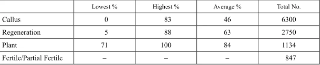
In addition to the significant differences in the transformation efficiency, there were significant visual differences between the calli growing in petri plates under hygromycin or bialaphos selection. During the 2×3 weeks of selection period using hygromycin as the selective antibiotic, most of the calli contained brown and necrotic sectors, indicating dy- ing non-transgenic section of the calli. The growing healthy and yellowish part of the calli which were transferred to regeneration media were regenerated to plantlets with the average efficiency of ~63%. Non-transformed calli sections were visibly brown or ne- crotic. In contrast, under bialaphos selection, some of the calli stopped growing or had a very slow growing pattern over 2×3 weeks of selection period with no, or few brown and necrotic sections. Furthermore, some of the calli which even continued to grow did not regenerate into plantlets when transferred to regeneration medium. This pattern of calli selection under the application of bialaphos was not as straightforward compared to hy- dromycin for identifying transgenic calli. Hygromycin B appeared to be a better plant selection agent compared to bialaphos. Overall, about 6300 B. distachyon Bd21 embryo- genic calli were used for transformation. Our data demonstrated variable transformation efficiency for regenerated plantlets that ranged from 5 and 88% (Table 1). Altogether, 1134 putative transgenic lines were obtained of which about 25% (287) of the small plant- lets died after transfer to the soil or were sterile and did not produce any seeds. The vari- ous steps from callus induction to transformation and regeneration are summarised in the schematic flow chart in Fig. S3.
Validation by PCR of the T-DNA insertions
PCR analysis targeting the hygromycin resistance gene (HPT) was used to ascertain the presence of T-DNA in genomic DNA isolated from a sub-set (9) of the transgenic plants.
The results showed the presence of a clear band at the expected size in 6 of the 9 putative transgenic plants, indicating the presence of the T-DNA in these plants. There was an- other fainter band of a slightly smaller molecular weight in all amplifications and this is
Table 1. Transformation efficiency of B. distachyon Bd21 from callus to mature plants
Lowest % Highest % Average % Total No.
Callus 0 83 46 6300
Regeneration 5 88 63 2750
Plant 71 100 84 1134
Fertile/Partial Fertile – – – 847
likely to be due to non-specific amplification (as it is also present in the positive control using the vector, pEU33AN as the template) (Fig. S4A). Similarly, 9 transgenic lines re- sistant to the bialaphos carrying the BAR gene as a selectable marker were confirmed by PCR (Fig. S4B) for the integration of pAM-PAT-P35S-TaFROG-YFP construct into transgenic Brachypodium distachyon Bd21 plants. Moreover, confocal laser scanning mi- croscopy showed the correct expression of TaFROG-YFP in Brachypodium distachyon cells within distinct nuclear bodies (Fig. S5) as described previously in wheat (Perochon et al. 2015). The similarity in subcellular localization of fluorescent protein-tagged wheat proteins in both wheat and B. distachyon further supports the usefulness of B. distachyon as an experimental system for determining the subcellular localization of temperate grass proteins.
Discussion
While B. distachyon has emerged as a plant model for temperate grasses, the development of an efficient transformation protocol for B. distachyon is highly significant for any func- tional genomics research. This includes optimization of protocols for callus induction and proliferation. Although methods for callus induction and proliferation have already been developed by several researchers (Draper et al. 2001; Vogel and Hill 2008; Alves et al.
2009), optimization is necessary due to potential inter-laboratory differences in optimal conditions for tissue culture.
As successful induction of compact embryogenic callus (CEC) from immature em- bryos from B. distachyon is dependent on auxin concentration (Draper et al. 2001; Chris- tiansen et al. 2005; Vogel et al. 2006b; Păcurar et al. 2008; Vain et al., 2008; Vogel and Hill 2008; Alves et al. 2009), different levels of 2,4-D, ranging from 0 to 9 mg l-1 were tested. The results showed that 2,4-D at a concentration of 2.5 mg l–1 is optimal for callus induction (Fig. S1), in agreement with previous studies that also reported an optimal 2,4- D concentration of 2.5 mg l–1 for callus induction in B. distachyon Bd21 (Vogel et al.
2006b; Vain et al. 2008; Alves et al. 2009; Lee et al. 2011), B. distachyon Bd21-3 (Vogel and Hill 2008; Alves et al. 2009), and B. distachyon BDR018 (Păcurar et al. 2008). Chris- tiansen et al. (2005) have also reported that a 2,4-D concentration of 2.5 mg l–1 is suitable for callus induction in 37 accessions of B. distachyon. Of the other concentrations of 2,4- D tested, some of the calli differentiated roots, while others developed necrosis (Fig. S1).
We were also guided by the protocol of Alves et al. (2009) by supplementing the medium with CuSO4 (0.6 mg l–1) to promote embryogenic callus growth and reduce the possibil- ity of regenerating albino plants. In addition, subculture of CECs longer than 6 weeks for transformation was not recommended due to unwanted somatic genetic variation and re- generation of albino plants (Alves et al. 2009). As such, CEC were periodically generated to ensure a constant supply for genetic transformation.
Additionally, 5-week old calli were grown and sub-cultured on different levels of 2,4- D (0, 2.5, 5.0, 7.5, and 9.0 mg l–1) to examine the quality and quantity of calli for subse- quent transformation. Our results showed that 2,4-D concentrations of 2.5 and 5.0 mg l–1 were suitable for calli propagation and that both these 2,4-D concentrations produced the
highest amounts of embryogenic calli (Fig. S2). However, it should be noted that some necrosis, evident from browning of calli, were observed when 5.0 mg l–1 of 2,4-D were used (Fig. S2C). Taking all these into consideration, it was decided that 2.5 mg l–1 of 2,4- D should be used for all subsequent experiments.
The results of our experiments highlighted the usefulness of hygromycin as a selecta- ble agent in Agrobacterium-mediated transformation of B. distachyon, and the use of bi- alaphos required further optimization. Another factor which needs to be considered more precisely in the future experiments is the type of promoter used for driving the selectable marker gene. Different constitutive promoters have been exploited to drive both HPT and BAR genes by researchers. It is noteworthy to state that other factors can also affect the transformation efficiency like the type of promoter driving the selectable agent and the type of vector. So, it would be valuable to use different types of vectors including Gate- way® vectors with different promoters and selectable agents for further experiments.
A gradual decrease (40:30:20 mg l–1) in concentration of hygromycin is recommended with the help of reporter genes such as gfp. However, screening for GFP fluorescence ap- pears to be of limited usefulness in our case because of the high level of autofluorescence in B. distachyon (data not shown). Consequently, we used a constant concentration of hygromycin (40 mg l–1) for selection of transformed calli and for regeneration of trans- genic plantlets. After 3 weeks of culturing calli in selection medium, healthy and yellow calli were dissected and transferred to fresh selection medium. For calli larger than 3-4 mm, the calli were divided into 2 or 3 smaller pieces. While this method was useful for maintaining transgenic calli, it should also be noted that this may lead to the regeneration of multiple identical transgenic lines.
In summary, the lowest and highest transformation efficiency for regenerated plantlets obtained in our experiments was 5 and 88% respectively with an average of 63%. Differ- ent transformation efficiencies have been reported. For example, Vogel and Hill (2008) reported an average transformation efficiency of 41%, while Păcurar et al. (2008), Lee et al. (2011), and Alves et al. (2009) reported average transformation efficiencies of 96%, 80%, and 90%, respectively. Regardless of the transformation efficiency, the ability to generate a transgenic population for B. distachyon opens up the possibility for the wider scientific community to use this T-DNA tagged population for screening experiments to identify phenotypes associated with biotic or abiotic stresses, and developmental pro- cesses. Additionally, we also showed that B. distachyon is a useful experimental model for determining the subcellular localization of fluorescent-protein tagged wheat proteins.
Acknowledgements
This work is supported by Science Foundation Ireland (SFI) Research Frontiers Pro- gramme grants 06/RFP/GEN034 and 08/RFP/EOB1087 (to C.K.-Y.N.) and project Nos.
10/IN.1/B3028 and 14/1A/2508 (to A. P. and F.M. D.), and an Iranian Government Re- search Scholarship (to A.B.).
References
Alves, S.C., Worland, B., Thole, V., Snape, J.W., Bevan, M.W., Vain, P. 2009. A protocol for Agrobacterium- mediated transformation of Brachypodium distachyon community standard line Bd21. Nat. Protoc. 4:638–
An, T., Cai, Y, Zhao, S., Zhou, J., Song, B., Bux, H., Qi, X. 2016. Brachypodium distachyon T-DNA insertion 649.
lines: a model pathosystem to study nonhost resistance to wheat stripe rust. Sci. Rep. 6:25510.
Bragg, J.N., Anderton, A., Nieu, R., Vogel, J.P. 2015. Brachypodium distachyon. In: Wang, K. (ed), Agrobacterium Protocols. Springer. New York, USA. pp. 17–33.
Bragg, J.N., Wu, J., Gordon, S.P., Guttman, M.E., Thilmony, R., Lazo, G.R., Gu, Y.Q, Vogel, J.P., 2012.
Generation and characterization of the western regional research center Brachypodium T-DNA insertional mutant collection. PLoS ONE 7:e41916.
Bablak, P., Draper, J., Davey, M.R., Lynch, P.T. 1995. Plant regeneration and micropropagation of Brachypodium distachyon. Plant Cell Tiss. Org. 42:97–107.
Cheng, M., Hu, T., Layton, J., Liu, C.N., Fry, J.E. 2003. Desiccation of plant tissues post-Agrobacterium infec- tion enhances T-DNA delivery and increases stable transformation efficiency in wheat. In Vitro Cell. Dev.- Pl. 39:595–604.
Cheng, M., Lowe, B.A., Spencer, T.M., Ye, X., Armstrong, C.L. 2004. Factors influencing Agrobacterium- mediated transformation of monocotyledonous species. In Vitro Cell. Dev.- Pl. 40:31–45.
Christiansen, P., Andersen, C.H., Didion, T., Folling, M., Nielsen, K.K. 2005. A rapid and efficient transforma- tion protocol for the grass Brachypodium distachyon. Plant Cell Rep. 23:751–758.
Collier, R., Bragg, J., Hernandez, B.T., Vogel, J.P., Thilmony, R. 2016. Use of Agrobacterium rhizogenes strain 18r12v and paromomycin selection for transformation of Brachypodium distachyon and Brachypodium sylvaticum. Front. Plant Sci. 7:716.
Draper, J., Mur, L.A., Jenkins, G., Ghosh-Biswas, G.C., Bablak, P., Hasterok, R., Routledge, A.P. 2001.
Brachypodium distachyon. A new model system for functional genomics in grasses. Plant Physiol.
127:1539–1555.
Eamens, A.L., Blanchard, C.L., Dennis, E.S., Upadhyaya, N.M. 2004. A bidirectional gene trap construct suit- able for T-DNA and Ds-mediated insertional mutagenesis in rice (Oryza sativa L.). Plant Biotechnol. J.
2:367–380.
Garvin, D., Gu, Y., Hasterok, R., Hazen, S., Jenkins, G., Mockler, T., Mur, L., Vogel, J. 2008. Development of genetic and genomic research resources for Brachypodium distachyon, a new model system for grass crop research. Crop Sci. 48:S69–S84.
Hanahan, D. 1983. Studies on the transformation of Escherichia coli with plasmids. J. Mol. Biol. 166:557–580.
Hunt, D., Chambers, J.P., Behpouri, A., Kelly, S.P., Whelan, L., Piettrzykowska, M., Downey, F., Mccabe, P.F., Ng, C.K.Y. 2014. Brachypodium distachyon cell suspension cultures: establishment and utilization. Cereal Res. Commun. 42:58–69.
Lee, M.B., Jeon, W.B., Kim, D.Y., Bold, O., Hong, M.J., Lee, Y.J., Park, J.H., Seo, Y.W. 2011. Agrobacterium- mediated transformation of Brachypodium distachyon inbred line Bd21 with two binary vectors containing hygromycin resistance and GUS reporter genes. J. Crop Sci. Biotechnol. 14:233–238.
Mohammadhassan, R., Kashefi, B., Shabanzadeh, Delcheh, K. 2014. Agrobacterium-based vectors: a review.
Intl. J. Farm. Allied Sci. 3:1002–1008.
Păcurar, D.I., Thordal-Christensen, H., Nielsen, K.K., Lenk, I. 2008. A high-throughput Agrobacterium- mediated transformation system for the grass model species Brachypodium distachyon L. Transgenic Res.
17:965–975.
Perochon, A., Jianguang, J., Kahla, A., Arunachalam, C., Scofield, S.R., Bowden, S., Wallington, E., Doohan, F.M. 2015. TaFROG encodes a pooideae orphan protein that interacts with SnRK1 and enhances resistance to the mycotoxigenic fungus Fusarium graminearum. Plant Physiol. 169:2895–906.
Steinwand, M.A., Young, H.A., Bragg, J.N., Tobias, C.M., Vogel, J.P. 2013. Brachypodium sylvaticum, a model for perennial grasses: transformation and inbred line development. PLoS ONE 8:e75180.
The International Brachypodium Initiative. 2010. Genome sequencing and analysis of the model grass Brachypodium distachyon. Nature 463:763–768.
Thole, V., Peraldi, A., Worland, B., Nicholson, P., Doonan, J.H., Vain, P. 2011. T-DNA mutagenesis in BrachyIpodium distachyon. J. Exp. Bot. 63:567–576.
Thole, V., Vain, P. 2012. Agrobacterium-mediated transformation of Brachypodium distachyon. Transg. Plants 847:137–149.
Vain, P., Thole, V. 2009. Gene Insertion Patterns and Sites. In: Jones, H.D., Shewry, P.R. (eds), Transgenic Wheat, Barley and Oats: Production and Characterization Protocols Humana Press. Totowa, NJ, USA. pp.
203–226.
Vain, P., Worland, B., Thole, V., McKenzie, N., Alves, S.C., Opanowicz, M., Fish, L.J., Bevan, M.W., Snape, J.W. 2008. Agrobacterium-mediated transformation of the temperate grass Brachypodium distachyon (genotype Bd21) for T-DNA insertional mutagenesis. Plant Biotechnol. J. 6:236–245.
Vogel, J.P., Gu, Y.Q., Twigg, P., Lazo, G.R., Laudencia-Chingcuanco, D., Hayden, D.M., Donze, T.J., Vivian, L.A., Stamova, B., Coleman-Derr, D. 2006a. EST sequencing and phylogenetic analysis of the model grass Brachypodium distachyon. Theor. Appl. Genet. 113:186–195.
Vogel, J.P., Hill, T. 2008. High-efficiency Agrobacterium-mediated transformation of Brachypodium distachy- on inbred line Bd21-3. Plant Cell Rep. 27:471–478.
Vogel, J.P., Garvin, D.F., Leong, O.M., Hayden, D.M. 2006b. Agrobacterium-mediated transformation and inbred line development in the model grass Brachypodium distachyon. Plant Cell Tiss. Org. 84:199–211.
Electronic Supplementary Material (ESM)
Electronic Supplementary Material (ESM) associated with this article can be found at the website of CRC at http://www.akademiai.com/content/120427/
Electronic Supplementary Table S1. Effects of different concentrations of 2,4-D on embryogenic callus induc- tion and propagation of B. distachyon Bd21
Electronic Supplementary Table S2. Transformation efficiency of B. distachyon Bd21 based on regenerated plantlets
Electronic Supplementary Table S3. Genetic transformation reports on Brachypodium distachyon Electronic Supplementary Figure S1. Immature embryos of B. distachyon grown on medium with different concentrations of 2,4-D. (A) no 2,4-D, (B) 2.5, (C) 5.0, (D) 7.5, and (E) 9.0 mg l–1. White arrows indicate
compact embryogenic callus (CEC). Scale bar = 1 cm
Electronic Supplementary Figure S2. Embryogenic callus of B. distachyon grown on medium with different concentrations of 2,4-D. (A) no 2,4-D, (B) 2.5, (C) 5.0, (D) 7.5, and (E) 9.0 mg l–1. White arrows indicate dif-
ferentiated or necrotic cells. Scale bar = 1 cm
Electronic Supplementary Figure S3. Schematic representation of Agrobacterium-mediated transformation of Brachypodium distachyon
Electronic Supplementary Figure S4. Molecular characterization of Brachypodium transgenic lines. Verification by PCR of the insertion of the T-DNA construction carrying (A) the hygromycin (HPT) or (B) Bialaphos (BAR) resistance genes in 9 putative transgenic lines (lanes 1–9). The wild type plant B. distachyon Bd21 (lane 10) and the vector utilised for the transformation (lane 11) were used as a PCR positive or negative control respec-
tively. M: molecular weight marker
Electronic Supplementary Figure S5. Confocal laser scanning microscopy of TaFROG-YFP in Brachypodium distachyon. Stable Brachypodium leaves overexpressing TaFROG-YFP were analysed. TaFROG-YFP showed a punctate distribution within the nucleus, consistent with the distribution of TaFROG-YFP in wheat cells.
Scale bar = 10 µm