- SOMFAI TAMÁS -
SYNCHRONIZATION OF IN VITRO MATURATION OF PORCINE OOCYTES
Supervisors:
Dr. Dr. habil. Iváncsics János † Dr. Farkasné Dr. Bali Papp Ágnes
Ph.D. School for the Biological, Technological, Ecological, Feeding and Economical Questions of Animal Production.
Chairman of the PhD School: Prof. Dr. Schmidt János
Sub program: Improvement and Breeding Technology Considerations of Animal Production
Sub program leader: Prof. Kovácsné Dr. Gaál Katalin
UNIVERSITY OF WEST-HUNGARY
FACULTY OF AGRICULTURE AND FOOD SCIENCES MOSONMAGYARÓVÁR
2004
CRYOPRESERVED OR FRESH SEMEN
Értekezés doktori (PhD) fokozat elnyerése érdekében Írta:
Somfai Tamás
Készült a Nyugat-Magyarországi Egyetem “Az Állati Termék Előállítás biológiai, technológiai, ökológiai, takarmányozási és ökonómiai kérdései ” Doktori Iskola
“Az állati termék termelés nemesítési és tartástechnológiai vonatkozása” (I jelû) alprogramja keretében
Témavezetõ: Dr. Farkasné Dr. Bali Papp Ágnes
Elfogadásra javaslom (igen / nem) (aláírás)
A jelölt a doktori szigorlaton ... % -ot ért el,
Sopron/Mosonmagyaróvár
...
a Szigorlati Bizottság elnöke
Az értekezést bírálóként elfogadásra javaslom (igen /nem)
Elsõ bíráló (Dr. ... ...) igen /nem
(aláírás)
Második bíráló (Dr. ... ...) igen /nem
(aláírás)
igen /nem
(aláírás)
A jelölt az értekezés nyilvános vitáján ...% - ot ért el
Sopron/Mosonmagyaróvár,
a Bírálóbizottság elnöke
A doktori (PhD) oklevél minõsítése ...
Az EDT elnöke
CONTENT
1 ABSTRACT ...7
KIVONAT……… ………..9
2 OVERVIEW OF LITERATURE... 11
2.1 Current status of in vitro embryo production in pigs... 11
2.2 Importance of meiotic synchronisation during in vitro maturation ... 13
2.3 Manipulation of MPF activity ... 15
2.4 Manipulation of intercellular cAMP level... 16
2.5 Fertilization and development of immature oocytes ... 18
2.6 The importance of somatic cells around the oocyte ... 20
3 EXPERIMENTS ... 22
3.1 Objectives ... 22
3.2 Synchronisation of meiotic maturation by high level of intercellular cAMP... 24
3.2.1 Materials and methods ... 24
3.2.1.1 Oocyte Collection and In Vitro Maturation... 24
3.2.1.2 In Vitro Fertilization (IVF) and In Vitro Culture (IVC)... 25
3.2.1.3 Oocyte and embryo evaluation with orcein staining... 26
3.2.1.4 Statistical analysis ... 35
3.2.2 Experimental Design ... 35
3.3 In vitro fertilization and development to blastocyst stage of immature porcine oocytes arrested before metaphase-II stage... 37
3.3.1 Materials and methods ... 37
3.3.1.1 Oocyte collection and in vitro maturation ... 37
3.3.1.2 IVF and IVC of porcine oocytes ... 38
3.3.1.3 Oocyte and embryo evaluation with orcein staining... 38
3.3.1.4 Blastocyst evaluation with differential staining ... 38
3.3.1.5 Statistical analysis ... 39
3.3.2 Experimental design... 40
3.4 Relationship between cumulus morphology and oocyte maturation... 42
3.4.1 Materials and methods ... 42
3.4.1.1 Oocyte collection and in vitro maturation ... 42
3.4.1.2 Classification of COCs... 43
3.4.1.3 Parthenogenetic Activation (PGA) of IVM Oocytes ... 45
3.4.1.4 IVF and IVC of porcine oocytes... 45
3.4.1.5 Oocyte and embryo evaluation with orcein staining... 46
3.4.1.6 Statistical Analysis ... 47
3.4.2 Experimental design... 47
4 RESULTS……… ………49
4.1 Synchronisation of meiotic maturation by high level of intercellular cAMP...49
Discussion………56
4.2 In vitro fertilization and development to blastocyst stage of immature porcine oocytes arrested before metaphase-II stage... 63
Discussion………..70
4.3 Effect of cumulus morphology on nuclear and cytoplasmic maturation... 79
Discussion………..85
5 SUMMARY………91
6 NEW SCIENTIFIC RESULTS ... 96
7 REFERENCES……….97
8 ACKNOWLEDGEMENTS ... 115 9 APPENDIX……….117 9.1 Nomenclature of abbreviations ... 117
1 ABSTRACT
In the present study, the effect of meiotic synchronisation by a transient meiotic arrest at GV stage on nuclear and cytoplasmic maturation, in vitro fertilization and subsequent embryonic development of in vitro matured (IVM) porcine oocytes was investigated.
Meiotic synchronisation was achieved by elevating the intercellular cAMP level of the oocyte during the first 22 h of the 46 h IVM period.
Supplementation of maturation medium with 1 mM dbcAMP succesfully inhibited meiosis during the first 22 of IVM and synchronised maturation of the cultured oocytes that resulted in a higher rate of mature (M-II) oocytes and a higher rate of monospermic fertilization and blastocysts after IVF and IVC of the treated oocytes than that of the control. This suggests that synchronization of maturation using dbcAMP enhances meiotic potential of oocytes and results in a high developmental competence by monospermic fertilization.
In the second part of the study, we compared the developmental capacity of mature and meiotically arrested porcine oocytes derived from IVM. The most of the oocytes that failed to resume meiosis during IVM were arrested at GV and M-I stage while a remarkable proportion of oocytes also remained at proM-I stage. After IVF there was no difference in polyspermy and pronucleus formation between the M-II and M-I arrested oocytes while GV arrested oocytes showed a significantly higher frequency of polyspermy and failed to form pronuclei. After IVC, there was no significant difference in blastocyst rates between the M-II and M-I arrested oocytes, however the
blastocyst derived from M-I oocytes had less cells than that of from M-II oocytes. The ratio of inner cell mass and trophectoderm cells did not differ between the two groups. These results prove the developmental ability of M-I arrested oocytes to blastocyst stage and reveal their decreased cleavage speed, probably due to their abnormal ploidy.
In the third part of the study, the effect of somatic cells surrounding the oocyte on nuclear progression and cytoplasmic maturation was investigated. During IVM different morphologic classes of porcine COCs can be distinguished regarding the status and expansion of the cumulus cells. It was found that the frequency of normal cumulus expansion is higher when granulose cells are attached to the COCs.
Nuclear progression of COCs was slightly accelerated without granulosa cells. Oocytes attached to the bottom of culture dish with dark, compact cumulus undergo precoccious nuclear and cytoplasmic maturation. The rate of monospermic fertilization after IVF of normal COCs showing normal cumulus expansion was higher than that of COCs attached to the dish. It was concluded, that diverse behavior of cumulus cells during in vitro culture affects both maturation and IVF of porcine oocytes. Granulosa cells promote normal cumulus expansion thus decrease heterogeneity in nuclear and cytoplasmic maturity amongst oocytes.
KIVONAT
Éretlen petesejtek in vitro érlelése nagy mennyiségű alapanyagot képes biztosítani különböző embrió-technológiai illetve manipulációs módszerekhez. Ismert jelenség azonban, hogy az in vitro maturáció (IVM) során a sertés petesejtek eltérő időben kezdik meg illetve fejezik be a meiotikus érést, így az IVM végére az érettség szempontjából a petesejtek jelentős szórást mutatnak, ami a manipulációs eljárások hatékonyságát jelentősen csökkenti.
Tanulmányunk első részében megvizsgáltuk, hogy sertés petesejtek érésének a meiózis átmeneti blokkolásával történő szinkronizálása milyen hatással van azok érési, termékenyülési illetve embrionális fejlődési képességére. A magi érés blokkolását a 46 órás maturáció első 22 órájában, a sejten belüli cAMP szint emelésével végeztük. A maturáció átmeneti blokkolását követően a kezelt csoport maturációs rátája, az in vitro fertilizációt (IVF) követően a monospermia, illetve a hólyagcsírák aránya magasabb volt, mint a kontrol csoportban, ahol jelentős mennyiségű petesejt rekedt meg M-I állapotban. Tehát a magi érés szinkronizálása javítja a petesejtek meiotikus és fejlődési potenciálját.
A tanulmány második részében az érett (M-II) petesejtek termékenyítést követő embrionális fejlődési képességét hasonlítottuk össze olyan petesejtekével, melyek a meiózis során éretlen állapotban megrekedtek. Az ilyen petesejtek főként GV vagy metafázis-I állapotban állnak meg de jelentős számú petesejt blokkolt le a prometafázis-I állapotában is. Az M-II és az M-I állapotú petesejtek között nem volt különbség a polispermia és a pronukleuszképződés tekintetében az IVF-et követően. Ezen felül, az IVF-et követő embriótenyésztés során nem különbözött a hólyagcsíra
állapotba jutott embriók aránya sem, mindamellett az M-I állapotú petesejtekből származó embriók alacsonyabb sejtszámmal rendelkeztek, mint az azonos korú, M-II állapotú petesejtekből származók. Az embrionális sejtcsomó és a trofektodermális sejtek aránya nem különbözött a két csoport között. Ezen eredmények egyértelműen bizonyítják, hogy a tartósan M-I állapotban megrekedt sertés petesejtek éppúgy képesek hólyagcsíra állapotig fejlődni, mint érett társaik, azonban osztódási sebességük alacsonyabb, feltehetően azok abnormális ploidiája miatt. Ez kihangsúlyozza az IVF-re használt petesejtek érettségi állapot alapján történő elbírálásának fontosságát.
A dolgozat harmadik részében azt vizsgáltuk, hogy milyen hatást fejtenek ki a petesejtet körülvevő testi sejtek a petesejtek spontán sejtmagi érésére az IVM során. Eredményeink azt mutatják, hogy a kumulusz sejtek tenyésztőedényhez történő kapcsolódása kiváltja a petesejtek spontán, idő előtti érését és a petesejtek öregedését is.
Ezzel jelentősen megnő a polispermiás megtermékenyülés gyakorisága. Amennyiben azonban a petesejtet körülvevő kumulusz réteghez granulóza sejtek is kapcsolódnak, az IVM során a normális kumulusz expanzió nagyobb gyakorisággal következik be, kevesebb sejt kapcsolódik a tenyésztő edényhez és ezzel párhuzamosan az idő előtti érés gyakorisága lecsökken, tehát a granulóza sejtek természetes úton szinkronizálják a petesejtek érését.
2 OVERVIEW OF LITERATURE
2.1 Current status of in vitro embryo production in pigs
In vitro production (IVP) of mammalian embryos is an important supporting technology not only for basic sciences and medicine, but also for advanced animal husbandry and biotechnology which enables us to generate a large number of viable embryos. IVP includes three major technological steps; the in vitro maturation (IVM) of immature oocytes, in vitro fertilization (IVF) and in vitro culture (IVC) of the fertilized oocytes.
In vitro embryo production has several advances; it makes us possible to utilize at least a significant proportion of the vast number of follicular oocytes that are normally lost through atresia using slaughterhouse ovaries as basic material for oocyte collection that are normally abattoir-vaste. Thus IVP enables us to produce a larger number of embryos with less cost and in less time compared to in vivo embryo production in pigs. Besides, in vitro maturation of oocytes provide mature eggs that can be materials as recipient oocytes for other reproductive technologies such as ICSI (Nakai et al., 2003) and cloning (Betthauser et al., 2000; Boquest et al., 2002;
Iwamoto et al., 2003). These technologies using IVM oocytes are now expected to be used to produce transgenic pigs. Moreover the successful embryo transfer in rare pig breeds (Rátky et al., 2001) and the cryopreservation of in vitro produced pig blastocysts was reported recently (Dinnyés et al., 2003) underlining the importance of in vitro technologies in gene banking of rare breeds, endangered species and precious individuals as well.
The improvement of IVP systems could enhance the efficiency of micromanipulation and gene transfer technologies in the porcine species. This may manifest its effect in pork production by creating new transgenic species with better body and meat characteristics or growth.
The history of in vitro reproduction in pigs started three decades ago.
Motlik and Fulka (1974) first reported the ability of in vitro matured (IVM) porcine oocytes to be fertilized. The first successful in vitro fertilization (IVF) of IVM oocytes in pigs was reported by Iritani et al.
(1978). Important steps towards were made by Nagai et al. (1988), who successfully used frozen-thawed pig spermatozoa for IVF. The ability of IVM/IVF oocytes to develop to blastocyst stage was first confirmed by Mattioli et al. (1989). Further, piglets were born after the transfer of IVM/IVF embryos that were cultured to the 2-4 cell stage (Yoshida et al., 1993; Funahashi et al., 1996) or to morula stage (Day et al., 1998). However the first successful transfers of IVP embryos at blastocyst stage were reported only recently (Marchal et al., 2001; Kikuchi et al., 2002).
In spite of the improvements that have been made in many aspects of IVF procedures, porcine IVP still struggles with problems that remained unsolved during the years such as the high incidence of polyspermy which is still the major problem that decreases the efficiency of in vitro embryo production in pigs (Nagai, 1994). The phenomenon of polyspermy can be affected by several factors such as the concentration of spermatozoa and the time period of in vitro fertilization (Nagai, 1996). Polyspermy in porcine IVM-IVF oocytes has also been considered to result from an irregular distribution of cortical granules in oocytes matured in vitro (Cran and Cheng, 1986) and oocyte aging caused by the asynchronous meiotic progression of
porcine oocytes during in vitro maturation (Grupen et al., 1997).
Therefore it can be stated, that one of the main reasons of polyspermy is the inadequate in vitro maturation culture of the oocytes. However, it must be noted, that high incidence of polyspermy is a typical characteristic of porcine species that ranges from 5-35% even in vivo (Hancock, 1959; Hunter RHF, 1967; Hunter RHF 1972; Hunter RHF, 1973) and that many of the polyspermic fertilized oocytes can develop to the blastocyst stage (Han et al., 1999a). Most of the fetuses resulted from the transfer of polyspermic embryos were found to be diploid (Han et al., 1999b) suggesting the existence of a mechanism(s) in porcine oocytes that can neutralize the effect of polyspermy on embryo ploidy.
2.2 Importance of meiotic synchronisation during in vitro maturation
The success of nuclear and cytoplasmic maturation of oocytes is a crucial point of efficiency in IVM/IVF systems. Maturation conditions can be upgraded by coculturing oocytes with oviduct epithelial cells and follicle cells (reviewed by Nagai, 1994), however the need for defined culture conditions has inceased recently in order to industrialize in vitro embryo production. The metabolism of nuclear and cytoplasmic maturation is not completely understood yet thus the present IVM systems can not completely represent the conditions that exist inside the follicle before and during the maturation of oocytes in vivo. Antral follicles are known to keep the oocytes arrested at germinal vesicle stage until the occurance of the meiosis activating signal (Pincus and Enzmann, 1935; Eppig and Downs, 1984). During oocyte collection and in vitro culture, spontaneous maturation can start since germinal vesicle (GV) oocytes resume meiosis
spontaneously when removed from the follicle (Pincus and Enzmann, 1935). A large variation in nuclear morphology of GV stage pig oocytes was found just after collection (Funahashi et al., 1997a;
Nagai et al., 1997) and after a certain time of culture (Funahashi et al., 1997a). It has been shown that there are differences in GV configuration and polypeptide synthesis between oocytes obtained from different size antral follicles causing heterogeneity in overall developmental competence within oocytes, collected for IVM/IVF (Mcgaughey et al., 1979). Since the source of oocytes may differ (from different individuals and different size follicles), the developmental competence of GV stage pig oocytes used for IVM/IVF can also vary within the population used in one experiment. Therefore some oocytes can start meiosis earlier than others causing heterogeneity in maturation status among the oocytes after IVM that may affect monospermic fertilization, and further embryonic development. Supporting this theory a large variation in morphology and cell number of IVM/IVF pig blastocysts was reported by Kikuchi et al. (2002). To overcome this phenomenon the synchronization of nuclear maturation is necessary which can be achieved by a transient inhibition of meiotic maturation during the first half of the 44-48 h of IVM. Inhibition of GVBD in oocytes prior to in vitro maturation may have another advantage since oocytes can increase their developmental competence during meiotic arrest (Downs et al., 1986; Funahashi et al., 1997; Hashimoto et al., 2002). A possible reason of this phenomenon might be the fact that protein synthesis in oocytes does not stop completely when arrested at GV stage temporarily (Marchal et al., 2001).The transient inhibition of GVBD can be acieved by supression of activity of metaphase (or maturation)
promoting factor (MPF) or by chemicals that elevate intercellular levels of cyclic adenosine monophosphate (cAMP).
2.3 Manipulation of MPF activity
Progression of meiosis is regulated by a certain fluctuation in the activity of metaphase promoting factor (MPF), a protein kinase that plays its key role in promoting M-phase in mammalian oocytes (Hashimoto and Kishimoto, 1988; Fulka Jr. et al., 1992; Motlik et al., 1998). MPF consists of a regulator subunit, cyclin B-1 and a catalytic subunit, p34cdc2 and plays an important role in the brakedown of the germinal vesicle, which is the first morphologic step of meiotic maturation. MPF shows its highest activity at metaphase-I and metaphase-II stages and turns inactive during anaphase-I and telophase-I (Hashimoto and Kishimoto, 1988; Fulka Jr. et al., 1992;
Motlik et al., 1998). Supression of MPF activity in isolated oocytes prevents meiotic maturation and keeps the oocytes at GV stage. The inhibition of protein synthesis is a possible way to achieve this, however the use of protein synthesis inhibitors such as puromycin (Motlik et al., 1991) or cycloheximide (Kubelka et al., 1988; Lonergan et al., 1998) block not only the synthesis of MPF proteins but stop protein synthesis in general inside the oocyte. This may cause side effects regarding the further meiotic and developmental competence of the oocyte (Lonergan et al., 1998). Considering this, the use of phosphorylation inhibitors such as 6-dimethylaminopurine (6-DMAP) (Avery et al., 1998) or specific protein-kinase inhibitors such as butyrolactone-I (BL-I), roscovitine (ROS) is more expedient. BL-I, a potent inhibitor of MPF is known to prevent the resumption of meiosis reversibly in bovine (Kubelka et al., 2000; Lonergan et al., 2000;
Imai et al., 2002) and porcine oocytes (Wu et al., 2002; Hirao et al.,
2003) via engaging the ATP binding sites of p34cdc2, the catalytic subunit of MPF, without affecting chromosome condensation activity, mitochondrial and microfilament dynamics. However a delay of cytoplasmic maturation in metaphase II stage porcine oocytes was observed when oocytes were arrested at germinal-vesicle stage using BL-I prior to in vitro maturation (Hirao et al., 2003). Besides, some reports reveal possible side effects of BL-I on oocyte quality. Using high (100-300 µM) concentrations of BL-I for parthenogenetic activation resulted in an elevated rate of activated porcine oocytes with two female pronuclei and only one polar body (showing a disability to extrude second polar body) compared to oocytes treated with lower doses of BL-I suggesting a (dose dependent) detrimental effect of BL-I on cytoskeleton probably via a non-specific effect on the MAP kinase system (Dinnyės et al., 2000). Recently a long term (40 h) cultivation of bovine COCs with a high concentration (100 µM) of BL-I was found to destroy the contact between cumulus cells and oocyte and have detrimental effects on cytoplasmic and nuclear morphology (Fair et al., 2002).
ROS, another specific inhibitor of CDC2 protein kinase can also be used for transient inhibition of GVBD in mammalian oocytes (Marchal et al., 2001) and recently a combination of BL-I and ROS at low concentrations was reported to be effective to inhibit GVBD in bovine, without any side effects (Ponderato et al., 2001).
2.4 Manipulation of intracellular cAMP level
A high level of intercellular cAMP is responsible for activating cAMP dependent protein kinase (PKA) that controls meiotic arrest of oocytes at GV stage (Bornslaeger et al., 1986; Cameron et al., 1987).
For elevating the level of cAMP within mammalian oocytes,
gonadotropins such as FSH and LH acting through follicular cells are responsible (Bornslaeger and Schultz, 1985; Mattioli et al., 1994;
Shimada et al., 2003). Resumption of meiosis, germinal vesicle breakdown (GVBD) is associated with an irreversibile cascade starting with the reduction in intraoocyte cAMP that is followed by PKA inactivation and the activation of mitogen activated protein (MAP) kinase (Schultz et al., 1983; Bornslaeger et al., 1986; Sun et al., 1999). Spontaneous maturation is supposed to occur by the interruption of metabolism between the follicle components (granulose cells and/or follicular fluid) and the oocyte in which cAMP is maintained at a high level.
The intercellular level of cAMP can be elevated artificially by different chemicals such as invasive adenylate cyclase (iAC), an enzyme that transfers ATP of the oocyte into cAMP or phosphodiesterase inhibitors like 3-isobutylmethyl-xanthine (IBMX) which prevents degradation of cAMP. Treatments with permeable and stabile compounds that are similar to cAMP can also be used. Addition of dibutyryl cyclic AMP (dbcAMP), a membrane permeable cAMP analogue, into IVM medium during the first 20 h of maturation inhibits GVBD and has a uniform effect on the nuclear stage of pig oocytes (Funahashi et al., 1997b).
The use of a combination of invasive adenylate cyclase (iAC) and 3- isobutyl 1-methylxanthine (IBMX) during oocyte collection increased the meiotic and subsequent embryonic developmental competence of IVM/IVF bovine oocytes (Luciano et al., 1999) suggesting that changes in the level of intracellular cAMP during collection might affect further meiotic or developmental competence of oocytes.
2.5 Fertilization and development of immature oocytes
During IVM, not all the cultured oocytes complete their nuclear maturation; some of them remain at germinal vesicle (GV) stage or can be arrested at metaphase-I (M-I) stage even by the end of the culture period needed for the full nuclear maturation (Bae and Foote, 1980; Bagger et al., 1987; Motlik and Fulka, 1986; Eppig et al., 1994; Polanski, 1995; Kikuchi et al., 1999).
The completion of nuclear maturation of porcine follicular oocytes is affected by cytoplasmic factors such as the capacity of oocytes to start or finish meiosis and the culture conditions. Therefore, after maturation culture, the nuclear status of oocytes results in various stages of meiosis. Some of the oocytes reach M-II stage, whereas the others remained at immature stages such as GV and M-I. Meiotic arrest can be caused by numerous factors such as insufficient meiotic competence affected by the follicle and oocyte diameter (Szybek, 1972; Sorensen and Wassarman, 1976; Motlik and Fulka, 1986;
Eppig et al., 1994) or stress caused by the inadequate culture conditions such as isolation and culture media (Bae and Foote, 1980;
Bagger et al., 1987; Kikuchi et al., 1999). The age and the strain (in mice) of the donor animals also affects meiotic competence of GV oocytes: in pigs oocytes obtained from adult sows have an advanced competence to resume in vitro maturation than that of obtained from prepuberal gilts (Marchal et al., 2001a). In mice oocytes of strain LT are known to exhibit a high incidence of arrest in the progression of meiosis at M-I stage (Hirao and Eppig, 1997).
In several IVM/IVF systems in pigs, cumulus-oocyte complexes are inseminated instead of denuded oocytes to enhance better acrosome reaction of the spermatozoa (and thus fertilization) by the presence
of cumulus cells which are known to initiate acrosome reaction (reviewed by Van Soom et al., 2002). In such systems, all the cultured oocytes are inseminated including ones that are arrested at the immature stage before the achievement of nuclear maturation to metaphase-II (M-II). However less is known about the embryonic development of fertilized immature oocytes.
It has been reported, that porcine oocytes fail to form both female and male pronuclei when they are penetrated by spermatozoa at GV stage (Wang et al., 1994; Wang and Niwa, 1997). In such situation the GV remains intact and the heads of the penetrating spermatozoa remain condensed. The rate of penetrating spermatozoa increases due to the lack of cortical granule distribution which is a characteristic of the immature porcine oocytes (Wang et al., 1997).
Maturing mammalian oocytes penetrated by spermatozoa at M-I stage are known to be able to complete their nuclear maturation to M-II (Chian et al., 1992; Polanski, 1995; Kikuchi et al., 1999).
However, formation of pronuclei can not be observed because due to the elevated activity of MPF at M-I and M-II stages the female chromatin remains at metaphase stage and the head of the penetrating sperm cells remain compact or the male chromatin form abnormal clusters or metaphase chromosomes that can be incorporated into the maternal metaphase plate (Kikuchi et al., 1999).
However it was reported that mouse (Eppig et al., 1994; Polanski, 1995) and porcine (Kikuchi et al., 1999) oocytes permanently arrested at M-I stage undergo cytoplasmic maturation after a certain period of in vitro culture needed for nuclear maturation and, in such oocytes, male and female pronuclei are formed after fertilization. Up
to our knowledge, - there is no study about the embryonic developmental ability of such M-I arrested porcine oocytes.
2.6 Importance of somatic cells around the oocyte
Cumulus cells play an important role on oocyte maturation since they provide and transfer several known and unknown factors that are essential for normal meiotic and cytoplasmic maturation and further embryonic development after fertilization such as glutathione (GSH), which is an important factor for subsequent formation of male pronucleus (Yoshida et al., 1993). Cumulus cells can incorporate cystine, the oxidised form of L-cysteine which can not be utilized by the oocyte, to
synthesize GSH.
Glutathione produced in the cumulus cells may enter the oocytes through the corona- oocyte complexes (reviewed by Nagai, 1994; Nagai, 2001) (Figure 1).
Another substrate which can not be metabolized by the oocyte itself is glucose. Cumulus cells metabolize glucose to
pyruvate that can pass to the oocyte and enhance its quality (Downs and Utecht, 1999). Moreover, cumulus cells are known to play an
Fig 1. Glutathione synthesis in the cumulus cells and the oocyte and its transport from the cumulus cells to the oocyte through gap junctions (Nagai, 2001).
important role in regulation of meiotic progression of oocytes. During the growth and capacitation of oocytes (before initiation of meiosis) cumulus cells are responsible for maintenance of oocyte nucleus in GV stage via elevating intercellular cAMP level of the oocyte (Dekel and Beers, 1980; Racowsky 1984; Eppig and Downs, 1984; Tanghe et al., 2002; Shimada et al., 2003) by transferring an inhibitor signal trough gap junctions. Initiation of meiosis is also related to cumulus- function, there are evidences that cumulus cells secrete a meiosis- inducing factor (Guliang et al., 1994; Xia et al., 2000; Downs, 2001).
However, the interruption of the meiosis-arresting signal, (e.g. the disruption or occlusion of gap junctions between the oocyte and the surrounding somatic compartment) also initiate the meiotic maturation in the oocytes (Larsen et al., 1986; Isobe et al., 1996;
Isobe and Terada, 2001). Regarding the somatic compartment of the follicle, not only the cumulus cells affect the oocyte nuclear maturation. The transient inhibitoric effect of granulose cells on nuclear maturation of oocytes has also been published (Motlik et al., 1991; De Loos et al., 1994), suggesting a possible role of granulose in regulation of oocyte maturation.
A relationship between the LH induced changes of COC morphology and the nuclear progression during in vivo maturation of porcine oocytes has already been reported (Torner et al., 1998). However, during the in vitro culture of porcine cumulus-oocyte complexes (COC) a different behavior of somatic cells can be observed enabling us to distinguish four morphological categories of COCs. Since there are morphological differences (colour, grade of expansion) between the somatic compartment of COCs from each categories we suggest a difference in the metabolic functions of such cells, that might affect nuclear and cytoplasmic maturation of the oocytes.
3 EXPERIMENTS
3.1 Objectives
The experiments presented in the present study were made according to three major objectives
1. The first objective was to examine the effect of intracellular cAMP during oocyte collection and in vitro culture on nuclear maturation, fertilization and subsequent embryonic development of porcine oocytes. Maturation media supplemented with or without IBMX and iAC were used for oocyte collection and following oocyte maturation culture was performed in the presence or absence of dbcAMP.
2. Without meiotic synchronisation a remarkable amount of oocytes remained arrested at M-I stage in our first study. The cytoplasmic maturation of such oocytes was reported by Kikuchi et al. (1999) but without any information about their developmental competence. The second objective of our experiments was to study the developmental potential of porcine oocytes that were permanently arrested before M-II stage during IVM. The nuclear status of oocytes with (PB+) and without (PB-) was investigated after 48 h of IVM. Pronuclear formation, monospermy rates and developmental ability to blastocyst stage after IVF and IVC of M-II stage and meiotically (GV or M-I stage) arrested oocytes was compared.
3. The third aim of the present study was to investigate the possible correlation between the morphology and functional acivity of somatic cells. The kinetics of nuclear and cytoplasmic
maturation in cumulus-oocyte complexes (COCs) and granulose- cumulus-oocyte complexes (GCOCs) was studied as well.
3.2 Synchronisation of meiotic maturation by high level of intercellular cAMP
3.2.1 MATERIALS AND METHODS
3.2.1.1 Oocyte Collection and In Vitro Maturation
Prepuberal porcine ovaries from cross-bred gilts (Landrace x Large White) were obtained from the local abattoir and carried to the laboratory in Dulbecco’s Phosphate Buffered Saline (PBS) within 2 h at 35°C. Dissection of follicles in 3-6 mm diameter and collection of cumulus oocyte complexes (COCs) were performed in a collection medium supplemented with or without iAC and IBMX: The basic collection medium (BCM) (used as control) was NCSU37 (Petters and Wells, 1993) supplemented with 50 µM β-mercaptoethanol (Sigma Chemical Co., St Luis, MO, USA, M-7522), 25 mM HEPES, 1 mg/ml polyvinyl alcohol (PVA) (Sigma, P-8136), 100 unit/ml penicillin G potassium (Sigma, P-7794), and 0.1 mg/ml streptomycin sulfate (Sigma, S-9137). The osmolarity was adjusted to 0.285 osmol/kg, the pH was regulated to 7.3. Complete collection medium (CCM) was BCM supplemented with 0.5 mM IBMX (Sigma, I-7018) and 0.1 µg/ml iAC (adenylate cyclase toxin; Alexis Biochemicals, Lausen, Switzerland, 630-088). The entire procedure of oocyte collection took about one hour each time.
COCs were cultured in a maturation medium, which was modified NCSU-37 containing 10% (v/v) pig follicular fluid (PFF), 50 µM β- mercaptoethanol, 0.6 mM cysteine, 10 IU/ml PMSG (PMS 1000 IU, Nihon Zenyaku Kogyo, Koriyama, Japan), 10 IU/ml hCG (Puberogen 500 unit, Sankyo, Tokyo, Japan) and 1mM dbcAMP (Sigma, D-0627).
Some COCs matured without dbcAMP were used as control. After the
first 22 hours of maturation, the COCs were transferred into 500µl maturation medium without any hormonal and dbcAMP supplement and cultured for additional 24 h. The COCs were cultured in batches of 20-30 in 500 µl of maturation medium (without covering by mineral oil) in four-well dishes at 39°C under 5% O2 (adjusting CO2
and N2 to 5% and 90%, respectively).
3.2.1.2 In Vitro Fertilization (IVF) and In Vitro Culture (IVC)
IVF and IVC were carried out as described previously (Kikuchi et al., 2002). After 46 h of maturation culture, COCs were transferred into 100 µl droplets of fertilization medium, which was Pig-FM (Suzuki et al., 2002) modified with 2 mM caffeine and 5 mg/mL bovine serum albumin (BSA, Fraction V, Sigma), covered by mineral oil. About 25 oocytes per 100 µl medium were fertilized by frozen-thawed (Kikuchi et al., 1998) and preincubated (for 15 min, Nagai et al., 1988) epididymal spermatozoa from a Landrace boar where the final concentration was 1 × 10⁵/ml.
After coincubation of gametes for 3 h, cumulus cells and attached spermatozoa were removed from the oocytes by pipetting through a fine glass pipette. They were transferred into IVC medium. Two types of IVC medium were prepared (Kikuchi et al., 2002). The basic IVC medium was NCSU-37 modified with addition of 0.4% (w/v) BSA and 50 µM β-mercaptoethanol. IVC-PyrLac (basic IVC medium plus 0.17 mM sodium pyruvate and 2.73 mM sodium lactate) was used from Day 0 (the day of IVF was defined as Day 0) to Day 2 and IVC-Glu (basic medium plus 5.55 mM glucose) was used from Day 2 to Day 6.
IVM-IVF oocytes were cultured at 38.5°C under 5% O2.
3.2.1.3 Oocyte and embryo evaluation with orcein staining
For evaluation of meiotic stage of oocytes, IVF results and total number of cells in blastocysts, oocytes or embryos were mounted on glass-slides and fixed with acetic ethanol (1:3) for at least three days and then stained with 1% orcein (in 45% acetic acid) and examined under a phase-contrast microscope.
Evaluation of oocytes: Meiotic progression starts with the chromatin condensation in GV stage oocytes and leads to the breakedown of the GV. According to the status of chromatin and the integrity of GV membrane four types of GV can be distinguished (Motlik and Fulka, 1976).
GV-I: The GV membrane is intact. The compact chromatin is arranged in ring or horseshoe shape around the nucleolus. The nucleoplasm is unstained, fine and granular (Figure 2 A)
GV-II: The GV membrane is still intact, the granulation of the nucleoplasm and the integrity nucleolus is still unaffected, however, a few orcein-positive zones (chromocenters) appeared on the nuclear membrane (Figure 2 B). In late GV-II stage the delocalisation of chromatin from the nucleolar part to the periphery of the nucleoplasm can be observed (Figure 2 C).
GV-III. This stage is still characterised by an intact GV membrane however the nucleoplasm loses its granulation. The chromatin is distributed in separate well-stained clumps localised mainly around the visible nucleolus in early GV-III (Figure 2 D) and later it forms a homogenous network of the decondensed chromatin filaments in the nucleoplasm (Figure 2 E).
GV-IV. The nuclear membrane becomes less distinct in the early GV- IV (Figure 2 F). Later, the nucleolus disappears completely. The chromatin can still form an irregular network (Figure 2 F) sometimes with distinguishable individual filamentous bivalents. Later, the chromatin shows an intensive condensation and reorganization
N
Fig 2. Nuclear progression of porcine oocytes during IVM: The breakdown of the germinal vesicle. A: GV-I; B,C: GV-II, arrow shows the chromocenters; D,E; GV-III, arrow shows decondesed chromatin filaments; F,G,H: GV-IV, arrow shows disrupting GV membrane; I:
GVBD. Scale bar represents 10 µm. Abbreviations: GV= germinal vesicle;
NC= nucleolus surrounded by chromatine; N= nucleolus without chromatin; CC= condensed chromatin.
GV
NC
NC NC
N
CC
around the centre of the GV, while the nuclear membrane shrinks (Figure 2 G) and starts to get damaged (Figure 2 H).
GVBD or diakinesis: The nuclear membrane is no longer visible. The chromatin is condensed into single lumps or discrete fragments.
Individual chromosomes and microtubules have not appeared yet (Figure 2 I). The brakedown of the germinal vesicle is the first major morphological step of the meiotic progression that leads to the condensation of chromosomes and the formation of meiotic spindle.
At prometaphase-I microtubuli of the future meiotic spindle appear and the chromosomes start to form the metaphase plate, however the chromosome pairs have not separated from each other completely as individuals, many of them are still attached (Figure 3 A and B). At the definite metaphase-I stage the chromosome pairs are completely separated as individuals and align on an equatorial plate of the meiotic spindle (Figure 3 C and D). As the division begins in anaphase-I, the chromosomes are more or less distinguishable (Figure 3 E), however, later at telophase-I, during the extrusion of the first polar body they tend to form compact masses of condensed chromatin (Figure 3 F). At the end of nuclear maturation, oocytes are at metaphase-II stage showing a meiotic spindle with metaphase chromosomes and the completely extruded first polar body (Figure 3 G and H).
Fig 3. Nuclear progression of porcine oocytes during IVM.
A: Prometaphase-I stage (lateral view) B: Prometaphase-I stage (frontal view) C: Metaphase-I stage (lateral view)
D: Metaphase-I stage (frontal view)
E: Anaphase-I stage (lateral view)
F: Telophase-I stage (lateral view)
G: Metaphase-II stage (lateral view)
H: Metaphase-II stage (frontal view).
1 PB = first polar body Scale bar represents 10 µm.
1 PB
1 PB
M: metaphase plate F: Frontal view of metaphase plate.
L: lateral view of metaphase plate.
Explanation for Fig 3:
Besides, abnormal nuclear morphology of oocytes can also be observed. The most common among them is the degeneration of oocytes which can be characterised by the damage of the germinal vesicle stage nucleus and the sponge-like texture of the usually small sized (approximately 90-100 µm in diameter) oocyte (Figure 4 A). Small sized oocytes in general remain arrested at GV-II or GV-III stage (Figure 4 B). The abnormality of an intact germinal vesicle is rarely seen such as ones having an extra nucleolus (Figure 4 C).
The meiotic and developmental potential of such oocytes is unknown.
Fig 4. Nuclear progression of porcine oocytes: abnormal nuclear morphology.
A: Degeneration B: Small oocyte arrested at GV stage.
C: GV with two nucleoli.
D: Chromosome plate at M-I stage. A single chromosome (marked with an arrow) failed to get incorporated into the M-I plate.
E: The complete failure of metaphase plate formation: scattered chromosomes.
F: The failure of first polar body extrusion:
an oocyte with two metaphase plates (marked with arrows).
Scale bar represents 50 µm in A, B and 10 µm in C,D,E,F.
GV
GV
Anomalies of metaphase plate formation can also occur such as the missing of chromosomes from the metaphase plate (Figure 4 D) or the complete failure of metaphase plate formation (Figure 4 E) which might be related to a suggested problem of meiotic spindle organization. The effect of these anomalies on the future developmental competence of these oocytes is not known, it is suggested, that the lack of single chromosomes from the metaphase plate might cause aneuploidy. In some cases, the nuclear division of the oocyte occurs without the extrusion of the first polar body resulting oocytes with two metaphase plates (Figure 4 F).
Fertilization of oocytes with two metaphase plates might result in digyny. The developmental ability of such oocytes has not been proved yet.
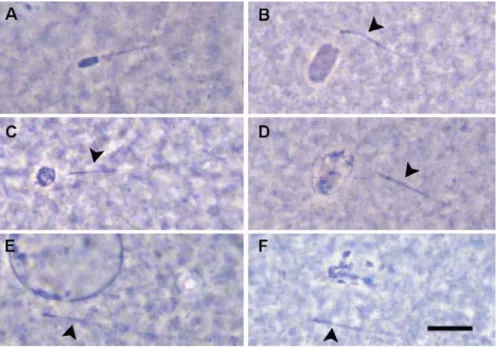
Evaluation of zygotes: To study the effect of different reatments on male pronucleus formation, fertilization rates and monospermic fertilization rates, inseminated oocytes were fixed 10 h after IVF and stained as described above. After staining, different stages in the transformation of sperm heads into male pronucleus can be distinguished. Right after penetration, the sperm head is compact with a more or less uniform dark coloration (Figure 5 A). In case of penetration of oocytes at GV stage or with high intercellular MPF activity, the sperm head remains compact even several hours later (Wang et al., 1994; Kikuchi et al., 1999). In case of optimal cytoplasmic maturity of the oocyte, the head of the fertilizing spermatozoa swells. Swollen sperm heads (Figure 5 B) are usually evenly stained and show a smooth, grey coloration. The next stage is the recondensation (Figure 5 C) of the swollen spermatozoa which is characterised by the uneven coloration of the shrunk, round- shaped sperm head after staining. Finally, this recondensed mass
start its decondensation (Figure 5 D) and develop into an unevenly stained enlarged membrane-surrounded vacuolum that leads to the development of the male pronucleus (Figure 5 E), an expanded,
completely round shaped vesicle including the decondensed filaments of the male genome and a nucleolus (or nucleoli). In case of inadequate cytoplasmic maturity (high MPF activity) of the oocyte the penetrated spermatozoon fails to form male pronucleus, remains condensed or under the influence of the active MPF, the sperm head transforms into metaphase chromosomes (Figure 5 F) (Kikuchi et al., 1999). In the present study, only the oocytes with male pronucleus(ei) and/or decondensed sperm head(s) with
Fig 5. Fertilization of porcine oocyte: male pronucleus formation. A:
condensed sperm head; B: swollen sperm head; C: re-condensed sperm head; D: decondensed sperm head; E: male pronucleus;F:
male chromatine transformed into metaphase chromosomes. Arrow heads show corresponding sperm tails. Scale bar represents 10 µm.
corresponding sperm tails were judged as fertilized. Normal monospermic fertilization is characterised by the extrusion of the second polar body (Figure 6 A) and the existance of the female pronucleus and a single male pronucleus (Figure 6 B; Figure 7 A). Oocytes with more than one male pronucleus were considered as polyspermic zygotes (Figure 7 B).
Fig 7. Fertilization of porcine oocyte: monospermy (A) with two (one ♂ and one ♀) and polyspermy (B) with three pronuclei.
Oocytes were fixed 10 h after IVF. Scale bar represents 10 µm.
PB 2
PB 1 S Fig 6. Fertilization of
porcine oocyte:
extrusion of the second polar body (A); male and female pronucleus formation (B). PB 1:
first polar body; PB2:
second polar body with the spindle (S).The arrow shows a sperm tail. Scale bar represents 10 µm.
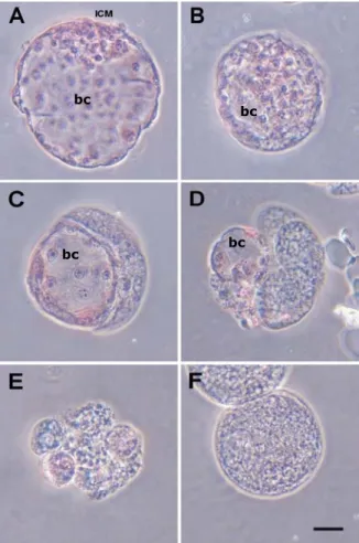
Evaluation of embryos: On Day 6 of IVC, IVM/IVF embryos were fixed and and stained as described above. Only embryos with a blastocoel and not less than ten blastomers were considered as blastocyst (Figure 8 A, B and C). In case of partially living embryos (Figure 8 C) only the embryos with not less than fifty percent living part were donsidered as normal blastocyst while embryos with blastocoel but with less than fifty percent living part and/or less than 10 live blastomers (Figure 8 D) were excluded form this category. Besides the blastocysts a remarkable proportion of embryos remains arrested at four cell stage, usually showing signs of fragmentation (Figure 8 E) or fail to divide (Figure 8 F).
Fig 8. Morphology of porcine IVP embrios on day 6 IVC after staining with orcein.
A: Expanded blastocyst with an explicit inner cell mass (ICM).
B: Expanding blastocyst.
C: Partially living (half) blastocyst with approximately 10 cells.
D: Partially living (quarter) embryo with a blastocoel but less than 10 cells.
E: Embryo arrested at an early stage (4-6 cells) showing the signs of fragmentation.
F: Undivided zygote.
bc: blastocoel
Scale bar means 50 µm.
bc bc bc
bc
3.2.1.4 Statistical analysis
Each treatment of each experiment was replicated at least three times. Statistical analyses of IVM data were subjected to analysis of variance (ANOVA) followed by Duncan’s multiple range test (P <
0.01) using GLM procedures of Statistical Analysis System (SAS Institute Inc., Cary, NC, USA). Data of IVF and IVC results were analysed by Chi-square test (P < 0.05). Data are expressed as mean
± SEM.
3.2.2 Experimental Design
Experiment 1: To evaluate the effects of IBMX and iAC in collection medium and dbcAMP in maturation medium, respectively, on nuclear progression and oocyte maturation, COCs were collected using BCM and CCM collection media, and were cultured in vitro with or without 1mM dbcAMP for 22 hours, then culture was followed in the medium without hormones and dbcAMP supplement. Nuclear progression during IVM was evaluated after fixation at 12, 22, 36 and 46 h of culture. Chromatin condensation (GV stage) in the oocytes was classified according to Motlik and Fulka (1976).
Experiment 2: To study the effect of IBMX and iAC in collection medium and dbcAMP in maturation medium on fertilization parameters, COCs were matured in the presence or absence of 1 mM dbcAMP, and then fertilized in vitro. The inseminated oocytes were fixed at 10h after the insemination. Only the oocytes with male pronucleus(ei) and/or decondensed sperm head(s) with corresponding sperm tails were judged as penetrated. Zygotes with one female and one male pronucleus (or decondensed sperm head) and with two polar bodies were classified as normally (monospermic) fertilized oocytes.
Experiment 3: Effect of IBMX and iAC in collection medium and dbcAMP in maturation medium for oocytes on their subsequent embryonic development after IVF. On Day 6, all of the IVM/IVF embryos were fixed and evaluated for the rate of blastocyst formation and their cell number in each blastocyst.
See results from page 48.
3.3 In vitro fertilization and development to blastocyst stage of immature porcine oocytes arrested before metaphase- II stage
3.3.1 MATERIALS AND METHODS
3.3.1.1 Oocyte collection and in vitro maturation
Ovaries from prepuberal cross-bred (Landrace × Large White) gilts (their body weigth was approximately 100 kg) were collected at the local slaughterhouse and were carried to the lab in PBS at 35-37 °C.
Cumulus-oocyte complexes (COCs) were collected by scraping of from 3-5 mm follicles in Medium 199 supplemented with 10% fetal bovine serum (Gibco, Life Technologies Inc., Grand Island, USA), 20mM HEPES, 100 unit/mL penicillin G potassium (Sigma Chemical Co., St. Louis, MO, USA, P-7794) and 0.1 mg/mL streptomycin sulfate (Sigma, S-9137). To obtain a remarkable number of oocytes arrested at M-I stage, a one-phase IVM was performed without the synchronisation of nuclear maturation. The basic medium was M199 which was reported to result better oocyte quality than NCSU 37 (which we used previously) regarding the cumulus expansion (Abeydeera et al., 1998; Abeydeera et al., 2000). Maturation culture was performed in Medium199 (prepared with 20mM HEPES; Gibco) supplemented with 10% porcine follicular fluid (pFF), 0.91 mM sodium pyruvate (Sigma, S-3362), 10 IU/ml PMSG, 10 IU/ml hCG, 20 ng/ml epidermal growth factor (EGF, Sigma), 150 µM cysteamine (Sigma) and antibiotics in 4-well dishes (Nunclon Multidishes, Nalge Nunc International, Denmark) in an atmosphere of 5% CO2, 5% O2
and 90% N2 at 39°C. The pH was adjusted to 7.3. At the end of the
culture period, COCs were denuded using a fine glass pipette after a brief treatment with 0.1% hyaluronidase.
3.3.1.2 IVF and IVC of porcine oocytes
IVF and in vitro culture (IVC) were carried out according to the previous report by Kikuchi et al. (2002) with slight modifications.
Briefly, after denuding of COCs, oocytes with and without a visible first polar body (PB) were separated under a stereo microscope into PB+ and PB- groups, respectively. About 20 oocytes were transferred into 100 µl Pig-FM (Suzuki et al., 2002) droplets covered by paraffin oil. They were coincubated then with 1 × 10⁵/ml frozen-thawed epididymal spermatozoa (Kikuchi et al., 1998) for 3 h at 39°C under 5% CO2, 5% O2 and 90%N2. The day of IVF was defied as Day 0.
After removal of spermatozoa attached to the surface of zona pellucida by gentle pipetting with a fine glass pipette, IVC was performed in IVC-PyrLac for Days 0-2 and in IVC-Glu for Days 2-6.
3.3.1.3 Oocyte and embryo evaluation with orcein staining
For evaluation of meiotic stage of oocytes, IVF results, pronucleus formation and total number of cells in blastocysts, oocytes or embryos were mounted, fixed, stained and evaluated as described in chapter 3.2.1.3.
3.3.1.4 Blastocyst evaluation with differential staining
Differential nuclear staining of inner cell mass (ICM) and trophectoderm (TE) cells were performed according to Macháty et al., (1998) with a small modification. Briefly, after digestion of zona pellucida with 0.5% (w/v) pronase, embryos were washed in IVC- PyrLac buffered with 20 mM-HEPES and supplemented with 4 mg/ml
Fig 9. Porcine IVM blastocyst after differential staining with Hoechst 33342 and propidium iodide. ICM nuclei are blue. TE nuclei are pink.
The photograph was taken at magnification 400 ×.
polyvinyl alcohol (IVC-PyrLac-HEPES), in which osmolairity was adjusted to 285 osmol/kg, and then they were exposed to 1:5 rabbit anti-pig whole serum (Sigma, P-3164) for 40 min at 39°C. Then they were washed three times in IVC-PyrLac-HEPES and incubated in 1:15 dilution of guinea pig complement (Sigma, S-1639) supplemented with 20 µg/ml propidium iodide (Sigma, P-4170) and 20 µg/ml Hoechst-33342 (Sigma, B-2261) for 30 min at 39°C. Then embryos were briefly fixed in 50% ethanol, and mounted individually in glycerol on a glass-slide covered with a coverslip. The blastocysts were examined under UV light using an Axioplan 2 epifluorescence microscope (Carl Zeiss Jena GmbH). The ICM nuclei labelled with Hoechst appeared blue
and TE nuclei labelled by both Hoechst and propidium iodide appeared pink to red (Figure 9). Numbers of ICM and TE nuclei were counted directly under the microscope.
3.3.1.5 Statistical analysis
Each treatment of each
experiment was replicated at least three
times. Statistical analyses of IVF and IVC
results were analysed by
ANOVA followed by Duncan’s multiple range test (p < 0.05). Percent data were transformed into arcsin before the statistical analysis. Data are expressed as mean ± SEM.
3.3.2 Experimental design
Experiment 1. To evaluate meiotic progression during maturation culture, COCs were cultured for 36, 48 and 60 h. They were denuded, fixed and stained then their nuclear status was evaluated.
Experiment 2. To evaluate meiotic potential of oocytes, COCs were cultured for 48 h then oocytes were denuded. PB+ and PB- oocytes were separated and their nuclear status was evaluated after fixation and staining.
Experiment 3. Following 48 h of maturation culture, PB+ and PB- oocytes were separated and IVF was performed. Inseminated oocytes were fixed 10 h after IVF and examined for fertilization status (sperm penetration and pronuclear formation). Oocytes were judged to be penetrated when they had one or more male pronuclei and/or sperm heads with corresponding sperm tails.
Experiment 4. After IVF, PB+ and PB- oocytes were cultured for 6 days to examine their ability to develop to the blastocyst stage (PB+
blastocyst and PB- blastocyst, respectively). Embryos with clear blastocoel and with at least ten living cells were considered to be blastocysts. In order to characterize embryo quality, the total number of cells (nuclei) in each blastocyst and blastocyst morphology were evaluated after fixation and staining. Blastocyst morphology was evaluated according to the proportion of living and dead/degenerated parts inside the zona. The following three types of blastocysts were distinguished: Type 1, live blastocyst, the proportion of
dead/degenerated part is less than 10 percent (Figure 10 A), Type 2, the proportion of dead part is between 10 and 30 percent (Figure 10 B), and Type 3, the proportion of dead part is between 30 and 50 percent (Figure 10 C).
Experiment 5. The number and ratio of ICM and TE cells of blastocysts from PB+
and PB- oocytes were evaluated after differential staining.
See results from page 62.
Fig 10. Porcine blastocysts produced in vitro after fixation on day 6 and staining with aceto- orcein. (A) Type 1 blastocyst, the proportion of dead/degenerated part is less than 10 percent.
(B) Type 2 blastocyst, 10-30% of the embryo is degenerated. (C) Type 3 blastocyst, 30-50% of the embryo is degenerated. All photographs were taken with a phase-contrast microscope at the same magnification. Arrows indicate the portion of the dead blastomeres less stained with orcein. Scale bar represents 50 µm.
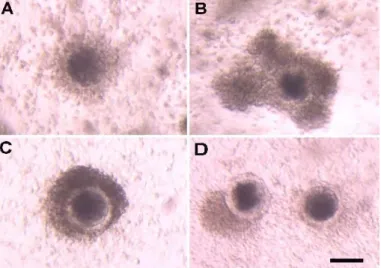
Fig 11. Sources of oocytes. (A): Granulosa-Cumulus- Oocyte Complexes (GCOCs); (B) Cumulus-Oocyte Complexes (COCs). Photographs were taken under a stereomicroscope at the same magnification. Scale bar represents 100 µm.
3.4 The relationship between cumulus morphology and oocyte maturation
3.4.1 MATERIALS AND METHODS
3.4.1.1 Oocyte collection and in vitro maturation Pig ovaries were
obtained from prepubertal
cross-bred gilt (Landrace × Large White, approx. 100 kg body weight) and transported
to the laboratory in
PBS at 35°C.
GCOCs (Figure 11 A) and COCs (Figure 11 B) of similar morphology were collected by dissection of 3-5 mm non-antral follicles in Medium 199 supplemented with 10% fetal bovine serum (Gibco), 20mM HEPES, 100unit/mL penicillin G potassium (Sigma Chemical Co., St.
Louis, MO, USA, P-7794) and 0.1mg/mL streptomycin sulfate (Sigma, S-9137). A two-phase IVM was performed: COCs and GCOCs were transferred to first maturation medium (IVM1), which was Medium199 (prepared with 20mM HEPES; Gibco) supplemented with 0.91 mM sodium pyruvate (Sigma, S-3362), 10 IU/ml PMSG, 10 IU/ml hCG, 20 ng/ml epidermal growth factor (EGF, Sigma), 150 µM cysteamine (Sigma) 100 unit/ml penicillin G potassium (Sigma), 0.1
mg/ml streptomycin sulfate (Sigma) and 0.1% polyvinyl alcohol (PVA, Sigma) and incubated separately, in groups of 25-30 (COC) and 15- 20 (GCOC) in 4-well dishes (Nunclon Multidishes, Nalge Nunc International, Denmark) in an atmosphere of 5% CO2 and 5% O2 in air at 39 °C. After 20 h of culture COCs and GCOCs were transferred into second maturation medium (IVM2) which was prepared in the absence of PVA but contained 10% porcine follicular fluid (pFF), otherwise identical to IVM1 and IVM was performed continuously under the conditions as described above. pFF was collected in advance by aspiration with a syringe and cetrifuged at 1,800 × g for 1.5 h and the supernatant was stored at –20 °C. Then enough amount of the stock were once thawed, mixed, centrifugated again and stored at –20 °C as the single batch until use.
3.4.1.2 Classification of COCs
Four types of COCs can be distinguished according to the characteristics of somatic compartment from 30 h of IVM.
Type 1: The COC is floating in the maturation medium, the oocyte is surrounded by a light coloured fully expanded cumulus mass (Figure 12 A).
Type 2: The COC is floating in the maturation medium, the oocyte is surrounded by a dark brown coloured compact or semicompact somatic compartment (Figure 12 B).
Type 3: The COC is attached to the bottom of the culture dish, the oocyte is surrounded by a dark brown coloured compact somatic compartment (Figure 12 C).
Type 4: The COC is attached to the bottom of the culture dish, the oocyte is partially denuded, the loss of cumulus cells ranges at least
Fig 12. Morphological classes of complexes at 30 h of IVM according to the behaviour of the somatic compartment. (A) Type 1: floating oocyte surrounded by expanded cumulus; (B) Type 2:
floating oocyte surrounded by dark, compact cumulus; (C) Type 3: the complex is attached to the bottom of the culture dish, the oocyte is surrounded by a dark, compact cumulus; (D) Type 4: the complexes are attached to the bottom of the culture dish, the oocyte is partially denuded, the loss of cumulus cells ranges at least 30% of the oocyte surface. The remaining cumulus cells are dark coloured and compact. Photographs were taken under a stereo microscope at the same magnification. Scale bar represents 100 µm.
the 30% of the oocyte surface. The remaining cumulus cells are dark coloured and compact (Figure 12 D).
3.4.1.3 Parthenogenetic activation (PGA) of IVM oocytes
At the end of IVM, oocytes were denuded using a fine glass pipette after a brief treatment with 0.1% hyaluronidase. Denuded oocytes with the first polar body -considered as matured oocytes- were harvested under a stereomicroscope. Matured oocytes were transferred into activation solution which consisted of 0.28 M d-mannitol, 0.05 mM CaCl2, 0.1 mM MgSO4, and 0.01% (w/v) BSA
and washed once. Then they were stimulated with direct current (D.C.) pulse of 1.0 kV/cm for duration of 100 µsec using a somatic hybridizer (SSH-2, Shimadzu, Kyoto, Japan). After electric pulse oocytes were transferred into 500 µl droplets of in vitro culture (IVC) medium which is a modified NCSU-37 medium containing 4 mg/ml BSA and 50 µM βmercaptoetanol, 2.73 mM sodium lactate and 0.165 mM sodium pyruvate. They were subsequently cultured for 8-10 h at 39°C under 5% O2 tension, and then fixed. After staining, activation status (pronuclear formation, extrusion of a second polar body or fragmentation) was evaluated under a phase-contrast microscope.
3.4.1.4 IVF and IVC of porcine oocytes
IVF and in vitro culture (IVC) were carried out as described above (chapter 2.2.1.2) with slight modifications. Briefly, after denuding of COCs, oocytes with a visible first polar body (PB) were selected under a stereo microscope and used for IVF. About 20 oocytes were transferred into 100 µl Pig-FM (Suzuki et al., 2002) droplets covered by paraffin oil. They were coincubated then with 1 × 10⁵/ml frozen- thawed epididymal spermatozoa (Kikuchi et al., 1998) for 3 h at 39°C under 5% CO2, 5% O2 and 90%N2. After removal of spermatozoa
attached to the surface of zona pellucida by gentle pipetting with a fine glass pipette, IVC was performed in IVC-PyrLac for 10 h.
3.4.1.5 Oocyte and embryo evaluation with orcein staining
For evaluation of meiotic stage of oocytes, parthenogenetic activation and IVF results, oocytes or embryos were mounted, fixed and stained as described in chapter 3.2.1.3.
Evaluation of nuclear status, fertilization rates and blastocyst formation was happened as described in chapter 3.2.1.3.
Evaluation of parthenogenetic activation: In the present study, the oocytes beyond the anaphase-II stage were defined as being activated and the percentages of activated oocytes; 1) normal activation (characterized by a female pronucleus formation with the first and second polar bodies Figure 13 A), 2) fragmentation (abnormal cleavage characterized by unequal blastomeres Figure 13 B), 3) Metaphase-III (M-III) stage oocytes (characterized by metaphase stage female chromosomes (Figure 13 D) - often
Fig 13. Nuclear morphology of porcine oocytes 8 hours after parthenogenetic
activation. A: normal activation; B:
fragmentation; C: M-III stage with abnormal chromosome
arrangement; D: M-III stage.
1 PB: first polar body;
2 PB: second polar body;FPN: female pronucleus;A: abromally arranged anaphase-like chromosomes;M:
metaphase plate
Scale bar represents 10 µm.
arranged abnormally (Figure 13 C) - with the first and second polar bodies), were scored. Unactivated oocytes (remaining at Metaphase- II: M-II, stage) were also scored.
3.4.1.6 Statistical analysis
Each treatment of each experiment was replicated at least three times. Statistical analyses of IVM data were subjected to analysis of variance (ANOVA) followed by Duncan’s multiple range test (P <
0.05) using GLM procedures of Statistical Analysis System (SAS Institute Inc., Cary, NC, USA). Data of PGA and IVF results were analysed by Chi-square test (P < 0.05).
3.4.2 Experimental design
Experiment 1: The relation between the morphology of somatic compartment and the kinetics of nuclear maturation was studied in case of COCs and GCOCs. COCs and GCOCs were classified according to the characteristics of their somatic compartment as described above at 30, 36, 42 and 48 h of IVM, and then oocytes were denuded using a fine glass pipette after a brief treatment with 0.1%
hyaluronidase. The denuded oocytes were fixed in acetic ethanol (1:3 v/v) for 3-5 days and stained with 1% aceto-orcein (Sigma), then nuclear status of oocytes was evaluated using a phase-contrast microscopy as described in chapter 3.2.1.3.
Experiment 2: The ability of oocytes to form a female pronucleus was assessed in order to estimate the capacity of the cytoplasm to potentiate oocyte activation. Oocytes from COCs and GCOCs of each morphologic type were collected separately at 42 h of IVM, and then parthenogenetic activation was performed as described above. Eight hours after the stimulation, oocytes were fixed in acetic ethanol (1:3
v/v) for 3-5 days and stained with 1% aceto-orcein (Sigma).
Activated status was evaluated using a phase-contrast microscopy as described in chapter 3.4.1.4.
Experiment 3: Oocytes from COCs and GCOCs of each morphologic types were subjected to IVF after 48 h of IVM. Inseminated oocytes were fixed 10 h after IVF and stained as described in chapter 3.2.1.3 to examine sperm penetration and pronuclear formation. The oocytes were considered to be penetrated when they had one or more male pronucleus(ei) and/or swollen sperm head(s) with a corresponding sperm tail(s). Zygotes with one female and one male pronucleus (or decondensed sperm head) and with two polar bodies were classified as normally (monospermic) fertilized oocytes.
See results from page 78.
4 RESULTS
4.1 Synchronisation of meiotic maturation by high level of intercellular cAMP
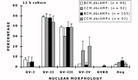
Experiment 1. No significant difference in chromatin condensation between oocytes collected and/or matured in the presence or absence of cAMP was observed at 12 h of culture (Figure 14).
Both in the dbcAMP- and dbcAMP+ groups almost all the oocytes remained at GV stage, where GV-II (48.6 ± 5.8% and 47.6 ± 8.0%, respectively) and GV-III (39.3 ± 4.6 and 38.6 ± 5.2%, respectively) were dominant. Only a very few remained at GV-I or reached more
Fig 14. Nuclear morphology (mean ± SEM) of oocytes after 12 h of culture of four different treatments. Abbreviations:
BCM= Basic Collection Medium; CCM= Complete Collection Medium; dbcAMP- = COCs cultured in the absence of 1 mM dbcAMP; dbcAMP+ = COCs cultured in the presence of 1 mM dbcAMP. Numbers of oocytes examined in different treatment groups are given in parentheses.
condensed stages of chromatin (GV-IV). The rate of degenerated oocytes was the same between the groups.
Significant differences in nuclear progression of oocytes matured with or without dbcAMP were detected at 22 h of culture (Figure 15).
By this time, GVBD occurred in a high rate (44.3 ± 8.1%) of oocytes that were matured in the absence of dbcAMP, the rest of them remained at GV stage and 9.6 ± 5.2 % of oocytes already reached metaphase-I (M-I) phase. The nuclear status of oocytes that were cultured with dbcAMP was at GV stage, which is similar to that at 12 h of culture with an unremarkable rate (1.0 ± 1.0%) of oocytes that underwent GVBD. The rate of degenerated oocytes in this period of culture was the same in the treatment groups. No difference in nuclear stage between oocytes collected with different levels of cAMP was observed at this period (data not shown).
Fig 15. Distribution (mean ± SEM) of meiotic stage of porcine oocytes after 22 h culture with or without 1 mM dbcAMP. Asterisk above the bars mean significant differences (p < 0.01). Numbers of oocytes examined in different treatment groups are given in parentheses.
By 36 h of culture, the most of oocytes underwent GVBD in both dbcAMP- and + groups (89.3 ± 2.4% and 93.6 ± 3.4%, respectively).
A considerable rate (38.0 ± 6.4%) of the oocytes matured in the absence of dbcAMP reached metaphase-II (M-II) by this time and a significant proportion showed M-I (16.6 ± 5.3%) (Figure 16).
The remaining oocytes that underwent GVBD were at prometaphase-I (proM-I) (5.0±1%), telophase-I (T-I) (14.3 ± 4.6%), or anaphase-I (A-I) (13.6 ± 2.6%). In contrast, in the dbcAMP+ group a significantly higher proportion of oocytes were at M-I (49.3±7.3%) or proM-I (32.6±2.3) stage but none of the oocytes showed M-II phase nucleus (Figure 16). No difference in nuclear stage between oocytes collected with different levels of cAMP was observed at this period of culture (data not shown).
Fig. 16. Distribution (mean ± SEM) of meiotic stage of porcine oocytes after an additional 14 h cultivation following 22 h culture (36 h in total) with or without 1 mM dbcAMP. Asterisk above the bars mean significant differences (p < 0.01). Numbers of oocytes examined in different treatment groups are given in parentheses.