medium improves the maturation, fertilisation and developmental competence of porcine
oocytes
ZHAO NAMULA
1,4, YOKO SATO
2p,
MANITA WITTAYARAT
3, QUYNH ANH LE
4, NHIEN THI NGUYEN
4, QINGYI LIN
1, MAKI HIRATA
1,4, FUMINORI TANIHARA
1,4and TAKESHIGE OTOI
1,41Faculty of Veterinary Science, Guangdong Ocean University, Zhanjiang, China
2School of Biological Science, Tokai University, Sapporo, Hokkaido 005-8601, Japan
3Faculty of Veterinary Science, Prince of Songkla University, Songkhla, Thailand
4Faculty of Bioscience and Bioindustry, Tokushima University, Tokushima, Japan
Received: December 28, 2019 • Accepted: April 21, 2020 Published online: November 20, 2020
ABSTRACT
This study was conducted to determine the effects of supplementing the maturation medium with the antioxidant curcumin on thein vitromaturation (IVM), fertilisation and development of porcine oocytes. Curcumin supplementation was performed at concentrations of 0, 5, 10, 20, and 40mM. At concentrations of 5–20mM, curcumin had significant positive effects (P < 0.05) on maturation and fertilisation rates compared to the non-treated group. Of the groups cultured with 5–20mM curcumin, the number of oocytes with DNA-fragmented nuclei after IVM was significantly lower than in groups matured without curcumin. Moreover, curcumin supplementation at 10mM also gave a significantly higher rate of blastocyst formation compared with oocytes matured without curcumin. Increasing the curcumin concentration to 40mM yielded negative effects on fertilisation and embryonic development compared with the groups treated with lower concentrations of curcumin. Supplementation with 10mM curcumin had beneficial effects on the oocyte maturation rate and DNA fragmentation index compared to the non-treated group both in the presence and absence of hydrogen peroxide. These results indicate that curcumin supplementation at a suitable concentration (10mM) is potentially useful for porcine oocyte culture systems, in terms of protecting oocytes from various forms of oxidative stress.
KEYWORDS
curcumin,in vitroculture, maturation, oocyte, oxidative stress, porcine
INTRODUCTION
Oocyte or embryo culture is widely utilised for various purposes, including overcoming post- fertilisation barriers and producing good-quality embryos for further usage (Borole et al., 2000). However, it should be recognised that oocytes and embryos in culture are not identical to thosein vivo(Halliwell, 2014). Exposure to environmental stress, mostly arising from a critical imbalance between the levels of reactive oxygen species (ROS) and antioxidant defence mechanisms in the culture medium, could have detrimental effects on the devel- opmental competence of embryos (de Lamirande et al., 1997; Halliwell, 2014). ROS have been reported to be associated with sperm–oocyte fusion inhibition, mitochondrial dysfunction, and damage to DNA, RNA, and protein (Comporti, 1989; Aitken et al., 1993).
Acta Veterinaria Hungarica
68 (2020) 3, 298–304 DOI:
10.1556/004.2020.00041
© 2020 Akademiai Kiado, Budapest
RESEARCH ARTICLE
*Corresponding author.
E-mail:yokos@tsc.u-tokai.ac.jp
Considerable evidence in animal studies indicates that the supplementation of antioxidants such as antioxidant flavonoids (quercetin and taxifolin) (Kang et al., 2016), melatonin (Do et al., 2015; Chen et al., 2017), selenium (Tareq et al., 2012), vitamin E (Tareq et al., 2012), resvera- trol (Kwak et al., 2012), chlorogenic acid (CGA) and caffeic acid (Nguyen et al., 2017) duringin vitromaturation (IVM) had beneficial effects and improved the production system of porcine embryos. Curcumin, a common dietary pigment and spice, is a potent antioxidant and anti-inflammatory agent extracted from turmeric (Curcuma longa), and is known to play an essential role against various pathological conditions such as cancer, atherosclerosis, and neurode- generative diseases (Menon and Sudheer, 2007). A previous study showed that the antioxidant activity of curcumin in mouse embryonic stem cells and blastocysts was due to its ability to attenuate ROS formation and subsequent apoptotic biochemical events by preventing methylglyoxal-induced oxidative stress (Hsuuw et al., 2005). However, treating fertilised zebrafish eggs and embryos with curcumin had adverse embryotoxic and teratogenic effects, inducing bent or hook-like tails, spinal column curving, oedema in the pericardial sac, retarded yolk sac resorption, and shorter body length (Wu et al., 2007). Since there is limited infor- mation as to whether curcumin supplementation improves the rates of maturation and fertilisation as well as the developmental competence of porcine oocytes, the present study was undertaken to evaluate such effects.
This study was designed to determine a suitable con- centration of curcumin supplementation during IVM culture of porcine oocytes for improving rates of IVM, fertilisation, and development to the blastocyst stage. The total cell number and DNA fragmentation in all blastocysts were analysed at the end of the experiment. The study was also performed to investigate whether curcumin would be able to protect porcine oocytes from hydrogen-peroxide- induced oxidative stress at a suitable concentration.
MATERIALS AND METHODS
In vitro maturation and assessment
IVM was performed according to methods described by Namula et al. (2013) with minor modifications. Briefly, cumulus–oocyte complexes (COCs) were collected from ovaries obtained from prepubertal crossbred gilts (Landrace3 Large White3 Duroc breeds) at a local slaughterhouse. The ovarian surface follicles were sliced on a sterilised dish using a surgical blade. Only COCs with a uniformly dark-pigmented ooplasm and intact cumulus cell masses were collected under a stereomicroscope. Approximately 50 COCs were then cultured in 500
m
L maturation medium, consisting of 25 mM HEPES tissue culture medium 199 with Earle’s salts (TCM 199;Invitrogen Co., Carlsbad, CA, USA) supplemented with 10%
(v/v) porcine follicularfluid, 50
m
M sodium pyruvate (Sigma- Aldrich), 2 mg/mL D-sorbitol (Wako Pure Chemical In- dustries Ltd.), 10 IU/mL equine chorionic gonadotropin(Kyoritu Seiyaku, Tokyo, Japan), 10 IU/mL human chorionic gonadotropin (Kyoritu Seiyaku) and 50
m
g/mL gentamicin (Sigma-Aldrich). These were cultured for 22 h in 4-well dishes (Nunc A/S, Roskilde, Denmark). The COCs were subsequently transferred into a maturation medium without hormone supplementation and cultured for an additional 22 h. COC incubation was conducted at 398C in a humidified incubator containing 5% CO2in air.To evaluate the effects of curcumin supplementation during IVM culture onin vitromaturation, fertilisation, and porcine oocyte development, the COCs were cultured in a maturation medium supplemented with curcumin (C7727, Sigma-Aldrich) dissolved in dimethyl sulphoxide and diluted to 0, 10, 20, and 40
m
M before use in each experiment. As a control, COCs were cultured in a maturation medium without curcumin and dilution solution (dimethyl sulphoxide).Analysis of the meiotic stage and DNA damage of oocytes
After maturation culture, the meiotic stage and DNA damage of oocytes were analysed with a combined technique for simultaneous nuclear staining and terminal deoxy- nucleotidyl transferase (TdT) nick-end labelling (TUNEL) (Otoi et al., 1999). Briefly, some oocytes were mechanically denuded from cumulus cells in Dulbecco’s PBS (DPBS;
Invitrogen Co.) supplemented with 150 IU/mL hyaluroni- dase (Sigma-Aldrich). Denuded oocytes werefixed overnight at 4 8C in 3.7% (w/v) paraformaldehyde diluted in DPBS.
After fixation, the oocytes were permeabilised in DPBS containing 0.1% (v/v) Triton-X100 for 40 min. They were subsequently incubated overnight at 4 8C in DPBS con- taining 10 mg/mL bovine serum albumin (Sigma-Aldrich).
The oocytes were then incubated influorescein-conjugated 20- deoxyuridine-50-triphosphate and terminal deoxynucleotidyl transferase (TUNEL reagent; Roche Diagnostics, Tokyo, Japan) for 1 h at 38.58C. After TUNEL staining, the oocytes were counterstained with 1
m
g/mL DAPI (Invitrogen Co.) for 10 min. They were then treated with an anti-bleaching so- lution (Slow-Fade; Molecular Probes Inc., Eugene, OR, USA), mounted on a glass slide, and sealed with clear nail polish.Labelled oocytes were examined using a microscope (Eclipse 80i, Nikon, Tokyo, Japan) with epifluorescence illumination.
Two standardfilter sets were used for detection offluorescein isothiocyanate (FITC) alone (465–495 nm as the excitation wavelength and 515–555 nm as the emission wavelength), and for detection of DAPI alone (340–380 nm as the exci- tation wavelength and 435–485 nm as the emission wave- length). Oocytes were determined to be in the germinal vesicle (GV), condensed chromatin (CC), metaphase I (MI), anaphase I to telophase I (AT), or metaphase II (MII) stage according to chromatin configuration, based on DAPI stain- ing. Those with diffusely stained cytoplasm characteristics of nonviable cells and those in which chromatin was un- identifiable or not visible were excluded from DNA damage analysis. To assess DNA damage in all oocytes after matu- ration culture, the nuclei that were labelled by TUNEL were counted.
In vitro fertilisation and assessment
In vitro fertilisation (IVF) was performed according to methods described by Namula et al. (2013) with minor modifications. Frozen-thawed spermatozoa were transferred into 6 mL of porcine fertilisation medium (PFM; Research Institute for the Functional Peptides Co., Yamagata, Japan) and washed by centrifuging at 5503gfor 5 min. The sperm pellet was resuspended in PFM to achieve a concentration of 2.03 106 cells/mL. The spermatozoa (250
m
L) were added to 250m
L PFM containing 50 matured oocytes in 4-well dishes. Thefinal sperm concentration was adjusted to 13 106cells/mL. The oocytes were co-incubated at 398C for 5 h in a humidified incubator containing 5% CO2, 5% O2, and 90% N2. After co-incubation, the inseminated zygotes were denuded from the cumulus cells and the attached sperma- tozoa by mechanical pipetting. Approximately 50 denuded zygotes were subsequently cultured in 500m
L PZM-5(Research Institute for the Functional Peptides Co.) overlaid by mineral oil in 4-well dishes.
To assess oocyte fertilisation, some zygotes were moun- ted on glass slides 10 h after insemination and fixed with acetic acid:ethanol (1:3 v/v) for 48–72 h. The fixed zygotes were stained with acetic orcein (1% orcein in 45% acetic acid) and examined by phase contrast microscopy. Oocytes containing both female and male pronuclei were considered fertilised, and were categorised as normal or polyspermic based on the number of swollen sperm heads and/or pro- nuclei in the cytoplasm (Do et al., 2015).
In vitro culture
The remaining zygotes were cultured continuouslyin vitro at 398C in a humidified incubator containing 5% CO2, 5%
O2, and 90% N2. All of the cleaved embryos were transferred into 500
m
L PBM (Research Institute for the Functional Peptides Co.) 72 h after insemination, and were cultured for an additional 4 days to evaluate their ability to develop to the blastocyst stage.Assessment of blastocyst quality
To evaluate the total cell number and DNA fragmentation in the blastocysts, all embryos at the blastocyst and expanded
blastocyst stages were fixed at the end of the culturing procedure. They were analysed using a combined technique for simultaneous nuclear staining with DAPI and TUNEL, as described above. The apoptotic index was calculated by dividing the number of cells containing apoptotic nuclei (labelled by TUNEL) by the total number of cells.
Assessment of the antioxidant effect of curcumin
To assess the protective effect of curcumin on hydrogen peroxide (H2O2)-induced DNA damage in porcine oocytes, the COCs were exposed to 1 mM H2O2 in a maturation medium either without or supplemented with curcumin (10
m
M) during IVM (Do et al., 2015). The concentration of curcumin found most suitable for the development of embryos (10m
M) was used in this experiment. After maturation at 39 8C for 44 h, the oocytes were denuded, fixed, and evaluated for nuclear status and DNA fragmen- tation, as described above.Statistical analysis
Experiments were repeated seven times for oocytes matured with curcumin, and four times for oocytes exposed to H2O2.
Statistical significance was inferred from analysis of variance (ANOVA) followed by Fisher’s protected least significant difference (PLSD) tests using STATVIEW (Abacus Con- cepts, Inc., Berkeley, CA, USA). Percentage data were sub- jected to arcsine transformation before statistical analysis.
Differences with a value ofP ≤ 0.05 were regarded as sig- nificant.
RESULTS
As shown in Table 1, the maturation rates of oocytes matured with 5, 10, and 20
m
M curcumin were significantly higher (P < 0.05) than those of oocytes matured without curcumin. However, supplementation with 40m
M curcumindecreased the proportions of oocytes that underwent germinal vesicle breakdown (GVBD) and reached meta- phase II (MII) compared with the other concentrations of curcumin studied. Curcumin supplementation decreased the
Table 1.Effects of curcumin supplementation in the maturation medium on the meiotic competence and DNA damage of porcine oocytes*
Concentration of curcumin (mM)**
No. of examined oocytes
No. (%) of oocytes with*** No. (%) of oocytes with DNA- fragmented nuclei
GVBD MII
Control 104 99 (94.6±2.2)a 57 (52.1±4.4)b,c 6 (7.9±2.8)a
0 106 101 (94.0±3.3)a 63 (57.3±4.4)b 7 (5.3±1.7)a,b
5 112 108 (95.7±1.9)a 90 (79.4±2.0)a 2 (1.5±1.0)b
10 109 108 (99.2±0.8)a 92 (81.4±3.8)a 1 (2.0±2.0)b
20 103 100 (96.1±2.3)a 78 (73.1±3.8)a 1 (2.4±2.4)b
40 116 91 (75.7±5.6)b 48 (42.8±4.8)c 1 (0.7±0.7)b
*All experiments were repeated 7 times. Data are expressed as the mean±SEM.
**As a control, oocytes were cultured in maturation medium without curcumin and dilution solution (dimethyl sulphoxide).
***GVBD; germinal vesicle breakdown, MII; metaphase II.
a–cValues with different superscript letters in the same column are significantly different (P< 0.05).
proportion of DNA-fragmented nuclei in oocytes after maturation culture compared with the control group.
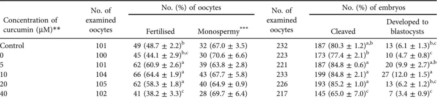
As shown in Table 2, the fertilisation rates of oocytes matured with 5, 10 and 20
m
M curcumin during IVM were significantly higher (P< 0.05) than those of oocytes matured without curcumin. Moreover, the blastocyst formation rate of oocytes matured with 10m
M curcumin was significantly higher (P < 0.05) than that of oocytes matured without curcumin. However, curcumin supplementation (5–20m
M)was observed to have no effect on the total cell number of blastocysts and their DNA fragmentation index (Fig. 1).
As shown in Fig. 2, exposure of oocytes to 1 mM H2O2 during IVM significantly reduced maturation rates and increased the proportions of DNA-fragmented nuclei compared to those of non-exposed oocytes (P < 0.05), irrespective of curcumin supplementation. Supplementing the maturation medium with 10
m
M curcumin significantly improved the maturation rate and reduced the proportion of DNA-fragmented nuclei compared with the control group (P < 0.05).DISCUSSION
Standard in vitroconditions fundamentally contain levels of oxygen about three times higher than those within the lumen of the female reproductive tract, resulting in higher free radical production (Mastroianni and Jones, 1965; Fowler and Call- ingham, 1978). The imbalance between ROS and the defence mechanism against them has an impact on the in vitro maturation and fertilisation ability of cells (de Lamirande et al., 1997). Under in vivo conditions, an efficient reduction– oxidation reaction (redox) system can ensure an ideal envi- ronment for embryonic development by maintaining the balance between the production of ROS and antioxidant en- zymes (Guerin et al., 2001; Agarwal and Majzoub, 2017).
However, thein vitroconditions of oocyte culture lack external non-enzymatic antioxidant protection, which leads to the increase of ROS (Guerin et al., 2001). Therefore, the supple- mentation of antioxidants as external protection against oxidative stress can be an effective strategy in vitro. Earlier studies have demonstrated that the mediation of antioxidants Table 2.Effects of curcumin supplementation in the maturation medium on the fertilisation and development of porcine oocytes*
Concentration of curcumin (mM)**
No. of examined
oocytes
No. (%) of oocytes No. of
examined oocytes
No. (%) of embryos
Fertilised Monospermy*** Cleaved
Developed to blastocysts Control 101 49 (48.7±2.2)b 32 (67.0±3.5) 232 187 (80.3±1.2)a,b 13 (6.1±1.3)b,c
0 100 45 (44.1±2.9)b,c 30 (70.6±6.6) 223 173 (77.4±2.1)b 10 (4.7±0.8)c
5 101 62 (60.9±2.6)a 39 (63.8±2.8) 221 187 (84.8±0.6)a 20 (9.9±2.7)a,b
10 104 66 (64.4±1.9)a 43 (67.7±5.8) 233 199 (84.8±2.1)a 27 (12.0±1.5)a
20 105 62 (58.3±1.8)a 40 (64.9±0.9) 226 193 (85.2±1.0)a 13 (6.2±1.2)b,c
40 102 41 (38.2±3.3)c 28 (69.7±6.4) 217 145 (65.0±7.0)c 7 (3.4±0.9)c
*All experiments were repeated 7 times. Data are expressed as the mean±SEM.
**As a control, oocytes were cultured in maturation medium without curcumin and dilution solution (dimethyl sulphoxide).
***The proportions of monospermic fertilisation were calculated by dividing the numbers of monospermic fertilised oocytes by the total number of fertilised oocytes.
a–cValues with different superscript letters in the same column are significantly different (P< 0.05).
Fig. 1.Effects of curcumin supplementation in the maturation medium on the total cell number (A) and DNA fragmentation index (B) of porcine blastocysts (mean±SEM). All blastocysts (7–27 embryos) that developed after IVF were used to estimate the cell number and the proportion of DNA-fragmented nuclei. Oocytes matured without curcumin served as the control group. Bars with different letters differ
significantly (P< 0.05)
(ascorbic acid and cysteine) to prevent damage to porcine oocytes caused by oxidative stress plays an important role in the acquisition of developmental competence after fertilisation (Tatemoto et al., 2001; Whitaker and Knight, 2010). In the present study, similarly, the results indicated that the supple- mentation of curcumin as an antioxidant at 5–20
m
M duringin vitro maturation had significant positive effects on the maturation and fertilisation rates of porcine oocytes. It is hence likely that curcumin may have certain positive effects on the developmental competence of porcine oocytes after IVF. It is known that curcumin and its derivatives exhibit antioxidant properties to scavenge free radicals (Jayaprakasha et al., 2006).
A previous study has reported evidence suggesting that the antioxidant action of curcumin potentially prevents oxidative stress and apoptosis by inhibiting methylglyoxal in embryonic stem cells and blastocysts in mice (Hsuuw et al., 2005).
Methylglyoxal is a metabolic by-product of glycolysis in mammalian cells and embryos. An excess of methylglyoxal could deplete specific thiol compounds such as glutathione (GSH) by spontaneously forming a covalent bond to GSH, leading to apoptosis-induced DNA fragmentation (Ankrah and Appiah-Opong, 1999; Kang, 2003; Hsuuw et al., 2005).
GSH has been reported to be synthesised in porcine COCs during in vitro maturation, reflecting its importance in protecting oocytes from oxidative damage (Yoshida et al., 1993; de Matos et al., 1997; Tatemoto et al., 2000). More- over, GSH is also a key factor participating in sperm decondensation, corresponding with oocyte activation at the time of fertilisation (Yoshida, 1993). Our results revealed that the number of oocytes with DNA-fragmented nuclei significantly decreased in the groups receiving cur- cumin supplementation (5–40
m
M) compared to the non- treated group. A remarkably high number of fertilised oocytes were observed when 5–20m
M curcumin was sup- plemented. In particular, the addition of 10m
M curcumindramatically improved oocyte development to the blasto- cyst stage. These results are in agreement with thefindings of Hsuuw et al. (2005), who demonstrated that at con- centrations higher than 10
m
M, curcumin significantly increased the rate of development to the blastocyst stage.This occurred alongside a reduction in the proportion of apoptotic cells in mouse blastocysts, possibly by completely blocking methylglyoxal production. This suggests that, at a suitable concentration (10
m
M), curcumin may play a crucial role in protecting oocytes against methylglyoxal- mediated oxidative stress and apoptosis through restoring GSH levels in porcine oocytes.Increasing the concentration of curcumin to 40
m
Mnegatively affected the rates of maturation, fertilisation, and cleavage in porcine oocytes. In a previous study by Jiang et al. (1996), it was found that treatment of mouse embryonic fibroblasts NIH 3T3 cells with 30–90
m
M curcumin for 30–48 h resulted in cell shrinkage, with DNA fragmentation, that subsequently detached from the culture plate. This study also reported a positive correlation between the degree of DNA fragmentation and curcumin concentration (Jiang et al., 1996).Moreover, curcumin has been shown to have a lethal effect on some vertebrates such as zebrafish, wherein all larvae died after exposure to 10
m
M curcumin for 24 h (Wu et al., 2007).These findings indicated that the beneficial antioxidant properties of curcumin were dose- and time-dependent, and its dose-response was also related to the animal species used.
To confirm the protective effect of curcumin against oocyte and embryo damage caused by excessive ROS, 1 mM of H2O2was included in this porcine oocyte culture system.
Hydrogen peroxide is a considerably harmful ROS, and can cross cell membranes due to being stable and small in size (Dekhuijzen et al., 1996). We found that 10
m
M curcuminsupplementation could not neutralise the detrimental effects of hydrogen peroxide because H2O2 exposure dramatically reduced the oocyte maturation rate, irrespective of the presence of curcumin. However, the supplementation of curcumin improved the oocyte maturation rate and DNA fragmentation index compared to the non-treated group both in the presence and absence of H2O2. These results indicate that curcumin could potentially exert antioxidant effects even under normal culture conditions without H2O2. Moreover, our results are consistent with previous obser- vations showing that in mouse neuroblastoma Neuro-2A cells and human intestinal epithelial cells, curcumin Fig. 2.Effects of curcumin (10mM) supplementation in the maturation medium on the maturation rate (A) and the proportion of DNA- fragmented nuclei (B) of porcine oocytes exposed to 1 mM H2O2during maturation culture. Oocytes matured without curcumin served as the control group. Each bar represents the mean value±SEM (n54 replications, each with 99–115 oocytes per treatment). Bars with
different letters differ significantly (P< 0.05)
treatment prior to H2O2 exposure significantly reduced cell apoptosis. This was done by suppressing the elevation of intracellular ROS, stabilising the mitochondrial membrane, and inducing the pathway by which curcumin stimulates heme oxygenase-1, thus leading to defence responses against various forms of oxidative stress (Zhao et al., 2011; Wang et al., 2012).
In conclusion, curcumin treatment at a suitable con- centration (10
m
M) has potential protective effects against oxidative stress, induced by either a high-oxygen-tension environment or H2O2 exposure, in porcine oocytes via multiple possible mechanisms. This may indicate that curcumin is a suitable antioxidant agent for preventing the downstream negative effects associated with oxidative stress caused by the current porcine oocyte culture system.ACKNOWLEDGEMENTS
We thank the Nippon Food Packer, K. K. Shikoku (Tokushima, Japan), for supplying pig ovaries. This study was supported in part by Japan Science and Technology Agency/Japan International Cooperation Agency, Science and Technology Research Partnership for Sustainable Development (JST/JICA, SATREPS). We acknowledge Tokushima University for their financial support of the Research Clusters program of Tokushima University (No.
1701001).
REFERENCES
Agarwal, A. and Majzoub, A. (2017): Role of antioxidants in assisted reproductive techniques. World J. Mens Health35, 77–
93.
Aitken, R., Harkiss, D. and Buckingham, D. (1993): Relationship between iron-catalysed lipid peroxidation potential and human sperm function. Reproduction98, 257–265.
Ankrah, N.-A. and Appiah-Opong, R. (1999): Toxicity of low levels of methylglyoxal: depletion of blood glutathione and adverse effect on glucose tolerance in mice. Toxicol. Lett.109, 61–67.
Borole, V., Dhumale, D. and Rajput, J. (2000): Embryo culture studies in interspecific crosses between arboreum and hirsutum cotton. Indian J. Genet. Pl. Br.60, 105–110.
Chen, Z., Zuo, X., Li, H., Hong, R., Ding, B., Liu, C., Gao, D., Shang, H., Cao, Z., Huang, W., Zhang, X. and Zhang, Y. (2017): Effects of melatonin on maturation, histone acetylation, autophagy of porcine oocytes and subsequent embryonic development.
Anim. Sci. J.88, 1298–1310.
Comporti, M. (1989): Three models of free radical-induced cell injury. Chem. Biol. Interact.72, 1–56.
de Lamirande, E., Jiang, H., Zini, A., Kodama, H. and Gagnon, C.
(1997): Reactive oxygen species and sperm physiology. Rev.
Reprod.2, 48–54.
de Matos, D. G., Furnus, C. C. and Moses, D. F. (1997): Glutathione synthesis duringin vitromaturation of bovine oocytes: role of cumulus cells. Biol. Reprod.57, 1420–1425.
Dekhuijzen, P., Aben, K., Dekker, I., Aarts, L., Wielders, P., Van Herwaarden, C. and Bast, A. (1996): Increased exhalation of hydrogen peroxide in patients with stable and unstable chronic obstructive pulmonary disease. Am. J. Res. Crit. Care154, 813–816.
Do, L. T., Luu, V. V., Morita, Y., Taniguchi, M., Nii, M., Peter, A. T.
and Otoi, T. (2015): Astaxanthin present in the maturation medium reduces negative effects of heat shock on the develop- mental competence of porcine oocytes. Reprod. Biol.15, 86–93.
Fowler, C. J. and Callingham, B. A. (1978): Substrate-selective activation of rat liver mitochondrial mono amine oxidase by oxygen. Biochem. Pharmacol.27, 1995–2000.
Guerin, P., El Mouatassim, S. and Menezo, Y. (2001): Oxidative stress and protection against reactive oxygen species in the pre- implantation embryo and its surroundings. Hum. Reprod.
Update7, 175–189.
Halliwell, B. (2014): Cell culture, oxidative stress, and antioxidants:
avoiding pitfalls. Biomed. J.37, 99.
Hsuuw, Y. D., Chang, C. K., Chan, W. H. and Yu, J. S. (2005):
Curcumin prevents methylglyoxal-induced oxidative stress and apoptosis in mouse embryonic stem cells and blastocysts.
J. Cell. Physiol.205, 379–386.
Jayaprakasha, G., Rao, L. J. and Sakariah, K. (2006): Antioxidant activities of curcumin, demethoxycurcumin and bisdemethoxy- curcumin. Food Chem.98, 720–724.
Jiang, M. C., Yang-Yen, H. F., Yen, J. J. Y. and Lin, J. K. (1996):
Curcumin induces apoptosis in immortalized NIH 3T3 and malignant cancer cell lines. Nutr. Cancer26, 111–120.
Kang, J. H. (2003): Oxidative damage of DNA induced by meth- ylglyoxalin vitro. Toxicol. Lett.145, 181–187.
Kang, J. T., Moon, J. H., Choi, J. Y., Park, S. J., Kim, S. J., Saadeldin, I. M. and Lee, B. C. (2016): Effect of antioxidant flavonoids (quercetin and taxifolin) on in vitro maturation of porcine oocytes. Asian-Australas. J. Anim. Sci.29, 352–358.
Kwak, S. S., Cheong, S. A., Jeon, Y., Lee, E., Choi, K. C., Jeung, E. B.
and Hyun, S. H. (2012): The effects of resveratrol on porcine oocyte in vitro maturation and subsequent embryonic devel- opment after parthenogenetic activation and in vitrofertiliza- tion. Theriogenology78, 86–101.
Mastroianni, L. and Jones, R. (1965): Oxygen tension within the rabbit fallopian tube. Reproduction9, 99–102.
Menon, V. P. and Sudheer, A. R. (2007): Antioxidant and anti- inflammatory properties of curcumin. In: Aggarwal, B. B., Sur, Y.-J. and Shishodia, Sh. (eds) The Molecular Targets and Therapeutic Uses of Curcumin in Health and Disease. Springer, Boston. pp. 105–125.
Namula, Z., Sato, Y., Kodama, R., Morinaga, K., Luu, V. V., Taniguchi, M., Nakai, M., Tanihara, F., Kikuchi, K., Nagai, T. and Otoi, T.
(2013): Motility and fertility of boar semen after liquid preservation at 5 degrees C for more than 2 weeks. Anim. Sci. J.84, 600–606.
Nguyen, T. V., Tanihara, F., Do, L., Sato, Y., Taniguchi, M., Takagi, M., Van Nguyen, T. and Otoi, T. (2017): Chlorogenic acid supplementation during in vitro maturation improves matu- ration, fertilization and developmental competence of porcine oocytes. Reprod. Domest. Anim.52, 969–975.
Otoi, T., Yamamoto, K., Horikita, N., Tachikawa, S. and Suzuki, T.
(1999): Relationship between dead cells and DNA fragmenta- tion in bovine embryos producedin vitroand stored at 4 de- grees C. Mol. Reprod. Dev.54, 342–347.
Tareq, K. M., Akter, Q. S., Khandoker, M. A. and Tsujii, H. (2012):
Selenium and vitamin E improve the in vitro maturation, fertilization and culture to blastocyst of porcine oocytes. J.
Reprod. Dev.58, 621–628.
Tatemoto, H., Ootaki, K., Shigeta, K. and Muto, N. (2001):
Enhancement of developmental competence after in vitro fertilization of porcine oocytes by treatment with ascorbic acid 2-O-alpha-glucoside duringin vitromaturation. Biol. Reprod.
65, 1800–1806.
Tatemoto, H., Sakurai, N. and Muto, N. (2000): Protection of porcine oocytes against apoptotic cell death caused by oxidative stress duringin vitro maturation: role of cumulus cells. Biol.
Reprod.63, 805–810.
Wang, N., Wang, G., Hao, J., Ma, J., Wang, Y., Jiang, X. and Jiang, H. (2012): Curcumin ameliorates hydrogen peroxide-induced epithelial barrier disruption by upregulating heme oxygenase-1 expression in human intestinal epithelial cells. Digest. Dis. Sci.
57, 1792–1801.
Whitaker, B. D. and Knight, J. W. (2010): Effects of N-acetyl- cysteine and N-acetyl-cysteine-amide supplementation on in vitro matured porcine oocytes. Reprod. Domest. Anim. 45, 755–759.
Wu, J.-Y., Lin, C.-Y., Lin, T.-W., Ken, C.-F. and Wen, Y.-D. (2007):
Curcumin affects development of zebrafish embryo. Biol.
Pharm. Bull.30, 1336–1339.
Yoshida, M. (1993): Role of glutathione in the maturation and fertilization of pig oocytesin vitro. Mol. Reprod. Dev.35, 76–
81.
Yoshida, M., Ishigaki, K., Nagai, T., Chikyu, M. and Pursel, V. G.
(1993): Glutathione concentration during maturation and after fertilization in pig oocytes: relevance to the ability of oocytes to form male pronucleus. Biol. Reprod.49, 89–94.
Zhao, X.-C., Zhang, L., Yu, H.-X., Sun, Z., Lin, X.-F., Tan, C. and Lu, R.-R. (2011): Curcumin protects mouse neuroblastoma Neuro-2A cells against hydrogen-peroxide-induced oxidative stress. Food Chem.129, 387–394.