Efficient treatment of a preclinical inflammatory bowel disease
model with engineered bacteria
Szilamer Ferenczi,
1,2Norbert Solymosi,
3István Horváth,
2Natália Sze} ocs,
2Zsuzsanna Grózer,
2Dániel Kuti,
1Balázs Juhász,
1Zsuzsanna Winkler,
1Tibor Pankotai,
4Farkas Sükösd,
5Anikó Stágel,
6Melinda Paholcsek,
6Dávid Dóra,
7Nándor Nagy,
7Krisztina J. Kovács,
1Ivan Zanoni,
8and Zoltan Szallasi
9,10,111Institute of Experimental Medicine, Laboratory of Molecular Neuroendocrinology, Budapest, Hungary;2Central European Biosystems, Gödöll}o, Hungary;3Centre for Bioinformatics, University of Veterinary Medicine Budapest, Budapest, Hungary; 4Department of Biochemistry and Molecular Biology, Faculty of Science and Informatics, University of Szeged, Szeged, Hungary;5Department of Pathology, Laboratory of Molecular Pathology, Faculty of Medicine, University of Szeged, Szeged, Hungary; 6Department of Human Genetics, Faculty of Medicine, University of Debrecen, Debrecen, Hungary; 7Department of Anatomy, Faculty of Medicine, Histology and Embryology, Semmelweis University, Budapest, Hungary;8Divisions of Immunology and Gastroenterology, Harvard Medical School, Boston Children’s Hospital, Boston, MA, USA; 9Danish Cancer Society Research Center, Copenhagen, Denmark; 10Computational Health Informatics Program, Boston Children’s Hospital, Boston, MA, USA; 112nd Department of Pathology, MTA-SE NAP, Brain Metastasis Research Group, Hungarian Academy of Sciences, Semmelweis University, Budapest, Hungary
We developed an orally administered, engineered, bacterium- based, RNA interference-mediated therapeutic method to significantly reduce the symptoms in the most frequently used animal model of inflammatory bowel disease. This bacte- rium-mediated RNA interference strategy was based on the ge- nomically stable, non-pathogenicE. coliMDS42 strain, which was engineered to constitutively produce invasin and the lister- iolysin O cytolysin. These proteins enabled the bacteriafirst to invade the colon epithelium and then degrade in the phago- some. This allowed the delivery of a plasmid encoding small hairpin RNA (shRNA) targeting tumor necrosis factor (TNF) into the cytoplasm of the target cells. The expression levels of TNF and other cytokines significantly decreased upon this treatment in dextran sulfate sodium (DSS)-induced colitis, and the degree of inflammation was significantly reduced.
With further safety modifications this method could serve as a safe and side effect-free alternative to biologicals targeting TNF or other inflammatory mediators.
INTRODUCTION
Inflammatory bowel disease (IBD) is an often severe, chronic disease of the colon and small intestine. It is characterized by recurrent inflammation of the gastrointestinal tract and has serious, potentially lethal consequences. Its exact pathomechanism is not known, but it is well established that cytokines play a central role in the development of the disease.1Several biologics and small-molecule inhibitors were developed to inhibit the pathological activity of the various cytokine pathways.2Of those, monoclonal antibodies targeting tumor necrosis factor (TNF) have become a cornerstone of IBD therapy.2,3This treat- ment option has several drawbacks that warrant the development of alternative treatments, such as high cost, severe potentially lethal side
effects, and the fact that up to 50% of patients do not respond to, or lose sensitivity to, this treatment over time.3 While some of the patients resistant to anti-TNF antibody therapy respond to other biologics, such as vedolizumab,4significant portions of the resistant cases are not responsive to these alternative therapies or become resis- tant to those with time.5
An alternative approach to the antibody-mediated inhibition of cytokine action is the use of genetically engineered bacteria that could deliver inhibitory molecules, such as small hairpin RNA (shRNA), directly into the target cells specifically against inflammatory media- tors. This approach was successfully applied for downregulating COX2 in experimental animals,6and bacteria were also used to pro- duce elafin inside the gut in order to reduce inflammation.7In order to directly target one of the cytokines centrally involved in the path- omechanism of IBD, we developed a method to downregulate TNF using a non-pathogenicEscherichia coliMDS42 strain with signifi- cantly reduced evolvability.8,9This strain was modified to constitu- tively expressinvasinthat allows bacteria to invade mammalian cells by abintegrin-dependent manner andlisteriolysin Othat destroys phagosomal membranes.6,10,11These proteins enabled the bacteria first to invade the colon epithelium and then degrade in the phago- some and thus deliver the plasmid encoding shRNA targeting TNF into the cytoplasm of the target cells. The shRNA silencing of TNF significantly reduced intestinal inflammation in a widely used preclin- ical model of IBD.
Received 4 November 2020; accepted 12 November 2020;
https://doi.org/10.1016/j.omtm.2020.11.010.
Correspondence: Zoltan Szallasi, Computational Health Informatics Program, Boston Children’s Hospital, Boston, MA, USA.
E-mail:zoltan.szallasi@childrens.harvard.edu
RESULTS
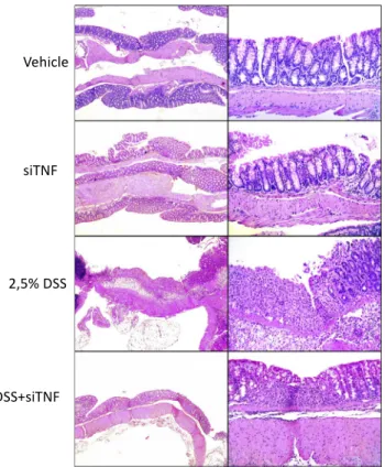
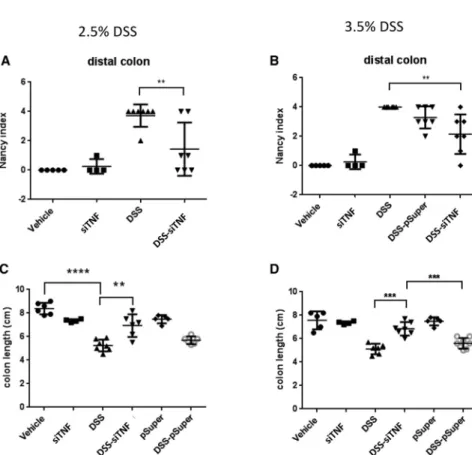
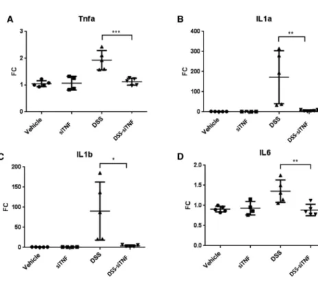
Dextran sulfate sodium (DSS)-induced colitis is one of the most commonly used models of ulcerative colitis (UC).12It induces inflam- matory infiltration, a reactive shortening of the colon, and an increase in the expression of inflammatory mediators. In our experiments, we used two different concentrations, 2.5% and 3.5% DSS, to induce co- lon inflammation of two different levels of severity. (For treatment protocols, see Figure S1A.) As expected, both concentrations of DSS induced a significant increase in the Nancy histological index of UC,13especially in the distal colon (Figures 1,2A, and 2B). (For inflammation scores and ulceration scores separately and changes in the distal colon, see Figure S2.) DSS-induced inflammation is accompanied by a reactive shortening of the colon. Both DSS concen- trations induced a significant, shortening of the colon (Figures 2C and 2D). Finally, we isolated RNA from the whole colon and measured by quantitative real-time PCR the expression level of several proinflam- matory cytokines known to be elevated in IBD.13.5% DSS induced a 2-fold induction of TNF, a 100-fold induction of interleukin (IL)-1a, an approximately 150-fold increase of IL-1b, and a 1.4-fold increase of IL-6 (Figure 3). We also investigated the spatial distribution of
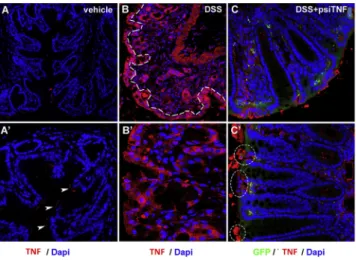
increased TNF production in the colon by immunohistochemistry (IHC). The diffuse pattern of increased expression suggested that TNF was present both in epithelial cells as well as the immune cells, such as macrophages, infiltrating the lamina propria (Figure 4).
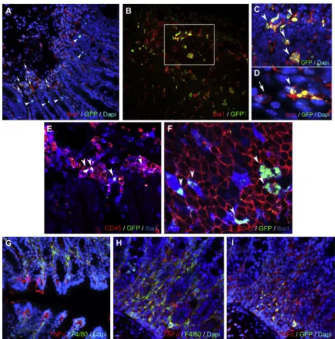
Next, we tested whether the engineered bacteria were able to deliver to the colon the pSuper plasmid and its derivative psiTNF vector, specif- ically designed to silence TNF. In order to do this, the plasmid was engineered to incorporate a mouse pgk promoter-drivengfpreporter whose expression was detected using both specific PCR primers (Fig- ure S3) and IHC (Figure 5). In particular, we assessed the capacity of the engineered bacteria to deliver their cargo to macrophages, a key inflammatory population that sustains colitis. Double staining with anti-GFP and intestinal macrophage marker anti-Iba1 antibodies shows the presence of GFP-expressing psiTNF plasmid in Iba1+mac- rophages in lymphoid aggregates and the lamina propria. GFP was also expressed in mucosal epithelial cells (Figure 5). In order to test the efficacy of the bacterium-delivered gene silencing system, the an- imals with the DSS-induced IBD were then administered theE. coli MDS42 bacteria specifically engineered to silence TNF. This treat- ment had a dramatic effect on the DSS-induced colitis. The increase of inflammatory mediators was significantly reduced. For each of the inflammatory mediators measured, the expression returned to base- line levels (Figure 3). Consequently, the Nancy index of UC was also significantly reduced (Figures 2A and 2B). In the case of 2.5%
DSS treatment the inflammation score returned to baseline or near baseline levels in the distal colon (Figure S2A). The ulceration score increase was also reduced by 64% (Figure S2C). For the higher DSS concentration (3.5%), both the inflammation and ulceration score were reduced by 50% in the distal colon (Figures S2B and S2D).
Finally, the colon length shortening was also significantly reduced to around the level seen in the control animals (Figures 2C and 2D).
As a control, we engineered the same bacteria carrying an empty vec- tor, without the shRNA for TNF. This treatment did not reduce the various measures of inflammation. Similarly, the colon length of the animals treated with only pSuper and psiTNF plasmids (with in- vasion plasmid) showed no significant alteration in comparison with the control (water only) (Figures 2C and 2D). The mucosa thickness did not show any significant change in the proximal or distal colon regions for any of the above listed treatments (data not shown).
We also determined the DAI (disease activity index) value indicating the severity of the inflammatory processes. Again, this was signifi- cantly reduced by the engineered bacteria delivering the TNF-specific shRNA responsible for TNF silencing (Figure 6).
We investigated changes induced by the various experimental condi- tions in the gut microbiome by sequencing the 16S RNA V3 and V4 hypervariable regions. DSS-induced colitis significantly altered the microbiome relative to control (Figures 7A and 7B). The microbiome composition of the DSS-treated and the DSS + pSuper-treated ani- mals were similar. The DSS-induced animals treated with the bacteria carrying the shRNA for TNF, however, showed differences relative to
Figure 1. Histological analysis of the colon obtained from the various experimental groups
Representative histological fields at two magnifications (50 and200) of the various treatment groups as indicated. Vehicle, tap water-administered animal group; siTNF, animals treated with modified E. coli MDS42 strains containing psiTNF and invasive plasmids; DSS, colon specimen of the 2.5% DSS-treated animals; DSS-siTNF, animals administered 2.5% DSS and treated by modified E. coliMDS42 strains containing psiTNF and invasive plasmids.
the disease model, as was demonstrated by a principal component analysis (Figure 7B). These changes were driven by a reduction in the abundance of the predominantly harmful, colitogenic Clostridia, Desulfovibrio, andParabacteroides(Figure 7A).
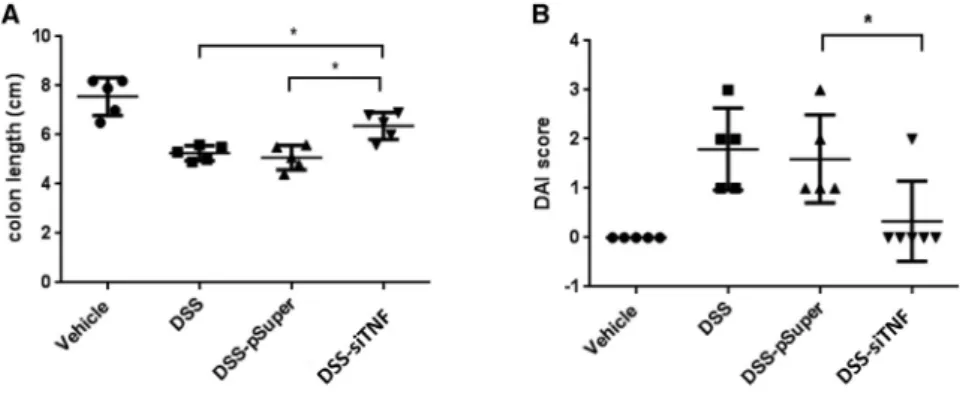
Encouraged by these results we also wanted to test whether the engi- neered bacteria have a similar therapeutic effect when they are applied after the disease had already developed. Animals were pretreated for 4 days with 2.5% DSS alone, and then the same treatment was continued in the presence or absence of the therapeutic administration of an engineeredE. colistrain for 10 days (Figure S1B). The 35%–40%
colon length reduction induced by the DSS treatment was significantly reduced to15% in this treatment regimen as well (Figure 8A). DAI measurements showed similar results. The DSS treatment alone induced a DAI of 1.8, which was reduced to 0.3 by the therapeutic bac- terial treatment (Figure 8B). Bacteria carrying the empty expression vector had no effect on the DSS-induced disease (Figures 8A and 8B).
The therapeutic effect of the engineeredE. coliwas also reflected by the reduced production of inflammatory mediators and increased production of some of the anti-inflammatory cytokines (Figure S4).
Consequently, the Nancy index, the inflammation score, and the ulceration score were significantly reduced (Figure S5).
DISCUSSION
During the past decade, engineered bacteria have emerged as a po- tential “living” vehicle delivering both diagnostic and therapeutic
molecules to the gastrointestinal tract.14 There are several ap- proaches to the therapeutic exploitation of such a biological system in terms of how directly the bacteria are interacting with the host biological system. For example, bacteria were engineered to express phenylalanine-metabolizing enzymes that, upon administration to the digestive tract, would reduce the symptoms of phenylketon- uria.15In this case, the bacteria exert their therapeutic effect inside the colon lumen by breaking down the phenylalanine introduced as part of the diet. Similarly, lactic acid bacteria exerted their therapeu- tic effect by producing elafin inside the gut, thereby reducing the inflammation in the same preclinical model that we used in this work.7In these cases, both the bacteria and the therapeutic effectors produced by them remained in the intestinal lumen and did not invade the colon wall. These approaches, therefore, did not change directly the intracellular levels of disease mediators. Considering the often aggressive and therapy resistant nature of IBD,16we hypoth- esized that suppressing the expression of TNF inside the target cells by delivering shRNA may exert a more dramatic therapeutic effect.
Efficient intracellular silencing of genes by engineered bacteria was first demonstrated by Xiang et al.17usingE. coli-expressing invasin, listeriolysin O cytolysin, and specific shRNA that could silence the b-1 catenin gene in the colon of experimental animals. Spisni et al.6used a strategy to silence cyclooxygenase-2 to treat experi- mental models of IBD by introducing the therapeutic bacteria in en- emas. In the present study, we further adjusted this approach to target one of the key mediators of IBD, TNF, using an orally appli- cable delivery method. Our results suggest that this minimally
Figure 2. The Nancy histological index and colon length of the 2.5% and 3.5% DSS-administered animals
Histological analysis was carried out on formalin-fixed, paraffin-embedded (FFPE) colon samples from experi- mental groups, and the levels of inflammation and ulcer- ation were scored as described inMaterials and Methods.
(A) The Nancy histological index (UC) of the distal colon for the various treatment groups with 2.5% DSS. (B) The Nancy histological index (UC) of the distal colon for the various treatment groups with 3.5% DSS. (C) Effect of the modifiedE. coliMDS42 strains containing psiTNF and invasive plasmids on the colon length in the treatment group with 2.5% DSS. (D) Effect of the modifiedE. coli MDS42 strains containing psiTNF and invasive plasmids on the colon length in the treatment group with 3.5% DSS.
Vehicle: tap water-administered animal group; siTNF, animals treated by modifiedE. coliMDS42 strains con- taining psiTNF and invasive plasmids; DSS, colon spec- imen of the DSS-treated animals; DSS-siTNF, animals administered DSS and treated by modifiedE. coliMDS42 strains containing psiTNF and invasive plasmids; pSuper, animals treated by modifiedE. coliMDS42 strains con- taining pSuper (empty vector) and invasive plasmids;
DSS-pSuper, animals administered 2.5% DSS and treated by modifiedE. coli MDS42 strains containing pSuper (empty vector) and invasive plasmids. Data represent the mean (SD). **p < 0.01, ***p < 0.001, ****p <
0.0001.
invasive direct delivery of therapeutic modulators can cause a signif- icant reduction of inflammation.
An appealing feature of this technology is itsflexibility to target other genes. There are rare forms of IBD, such as the very early onset IBD (VEOIBD), in children.18These patients often fail to respond to stan- dard therapy, but the disease can be linked to well-defined mutations or gene dysfunctions, which could form the basis of alternative ther- apeutic approaches. Since the bacterial delivery technology presented here can either suppress or overexpress specific genes, our delivery method can be engineered to correct specific gene malfunctions.
There are serious safety concerns about the use of live bacteria in the therapeutic setting. Bacteria may exchange genetic material with other bacteria in the colon, which may lead to uncontrollable or diffi- cult to control consequences.19Therefore, in contrast to earlier ef- forts, we used the genetically stable E. coli MDS 42 strain. More than 20% of the genome of this strain, including transposons, was deleted, and the recombination and conjugation abilities of the bacte- ria were eliminated. Thus, the risk of horizontal gene transfers was minimized. This risk could be further reduced by strategies that would introduce mechanisms into the bacterial vectors leading to their self-destruction within a few days in the colon but still able to deliver the therapeutic effect.
MATERIALS AND METHODS
Animals
Adult (8–10 weeks old) male FVB/Ant mice were obtained from the local colony bred at the Medical Gene Technology Unit (specific pathogen-free [SPF] level) at the Institute of Experimental Medicine
Figure 3. mRNA expression levels of proinflammatory cytokines in vehicle treated controls and 3,5% DSS induced colitis with and without treatment with the modified E. coli MDS42 strains containing psiTNF
(A) TNF mRNA expression measured on distal colon samples. (B) IL-1a mRNA expression measured on distal colon samples. (C) IL-1b mRNA expression measured on distal colon samples. (D) IL-6 mRNA expression measured on distal colon samples. Vehicle, tap water- administered animal group; DSS, colon specimen of the 3.5% DSS-treated animals; DSS-siTNF, animals admin- istered 3.5% DSS and treated by modifiedE. coliMDS42 strains containing psiTNF and invasive plasmids. Data represent the means (SD). *p < 0.05, **p < 0.01, ***p <
0.001.
(Budapest, Hungary). Animals were housed at the minimal disease (MD) level, three to five mice per cage, under controlled environmental conditions: temperature, 21C±1C; humidity, 65%; 12-h light/12-h dark cycle; lights on at 7:00
AM. Mice had free access to rodent food and water. All procedures were conducted in accor- dance with the guidelines set by the European Communities Council (86/609/EEC/2 and 2010/63 Directives of European Community), and the protocol was approved by the Institutional Animal Care and Use Committee of the Institute of Experimental Medicine (Buda- pest, Hungary) (permit no. PEI/001/29-4/2013).
UC model generation and animal treatment
In animal models, UC can be induced by DSS administration. The experimental animals were 10-week-old FVB/Ant male mice. Mice were treated with drinking water mixed with 2.5% and 3.5% DSS, which was administered for 8 days or 14 days. The engineered bacte- rial cells were grown in Luria-Bertani (LB) liquid medium at 37C.
The overnight bacterial culture was harvested by centrifugation and washed twice with sterile tap water to eliminate the antibiotic contam- ination (ampicillin and spectinomycin). Finally, the cell pellet was suspended in sterile tap water and administered to the animals. The engineered bacterial cells (2–5 108colony-forming units [CFU]) and vehicle (sterile tap water) were administered daily by oral gavages. During treatment, DAI values were observed. At the end of the experiment, the animals were anesthetized and decapitated.
The length of the colon samples was measured by a digital caliper.
Colon samples were frozen andfixed for histological processing. In- testinal contents were frozen.
Cloning procedures
The nucleotide sequences encoding shRNAS for efficiently silencing the TNF were designed using Serial Cloner 2.6.1 software.
The forward (50-GATCCCCGACAACCAACTAGTGGTGCTTTTCA AGAGAAAGCACCACTAGGTGGTTGTCTTTTTG-30) and reverse
(50-AGCTCAAAAAGACAACCAACTAGTGGTGCTTTCTCTTGA AAAGCACCACTAGTTGGTTGTCGGG-30) synthetic DNA oligonu- cleotides (Microsynth Seqlab, Germany) were heated in annealing buffer (100 mM NaCl, 50 mM HEPES [pH 7.4]) at 90C for 5 min and allowed to cool to room temperature. The annealed double- stranded synthetic DNA oligonucleotides were ligated into the pSUPER.retro.neo GFP vector system (Oligoengine, USA) after linearization with BglII and HindIII enzymes. During the cloning steps, the ligation was heated to 90C for 5 min and allowed to cool to room temperature. Subsequently, conventional heat shock transformation was introduced into the prepared E. coli MDS42 strain. Recombinant bacteria were selected on LB medium containing ampicillin. Plasmid constructs encoding the resulting psiTNF-a (shRNA) precursors were verified by nucleotide sequence assay and restriction enzyme digestion. Using the high-speed plasmid mini kit (Geneaid), a plasmid was isolated from recombinant colonies and digested with EcoRI and HindIII enzymes. The digested plasmids were checked by agarose gel electrophoresis (1% agarose [Sigma], 1Tris-boric acid EDTA buffer [pH 8.5]). The empty vector carries a 1,000-bp-long insert, while the plasmids containing the ligated synthetic oligonucleotides are 281-bp-long inserts that result after double digestion. Plasmids from positive colonies were analyzed by Sanger’s nucleotide assay using a specific sequencing primer (50-GGAAGCCTTGGCTTTTG-30). Theinvasingene of the pGB2-U plasmid was verified by PCR amplification and restriction enzymatic digestion PstI. PstI removes the fragment from the plasmid that con- tains theinvasinandlisteriolysin.
Quantitative real-time PCR
Frozen tissue samples were homogenized in TRI reagent solution (Am- bion, USA), and total RNA was isolated with a QIAGEN RNeasy mini kit (QIAGEN, Valencia, CA, USA) according the manufacturers’in- structions. To eliminate genomic DNA contamination, DNase I treat- ment was used and 100 mL of RNase-free DNase I (1 U of DNase) (Thermo Scientific, USA) solution was added. Sample quality control and the quantitative analysis were carried out by NanoDrop 2000 (Thermo Scientific, USA). Amplification was not detected in the RT- minus controls. The cDNA synthesis was performed with a high-capac- ity cDNA reverse transcription kit (Applied Biosystems, USA). Primers for the comparative Ct experiments were designed by the Primer Ex- press 3.0 program and Primer-BLAST software. The primers (Micro- synth, Balgach, Switzerland) were used in the real-time PCR reaction with Fast EvaGreen qPCR master mix (Biotium, USA) on an ABI Ste- pOnePlus instrument (Applied Biosystems, USA) and are listed inTable S1. The gene expression was analyzed by the ABI StepOne 2.3 program.
The amplicon was tested by melt curve analysis on an ABI StepOnePlus instrument. Experiments were normalized togapdhexpression.
Library construction for Illumina MiSeq sequencing of the 16S rRNA amplicons
Standard library preparation was performed according to the Illumina (San Diego, CA, USA) 16S metagenomic sequencing library preparation protocol (15044223 rev. B). The V3 and V4 hypervariable regions of the bacterial 16S rRNA gene were sequenced with Illumina MiSeq benchtop sequencer generating amplicons of460 by using the universal primer set as follows: 341F (50-CCTACGGGNGGCWGCAG-30) and 785R (50-GACTACHVGGGTATCTAATCC-30) primers flanked by Illu- mina overhang adaptor sequences (forward overhang, 50-TCGTC GGCAGCGTCAGATGTGTATAAGAGACAG-30; reverse overhang, 50-GTCTCGTGGGCTCGGAGATGTGTATAAGAGACAG-30). Af- ter completion of the amplicon PCR with 2KAPA HiFi HotStart ReadyMix, dual indexing of the 96 with adaptor sequences (i7-N7xx, 12 items; i5-S5xx, 8 items) was performed using the Illumina Nextera XT index kit (FC-131-1001/2). PCR cleanups and amplicon size selec- tions were carried out with KAPA Pure Beads (KAPA Biosystems) based on the technical data sheet (KR1245, v3.16) of the manufacturer, resulting infinal580- to 630-bp libraries. Every time, verifications were done with PCR Agilent D1000 screen tapes (5067-5582) and D1000 reagents (5067-5583). The 16S amplicon libraries for each sam- ple were quantified with qPCR, normalized with respect to amplicon sizes, and pooled into a single library in equal molar quantities. Finally, 5mL of the pooled 4 nM DNA library pool was prepared for sequencing on the Illumina MiSeq platform. The library pool was denatured with 0.2 M NaOH and diluted to 8 pMfinal concentration. Sequencing was carried out with MiSeq reagent kit v3 (618 cycle; MS-102-3003) following the manufacturer’s protocols (Illumina, San Diego, CA, USA). All data are publicly available and can be accessed through the PRJNA687617 from the NCBI Sequence Read Archive (SRA).
Sequencing read preparation for downstream analysis
Paired-end reads were demultiplexed by the integrated software of the Illumina MiSeq sequencing machine. The
Figure 4. Epithelial expression of TNF in DSS- and psiTNF-treated mice (A and A0) Mice treated with vehicle show minimal expression of TNF-ain the distal colon, limited to the apical surface of mucosal epithelial cells (A0, arrowheads).
(B and B0) After 7 days of DSS treatment, TNF production increases significantly, where every cell in the epithelial lining of the intestinal mucosa (B, dashed line) shows strong expression of TNF. (C and C0) Animals treated simultaneously with psiTNF plasmid (GFP) and DSS exhibit moderate levels of TNF expression. In contrast to animals not treated with the plasmid, distal colon of psiTNF-treated mice display only non-continuous patches of epithelial TNF expression (C0, en- circled areas).
FastQ files were imported into the QIIME 2 pipeline (https://
qiime2.org/) according to the “Atacama soil microbiome”
tutorial. Residual adaptor sequences (50-CTGTCTCTTATAC ACATCT-30) were trimmed from the 30 end of the reads with Cutadapt software integrated in the QIIME 2 pipeline.
Quality trimming was performed by using DADA2 software,20 and the denoising parameters were set as follows: forward read length was set to 299 bases; for the reverse reads, the length was
Figure 5. Immunohistochemical analysis of the distal colon of DSS-administered (8 days), psiTNF- treated (4 days) animals
(A) Distal colon of DSS-administered (8 days), psiTNF- treated (4 days) animals (mucosa, submucosa): psiTNF plasmid (GFP), Iba1+mucosal macrophages (arrows), and epithelial cells (arrowheads). (B–D) Distal colon of DSS- administered (8 days), psiTNF-treated (4 days) animals (lymphatic aggregate in mucosa): psiTNF plasmid (GFP);
GFP/IBA double-positive macrophages (arrowheads). (C and D) High-magnification images: Iba1+GFP+ (arrow- heads) and Iba1+GFPmacrophages (arrow in D). (E and F) Distal colon of DSS-administered (8 days), psiTNF-treated (4 days) animals (lymphatic aggregate in mucosa): psiTNF plasmid (GFP); GFP is expressed in CD45+Iba1+mucosal macrophages (arrowheads in E); only pycnotic and ramified CD45/Iba1 double-positive macrophages express GFP, whereas CD45+Iba1lymphocytes of round morphology show no plasmid incorporation (F). (G–I) Distal colon of DSS-administered (8 days), psiTNF-treated (4 days) ani- mals (mucosa); psiTNF plasmid (GFP); F4/80+mucosal macrophages and epithelial cells show strong TNF pro- duction (G). (H and I) Expression of TNF and GFP of F4/80+ mucosal macrophages in consecutive sections.
set to 249 bases. No trimming from the start of the reads was applied.
Operational taxonomic unit (OTU) generation Multiple sequence alignment was performed with MAFFT software,21 and reads were taxonomically classified using a naive Bayesian classifier trained with the Greengenes reference database (v13_8) by selecting mapping points according to the forward-reverse primer set that was used for amplifying the 16S V3–V4 regions of the bacterial community (341F, 806R). Aligned sequences were clustered into OTUs with a threshold level of 99% sequence identity. Singletons were discarded in order to reduce the likelihood of sequence artifacts interfering with farther
Figure 6. Effects of DSS-induced colitis on the disease activity index (DAI) of colitis
(A) Effect of the modifiedE. coliMDS42 strains containing psiTNF and invasive plasmids on the DAI. The animals were administered 2.5% DSS. (B) Biological effect of modifiedE. coliMDS42 strains containing psiTNF and invasive plasmids were evaluated considering the DAI of 3.5% DSS-induced colitis. DSS, colon specimen of the DSS-treated animals; DSS-siTNF, animals administered DSS and treated by modifiedE. coliMDS42 strains con- taining psiTNF and invasive plasmids; DSS-pSuper, ani- mals administered DSS and treated by modifiedE. coli MDS42 strains containing pSuper (empty vector) and invasive plasmids. Data represent the mean (SD). *p <
0.05, **p < 0.01.
downstream analysis. A phylogenetic tree was constructed with a FastTree plugin.22
Biodiversity analysis
For sample normalization, 8,300 sequencing (read) depths were set. Beta diversity analysis involved measuring weighted/un- weighted UNIFRAC distances.23For visualization of beta diversity
matrices, principal coordinate analysis (PCoA) plots were gener- ated using the EMPeror24plugin.
Data visualization
For data mining (preparation), the QIIME 2 pipeline was used.
QIIME artifact files were exported from the pipeline resulting BIOMfiles that were converted to TSVfiles, which were used with
Figure 7. Sequencing of the 16S RNA V3 and V4 hypervariable regions
Characterizing microbiome changes by sequencing the bacterial 16S RNA V3 and V4 hypervariable regions in the vehicle treated control, DSS induced colitis, and the DSS induced colitis that were treated with the modifiedE. coliMDS42 strain that either carried an empty plasmid or the psiTNF. (A) the relative abundance of the various bacterial genera; (B) Differences in the microbioime composition as expressed by the weighted principal component analysis (wPcoA).
Figure 8. Effect of the 4 + 10 days of 2.5% DSS- induced colitis on colon length and the DAI of colitis (A) Effect of the modifiedE. coliMDS42 strains containing psiTNF and invasive plasmids on colon length. The animals were administered 2.5% DSS. (B) Biological effect of modifiedE. coli MDS42 strains containing psiTNF and invasive plasmids were evaluated considering the DAI of 2.5% DSS-induced colitis. DSS, colon specimen of the DSS-treated animals; DSS-siTNF, animals administered DSS and treated by modifiedE. coliMDS42 strains con- taining psiTNF and invasive plasmids; DSS-pSuper, animals administered DSS and treated by modifiedE. coliMDS42 strains containing pSuper (empty vector) and invasive plas- mids. Data represent the mean (SD). *p < 0.05, **p < 0.01.
external visualization packages. Pie charts were constructed with R programming language (https://www.R-project.org)25 using the ggplot2 package (http://ggplot2.org).26
GFP tracing in the colon
Total RNA was isolated and cDNA synthesis was performed from the frozen colon samples. The gfpexpression in the colon tissue was demonstrated by PCR using specific oligonucleotide primers (for- ward, 50-GGA CGA CGG CAA CTA CAA GA-30; reverse, 50-AAG TCG ATG CCC TTC AGC TC-30). The PCR product was separated and stained on agarose gel.
Pathology (histology)
Formalin-fixed colon samples were processed and embedded in paraffin using the standard protocol. Sections of 4mm were stained with hematoxylin and eosin. Slides were analyzed by an expert pathologist in a blinded manner using the Nancy histological index for UC.13
Histological procedures
For immunofluorescence (IF), distal colon samples of adult (8- to 10-week-old) FVB/Ant mice, treated with 2.5% DSS and psiTNF-a plasmid, werefixed in 4% paraformaldehyde (PFA) in PBS for 24 h.
After extensive washing in PBS, the samples were infiltrated with 15% sucrose/PBS overnight at 4C. The medium was changed to 7.5% gelatin containing 15% sucrose at 37C for 2 h, and the tissues were rapidly frozen at60C in isopentane (Sigma). Frozen sections were cut at 10mm with a Shandon cryostat for confocal laser-scanning microscopy, collected on poly-L-lysine-coated slides (Sigma).
Immunofluorescence and image analysis
IF was performed as previously described.27Briefly, primary anti- bodies anti-GFP (R&D Systems), anti-TNF-a (Abcam), anti-Iba1 (Invitrogen), and anti- F4/80 (Invitrogen) were diluted 1:200 in 1%
PBS-BSA. For IF, frozen sections were incubated with primary anti- bodies overnight at 4C, followed by relevant secondary antibodies (Alexa Fluor 647-, 546-, and 488-conjugated anti-rabbit immuno- globulin G [IgG] and Alexa Fluor 488-conjugated anti-goat IgG, from Invitrogen) used for 1 h at room temperature (RT). Cell nuclei were visualized by DAPI (40,6-diamidino-2-phenylindole; Vector Laboratories). Sections were covered with aqueous Poly/Mount (Pol- ysciences, Warrington, PA, USA) and examined with a Zeiss LSM 780 laser-scanning confocal microscope (Zeiss). The ZEN software pack- age (black edition, Zeiss) was used for the scanning of 4-mm and 1.8-mm-thick optical slices of 10 megapixel (MP) resolution for low- and high-magnification images, respectively. Images were compiled with ImageJ and Adobe Photoshop CS6 software packages.
Statistical analysis
Data are expressed as means±SD. The data werefirst subjected to a Kolmogorov-Smirnov normality test. Data passing this test were analyzed by one-way ANOVA followed by the Tukey’s post hoc test. When the data showed non-Gaussian distribution, the Krus- kal-Wallis test was used. Statistical analysis was performed using
GraphPad Prism version 6 software (GraphPad, USA). p %0.05 was considered significant.
SUPPLEMENTAL INFORMATION
Supplemental Information can be found online athttps://doi.org/10.
1016/j.omtm.2020.11.010.
ACKNOWLEDGMENTS
This work was supported by the National Research Development and Innovation Office Hungary (grants KFI_16-1-2017-0245, 109622 and 124424 to K. J. K., and 109744 to S. F.).
AUTHOR CONTRIBUTIONS
Conception and design: S.F., Z.S., and K.J.K. Development of method- ology: S.F., D.K., Z.W., and D.D. Acquisition of data: S.F., N. Sze}ocs, Z.G., D.K., B.J., T.P., F.S., and D.D. Analysis and interpretation of data (e.g., statistical analysis, biostatistics, computational analysis): S.F., N.
Solymosi, Z.S., I.H., Z.G., A.S., M.P., D.D., N.N, K.J.K., and F.S.
Writing, review, revision of the manuscript: S.F., N. Solymosi, I.Z., Z.S., D.D., and K.J.K.
DECLARATION OF INTERESTS The authors declare no competing interests.
REFERENCES
1.Neurath, M.F. (2014). Cytokines in inflammatory bowel disease. Nat. Rev. Immunol.
14, 329–342.
2.Lamb, C.A., Kennedy, N.A., Raine, T., Hendy, P.A., Smith, P.J., Limdi, J.K., Hayee, B., Lomer, M.C.E., Parkes, G.C., Selinger, C., et al.; IBD guidelines eDelphi consensus group (2019). British Society of Gastroenterology consensus guidelines on the man- agement of inflammatory bowel disease in adults. Gut68(Suppl 3), s1–s106.
3.Papamichael, K., Lin, S., Moore, M., Papaioannou, G., Sattler, L., and Cheifetz, A.S.
(2019). Infliximab in inflammatory bowel disease. Ther. Adv. Chronic Dis.10, 2040622319838443.
4.Pagnini, C., Pizarro, T.T., and Cominelli, F. (2019). Novel pharmacological therapy in inflammatory bowel diseases: beyond anti-tumor necrosis factor. Front. Pharmacol.
10, 671.
5.Feagan, B.G., Rutgeerts, P., Sands, B.E., Hanauer, S., Colombel, J.-F., Sandborn, W.J., Van Assche, G., Axler, J., Kim, H.-J., Danese, S., et al.; GEMINI 1 Study Group (2013).
Vedolizumab as induction and maintenance therapy for ulcerative colitis. N. Engl. J.
Med.369, 699–710.
6.Spisni, E., Valerii, M.C., De Fazio, L., Cavazza, E., Borsetti, F., Sgromo, A., Candela, M., Centanni, M., Rizello, F., and Strillacci, A. (2015). Cyclooxygenase-2 silencing for the treatment of colitis: a combined in vivo strategy based on RNA interference and engineeredEscherichia coli. Mol. Ther.23, 278–289.
7.Motta, J.-P., Bermúdez-Humarán, L.G., Deraison, C., Martin, L., Rolland, C., Rousset, P., Boue, J., Dietrich, G., Chapman, K., Kharrat, P., et al. (2012). Food-grade bacteria expressing elafin protect against inflammation and restore colon homeostasis. Sci.
Transl. Med.4, 158ra144.
8.Umenhoffer, K., Fehér, T., Balikó, G., Ayaydin, F., Pósfai, J., Blattner, F.R., and Pósfai, G. (2010). Reduced evolvability ofEscherichia coliMDS42, an IS-less cellular chassis for molecular and synthetic biology applications. Microb. Cell Fact.9, 38.
9.Pósfai, G., Plunkett, G., 3rd, Fehér, T., Frisch, D., Keil, G.M., Umenhoffer, K., Kolisnychenko, V., Stahl, B., Sharma, S.S., de Arruda, M., et al. (2006). Emergent properties of reduced-genomeEscherichia coli. Science312, 1044–1046.
10.Young, V.B., Falkow, S., and Schoolnik, G.K. (1992). The invasin protein of Yersinia enterocolitica: internalization of invasin-bearing bacteria by eukaryotic cells is asso- ciated with reorganization of the cytoskeleton. J. Cell Biol.116, 197–207.
11.Grillot-Courvalin, C., Goussard, S., Huetz, F., Ojcius, D.M., and Courvalin, P. (1998).
Functional gene transfer from intracellular bacteria to mammalian cells. Nat.
Biotechnol.16, 862–866.
12.Perse, M., and Cerar, A. (2012). Dextran sodium sulphate colitis mouse model: traps and tricks. J. Biomed. Biotechnol.2012, 718617.
13.Marchal-Bressenot, A., Salleron, J., Boulagnon-Rombi, C., Bastien, C., Cahn, V., Cadiot, G., Diebold, M.-D., Danese, S., Reinisch, W., Schreiber, S., et al. (2017).
Development and validation of the Nancy histological index for UC. Gut66, 43–49.
14.Riglar, D.T., and Silver, P.A. (2018). Engineering bacteria for diagnostic and thera- peutic applications. Nat. Rev. Microbiol.16, 214–225.
15.Isabella, V.M., Ha, B.N., Castillo, M.J., Lubkowicz, D.J., Rowe, S.E., Millet, Y.A., Anderson, C.L., Li, N., Fisher, A.B., West, K.A., et al. (2018). Development of a syn- thetic live bacterial therapeutic for the human metabolic disease phenylketonuria.
Nat. Biotechnol.36, 857–864.
16.Nielsen, O.H., and Ainsworth, M.A. (2013). Tumor necrosis factor inhibitors for in- flammatory bowel disease. N. Engl. J. Med.369, 754–762.
17.Xiang, S., Fruehauf, J., and Li, C.J. (2006). Short hairpin RNA-expressing bacteria elicit RNA interference in mammals. Nat. Biotechnol.24, 697–702.
18.Moran, C.J., Klein, C., Muise, A.M., and Snapper, S.B. (2015). Very early-onset in- flammatory bowel disease: gaining insight through focused discovery. Inflamm.
Bowel Dis.21, 1166–1175.
19.Wegmann, U., Carvalho, A.L., Stocks, M., and Carding, S.R. (2017). Use of genetically modified bacteria for drug delivery in humans: revisiting the safety aspect. Sci. Rep.7, 2294.
20.Callahan, B.J., McMurdie, P.J., Rosen, M.J., Han, A.W., Johnson, A.J.A., and Holmes, S.P. (2016). DADA2: high-resolution sample inference from Illumina amplicon data.
Nat. Methods13, 581–583.
21.Katoh, K., and Standley, D.M. (2013). MAFFT multiple sequence alignment software version 7: improvements in performance and usability. Mol. Biol. Evol.30, 772–780.
22.Price, M.N., Dehal, P.S., and Arkin, A.P. (2010). FastTree 2—approximately maximum-likelihood trees for large alignments. PLoS ONE5, e9490.
23.Lozupone, C., and Knight, R. (2005). UniFrac: a new phylogenetic method for comparing microbial communities. Appl. Environ. Microbiol.71, 8228–8235.
24.Vázquez-Baeza, Y., Pirrung, M., Gonzalez, A., and Knight, R. (2013). EMPeror: a tool for visualizing high-throughput microbial community data. Gigascience2, 16.
25.R Core Team (2014). R: A Language and Environment for Statistical Computing (R Foundation for Statistical Computing).
26.Wickham, H. (2016). ggplot2: Elegant Graphics for Data Analysis, Second Edition (Springer).
27.Dora, D., Arciero, E., Hotta, R., Barad, C., Bhave, S., Kovacs, T., Balic, A., Goldstein, A.M., and Nagy, N. (2018). Intraganglionic macrophages: a new population of cells in the enteric ganglia. J. Anat.233, 401–410.