Ductular reaction correlates with fibrogenesis but does not contribute to liver regeneration in experimental fibrosis models
Andra´s Ro´ kusz1, Da´niel Veres2, Armanda Szu¨ cs1, Edina Bugyik1, Miklo´ s Mo´ zes3, Sa´ndor Paku1,4, Pe´ter Nagy1☯*, Katalin Dezső1☯
1 First Department of Pathology and Experimental Cancer Research, Semmelweis University, Budapest, Hungary, 2 Department of Biophysics and Radiation Biology, Semmelweis University, Budapest, Hungary, 3 Institute of Pathophysiology, Semmelweis University, Budapest, Hungary, 4 Tumor Progression Research Group, Joint Research Organization of the Hungarian Academy of Sciences and Semmelweis University, Budapest, Hungary
☯These authors contributed equally to this work.
*pdrnagy@gmail.com
Abstract
Background and aims
Ductular reaction is a standard component of fibrotic liver tissue but its function is largely unknown. It is supposed to interact with the matrix producing myofibroblasts and compen- sate the declining regenerative capacity of hepatocytes. The relationship between the extent of fibrosis—ductular reaction, proliferative activity of hepatocytes and ductular reaction were studied sequentially in experimental hepatic fibrosis models.
Methods
Liver fibrosis/cirrhosis was induced in wild type and TGFβoverproducing transgenic mice by carbon tetrachloride and thioacetamide administration. The effect of thioacetamide was modulated by treatment with imatinib and erlotinib. The extent of ductular reaction and fibro- sis was measured by morphometry following cytokeratin 19 immunofluorescent labeling and Picro Sirius staining respectively. The proliferative activity of hepatocytes and ductular reac- tion was evaluated by BrdU incorporation. The temporal distribution of the parameters was followed and compared within and between different experimental groups.
Results
There was a strong significant correlation between the extent of fibrosis and ductular reac- tion in each experimental group. Although imatinib and erlotinib temporarily decreased fibro- sis this effect later disappeared. We could not observe negative correlation between the proliferation of hepatocytes and ductular reaction in any of the investigated models.
Conclusions
The stringent connection between ductular reaction and fibrosis, which cannot be influenced by any of our treatment regimens, suggests that there is a close mutual interaction between a1111111111
a1111111111 a1111111111 a1111111111 a1111111111
OPEN ACCESS
Citation: Ro´kusz A, Veres D, Szu¨cs A, Bugyik E, Mo´zes M, Paku S, et al. (2017) Ductular reaction correlates with fibrogenesis but does not contribute to liver regeneration in experimental fibrosis models. PLoS ONE 12(4): e0176518.
https://doi.org/10.1371/journal.pone.0176518 Editor: Matias A Avila, University of Navarra School of Medicine and Center for Applied Medical Research (CIMA), SPAIN
Received: March 3, 2017 Accepted: April 12, 2017 Published: April 26, 2017
Copyright:©2017 Ro´kusz et al. This is an open access article distributed under the terms of the Creative Commons Attribution License, which permits unrestricted use, distribution, and reproduction in any medium, provided the original author and source are credited.
Data Availability Statement: All relevant data are within the paper and its Supporting Information files.
Funding: This work was supported by the Hungarian Scientific Research Fund (OTKA K116301 and PD109201), Ja´nos Bolyai Research Scholarship of the Hungarian Academy of Sciences, also by the European Union and the State of Hungary, co-financed by the European Social
them instead of a unidirectional causal relationship. Our results confirm a close connection between DR and fibrogenesis. However, since the two parameters changed together we could not establish a causal relationship and were unable to reveal which was the primary event. The lack of inverse correlation between the proliferation of hepatocytes and ductular reaction questions that ductular reaction can compensate for the failing regenerative activity of hepatocytes. No evidences support the persistent antifibrotic property of imatinib or erlotinib.
Introduction
Chronic damage of liver tissue causes gradual accumulation of extracellular matrix (ECM), fibrosis, which can eventually progress to complete architectural reconstruction termed cirrho- sis. Hepatic fibrosis/cirrhosis causes organ dysfunction and other complications resulting in common clinical problems. Yet, there are numerous unresolved questions regarding the path- ogenesis of this severe disease, e.g. the role and function of hepatic progenitor cells in fibrogen- esis and regeneration. Small epithelial tubules called “bile duct proliferation” were observed along the fibrotic septa of cirrhotic livers a long time ago but no special attention was paid to them. Now, these tubules are referred to as ductular reaction and they are thought to represent hepatic progenitor cells [1]. Their closed spatial and functional relationship with myofibro- blasts put them in the limelight [2–5], since myofibroblasts are the major source of the depos- ited collagenous matrix. The correlation between the extent of ductular reaction and fibrosis across a range of liver pathologies raises the question if there is a causal relationship between them [6]. Ductular reaction may also play a favourable role in liver cirrhosis. Originally Falk- owski et al. [7] proposed that the ductular reaction may be an alternative regenerative pathway, which is activated when the replicative capacity of the senescent hepatocytes is compromised.
This view was later supported by the description of hepatocytic differentiation of ductular pro- genitor cells [8,9]. This simple and attractive model, however, is mostly based on static tissue analysis and contradictory observations have recently emerged. The progenitor cell origin of regenerating hepatocytes was excluded in several experimental models applying the cre-lox based lineage tracing technique [10–12]. Others and we [13,14] failed to determine inverse relationship between the proliferative activity of hepatocytes and ductular reaction in human cirrhotic livers. These considerations led us to perform experiments where the dynamics of fibrosis, ductular reaction and hepatocyte proliferation can be continually monitored through- out the development of cirrhosis. Furthermore, our aim was to examine if the potentially favourable and unfavourable consequences of ductular reaction can be separated.
Liver fibrosis was induced in wild type C57Bl/6 mice by chronic administration of thioace- tamide (TA) and carbon tetrachloride/phenobarbital (CCl4/PhB). The TA experiments were also performed on transgenic mice overexpressing active transforming growth factor beta 1 (TGFβ1) in the liver [15]. TGFβis probably the most pleiotropic growth factor with major influence on hepatocyte and ductular proliferation, as well as on liver fibrosis [16].
The TA administration was also combined with two drugs. Imatinib and erlotinib are widely used tyrosine kinase inhibitors. The primary target of erlotinib is epidermal growth fac- tor receptor (EGFR) while imatinib has a broader spectrum. It was originally designed for the treatment of chronic myelogenous leukaemia by blocking the activity of bcr/abl tyrosine kinase but turned out to be an efficient inhibitor of c-kit and platelet-derived growth factor receptor (PDGFR) as well [17]. Both compounds are used for the treatment of different
Fund in the framework of TA´MOP 4.2.4. A/1-11-1- 2012-0001 ‘National Excellence Program’.
Competing interests: The authors have declared that no competing interests exist.
Abbreviations: BrdU, bromodeoxyuridine; CCl4, carbon tetrachloride; CDE, choline-deficient, ethionine-supplemented diet; DEN,
diethylnitrosamine; DMSO, dimethyl sulfoxide;
ECM, extracellular matrix; EGFR, epidermal growth factor receptor; EMT, epithelial-to-mesenchymal transition; PDGFR, platelet-derived growth factor;
PhB, phenobarbital; TA, thioacetamide; TGFβ, transforming growth factor beta.
malignant tumors and imatinib has been successfully applied in human patients as an antifi- brotic agent [18]. They have also shown antifibrotic activity in several experimental hepatic fibrosis models [19–22], but little is known about their effect on the behavior of ductular reac- tion and hepatocytes.
All fibrogenic protocols induced progressive hepatic fibrosis. There was a strong positive correlation between the extent of ductular reaction and fibrosis in each model. However, the compensatory growth function of ductular reaction could not be observed. Both imatinib and erlotinib had temporary antifibrotic effect but eventually, and under adverse conditions (TGFβoverexpression), they were inefficient. Neither drug had antifibrotic effect on estab- lished cirrhosis induced by TA treatment.
Materials and methods Animal experiments
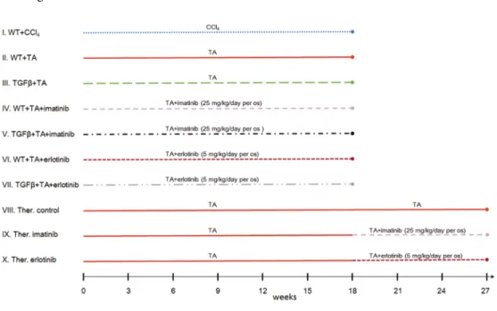
All experiments were conducted on 8 weeks old male C57Bl/6 mice inbred in our Institute. All animals were housed under controlled temperature with a 12 hour light-dark cycle, and had access to drinking water ad libitum. Ten experimental groups were formed (Fig 1):
Basic models:
I. Hepatic fibrosis was induced by carbon tetrachloride/phenobarbital treatment (CCl4
0,2ml/kg, dissolved in sunflower oil, twice a week per os/PhB 0,5g/l in drinking water) in wild type mice (WT+CCl4; n = 49),
II. or by TA administration (300 mg/l in drinking water) in wild type (WT+TA; n = 50) and
III. transgenic mice overexpressing active TGFβin hepatocytes [15] (TGFβ+TA; n = 61) (the mice were a kind gift from Snorri S. Thorgeirsson).
Drug treated models:
Fig 1. Schematic representation of the different experimental groups.
https://doi.org/10.1371/journal.pone.0176518.g001
IV. Imatinib (Glivec, Novartis, Basel; 25 mg/kg, dissolved in water, per os) was given daily to TA treated wild type mice (WT+TA+imatinib; n = 54) or
V. TGFβtransgenic mice (TGFβ+TA+imatinib; n = 38).
VI. Erlotinib (Tarceva, Roche, Basel; 5 mg/kg, dissolved in dimethyl sulfoxide (DMSO) then water) was given daily, beside TA treatment, to both wild type (WT+TA+- erlotinib; n = 48) and
VII. TGFβtransgenic mice (TGFβ+TA+erlotinib; n = 38).
The animals were sacrificed after 3, 6, 9, 12, 15 and 18 weeks of treatment. Each time point represents 5–16 animals.
Therapeutic models:
VIII. TA (300 mg/l in drinking water) was given to wild type mice for a maximum of 27 weeks (Ther. control; n = 18).
IX. Imatinib treatment (as described above) was started on the 19th week in addition to TA (Ther. imatinib; n = 18).
X. Erlotinib treatment was started on the 19th week (as described above) in addition to TA (Ther. erlotinib; n = 17).
Animals were sacrificed at 3, 6 and 9 weeks after the initiation of pharmacological treatment (in other words on the 21st, 24thand 27thweek of the experiment). Each time point represents 5–7 animals.
Each animal received three doses of bromodeoxyuridine (BrdU; 500 mg/kg) intraperito- neally, 20, 2 and 1 hour before termination. After humanely sacrificing the animals using cervical dislocation, samples from the liver were fixed for histological analysis, the rest was snap-frozen in liquid nitrogen. The animal study protocols were conducted according to National Institute of Health (NIH) guidelines for animal care and were approved by the Institutional Animal Care and Use Committee of Semmelweis University (Permit Number:
PEI/001/1730-12/2015).
Morphometric analysis
From each animal four parameters were measured/counted: the extent of fibrosis and ductular reaction, as well as the proliferative activity of hepatocytes and ductular reaction.
For the morphometric analysis of fibrosis three images from Picro Sirius stained sections were captured with a Zeiss Axioskop 2 plus microscope (Zeiss, Oberkochen, Germany) using a 5x objective and evaluated by the Quick PhotoMicro 2.2 software (Promicra, Prague, Czech Republic).
The area occupied by ductular reaction was measured on cytokeratin 19 (CK19) immunos- tained frozen sections (rat monoclonal anti-CK 19 antibody; cat. no. TROMA-III; Develop- mental Studies Hybridoma Bank, Iowa City, IA; dil.:1:200). From each liver three images were captured with a Bio-Rad confocal system (MRC 1024; Bio-Rad, Richmond, CA) and evaluated by the ImageJ 1.49k program (NIH, Bethesda, MD).
The incorporated BrdU was immunostained (mouse monoclonal anti-BrdU antibody; cat.
no.: 347580; BD Biosciences, Franklin Lakes, NJ; dil.: 1:20) as described before [23]. 5000 hepa- tocytes and 500 ductular cells were counted, the percentage of BrdU-positive cells was given as a result.
Statistical analysis
Statistical analysis was performed with StatSoft Statistica software (StatSoft Inc., Tulsa, OK;
version 8.0). The deviation from Gaussian distribution of variables was tested with Kolmogo- rov-Smirnov and Lilliefors’ method. The normality condition was fulfilled for all the 4 vari- ables in each group, therefore 2-way factorial ANOVA was performed with a Tukey-Kramer HSD test (with unequal sample size). Correlation between variables was tested using Spearman rank correlation test (a few equal values were in the dataset) and Spearman correlation coeffi- cients were determined. Results were considered significant at a p value less than or equal to 0,05. For characterising the differences between groups both means and medians were calcu- lated and data were visualized on scatter and box plots.
Results Basic models
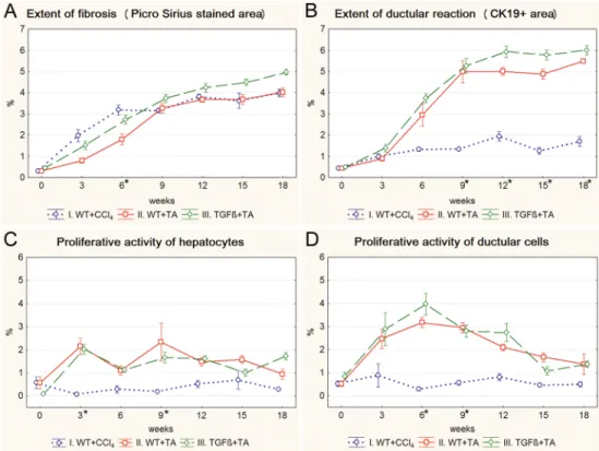
The fibrosis was progressive in each model and completely circumscribed pseudolobules were formed by the end of the experiment (S1 Fig). The advancement was faster at the begin- ning and then slowed down, especially in the CCl4treated mice (Fig 2A). As it was expected, the fibrosis was more severe at each time point in the TGFβ+TA group compared to wild type mice (WT+TA group) (Fig 2A). The extent of ductular reaction, quantitated by the mea- surement of CK19-positive area, was in line with the Picro Sirius data in the TA models
Fig 2. Results of the basic models (I. WT+CCl4, II. WT+TA, III. TGFβ+TA). Data are represented as means±standard error of the mean (SEM).*marks time points, where there was a significant difference between the results of the WT+CCl4and WT+TA groups. The extent of fibrosis is significantly higher in the WT+CCl4group on the 6thweek of treatment (A), while the extent of ductular recation is significantly lower from the 9thweek until the end of the experiment (B). The proliferative activity of hepatocytes (C) and ductular reaction (D) is significantly lower on the 3rdand 9th, or on the 6thand 9th week of the experiment, respectively.
https://doi.org/10.1371/journal.pone.0176518.g002
(Fig 2A and 2BandS2 Fig). It showed a slightly increasing trend in the CCl4treated mice as well but from the 9thweek it was significantly lower compared to the TA groups. Interest- ingly, the extent of ductular reaction was similar in the CCl4and TA treated wild type mice on the 3rdweek, despite the difference in the amount of fibrosis. There was a divergence between the TA and CCl4treated animals in the cell proliferation data as well. In the TA groups, a temporary rise in both the proliferation of hepatocytes and ductular reaction was followed by a gradual decline. CCl4induced much lower levels of cell proliferation with a rel- atively stable trend in both cell types (Fig 2C and 2D).
Drug treated models
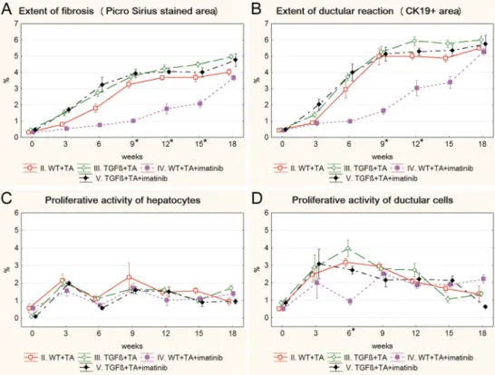
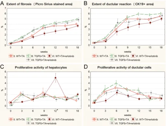
Both imatinib and erlotinib suppressed the progression of fibrosis and ductular reaction tem- porarily in wild type mice (Figs3A, 3B,4A and 4B). Imatinib treatment resulted in a signifi- cant decrease in the extent of fibrosis on the 9th, 12thand 15thweek (Fig 3A), and in the extent of ductular reaction on the 9thand 12thweek of the experiment (Fig 3B) (S3andS4Figs). The effect of erlotinib did not reach significant levels in any of the time points (Fig 4A and 4B).
However, at the end of the experiment, due to a compensatory growth, there was no difference in the degree of fibrosis or ductular reaction between the three groups of TA treated wild type mice. Not even a temporary inhibition as described above could be observed in the TGFβ transgenic mice (S5andS6Figs). None of the applied drugs had a lasting influence on the pro- liferative activity of hepatocytes or ductular reaction (Figs3C, 3D,4C and 4D).
Fig 3. Results of the imatinib treated groups (IV. WT+TA+imatinib, V. TGFβ+TA+imatinib) and their control groups (II. WT+TA, III. TGFβ+TA). Data are represented as means±standard error of the mean (SEM).*marks time points, where there was a significant difference between the results of the WT+TA and WT+TA+imatinib groups. Imatinib treatment temporarily resulted in significantly lower extent of fibrosis (A, 9th, 12th, 15thweek) and ductular reaction (B, 9thand 12thweek) in wild type mice. The proliferative activity of ductular reaction was also significantly lower on the 6thweek of the experiment in imatinib treated wild type mice (D). Imatinib treatment did not have any significant effect on TGFβtransgenic mice.
https://doi.org/10.1371/journal.pone.0176518.g003
Therapeutic models
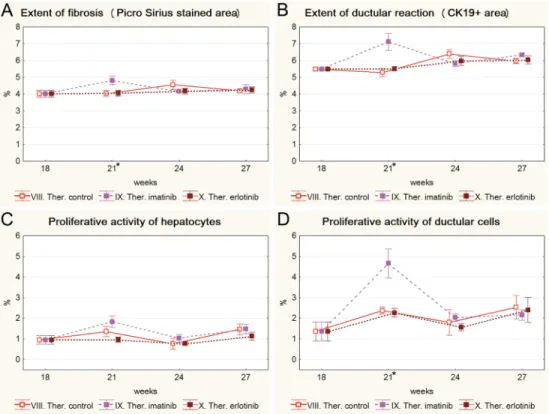
Both imatinib and erlotinib were used in a “therapeutic” experiment to simulate a clinical situ- ation and test if these drugs can influence preexistent fibrosis. The administration of both tyro- sine kinase inhibitors was started after 18 weeks of TA treatment and was continued further on (S7andS8Figs). Neither of the drugs were able to block the progression of fibrosis (Fig 5A).
Interestingly, imatinib even induced a significant temporary increase in the extent of Picro Sir- ius staining, and the extent and proliferative activity of ductular reaction (Fig 5A, 5B and 5D).
Correlation analysis
To investigate the relationship between the studied parameters without considering the time- wise distribution of the data, a correlation analysis was performed between each pair of param- eters in the different experimental groups. The results are shown inTable 1. The extent of fibrosis showed a strong positive correlation with the extent of ductular reaction in each model (S9 Fig). This correlation was the weakest in the CCl4group. Surprisingly, the proliferative activity of ductular reaction correlated negatively in three groups with Picro Sirius (S10A, S10B and S10D Fig) and in one group with CK19 staining (S11A Fig) indicating an inverse relationship between the extent of fibrosis/ductular reaction and the proliferative activity of ductular reaction. Contrary to our expectations, the proliferation of hepatocytes correlated positively in two groups with Picro Sirius (S10E and S10F Fig) and CK19 (S11C and S11D Fig)
Fig 4. Results of the erlotinib treated groups (VI. WT+TA+erlotinib, VII. TGFβ+TA+erlotinib) and their control groups (II. WT+TA, III. TGFβ+TA). Data are represented as means±standard error of the mean (SEM).*marks the time point, where there was a significant difference between the results of the WT+TA and WT+TA+erlotinib groups. Erlotinib treatment resulted in significantly higher hepatocyte proliferation on the 12thweek of the experiment (C). Erlotinib treatment did not have any significant effect on TGFβtransgenic mice.
https://doi.org/10.1371/journal.pone.0176518.g004
Fig 5. Results of the therapeutic experiment (VIII. Ther. control, IX. Ther. imatinib, X. Ther. erlotinib).
Data are represented as means±standard error of the mean (SEM).*marks the time points, where there was a significant difference between the results of the Ther. imatinib and Ther. control groups. Imatinib treatment resulted in significantly higher extent of fibrosis (A); extent (B), and proliferative activity (D) of ductular reaction on the 21stweek of the experiment (after 3 weeks of imatinib treatment). Erlotinib treatment did not have any significant effect on livers with established fibrosis.
https://doi.org/10.1371/journal.pone.0176518.g005
Table 1. Correlation between the analysed parameters.
Picro- CK19
p Picro-
hep
p Picro-
duct
p CK19-hep p CK19-duct p hep-
duct p
I. WT+CCl4(n = 49) 0.66 <0.001 0.48 <0.001 0.22 ns 0.37 0.009 0.24 ns 0.23 ns
II. WT+TA (n = 50) 0.83 <0.001 0.13 ns -0.33 0.018 0.18 ns -0.12 ns 0.02 ns
III. TGFβ+TA (n = 61) 0.81 <0.001 0.03 ns -0.56 <0.001 0.01 ns -0.41 <0.001 0.09 ns
IV. WT+TA+imatinib (n = 54)
0.94 <0.001 0.19 ns 0.31 0.025 0.17 ns 0.33 0.015 0.32 0.002
V. TGFβ+TA+imatinib (n = 38)
0.81 <0.001 -0.11 ns -0.46 0.004 -0.09 ns -0.32 ns 0.12 ns
VI. WT+TA+erlotinib (n = 48)
0.92 <0.001 0.53 <0.001 0.03 ns 0.55 <0.001 0.13 ns 0.09 ns
VII. TGFβ+TA+erlotinib (n = 38)
0.89 <0.001 -0.01 ns -0.2 ns -0.01 ns -0.28 ns -0.29 ns
VIII. Ther. control (n = 18) 0.61 <0.001 -0.33 ns 0.03 ns -0.34 ns -0.37 ns 0.3 ns
IX. Ther. imatinib (n = 18) 0.63 <0.001 0.2 ns 0.37 ns 0.4 ns 0.26 ns 0.47 0.048
X. Ther. erlotinib (n = 17) 0.81 <0.001 0.02 ns 0.09 ns -0.02 ns -0.13 ns 0.33 ns
The values represent Spearman’s correlation coefficients (rs). ns—not significant; Picro—extent of fibrosis; CK19 –extent of ductular reaction; hep—
proliferative activity of hepatocytes, duct—proliferative activity of ductular reaction.
https://doi.org/10.1371/journal.pone.0176518.t001
staining or the proliferative activity of ductular reaction (S11E and S11F Fig). Furthermore, no negative correlation was found in any of the groups between the proliferation of hepatocytes and the extent or proliferation of the ductular reaction.
Discussion
Liver fibrosis/cirrhosis was induced in mice by the two widely used fibrogenic agents, TA and CCl4. The TA model was diversified to modulate the histological reaction. Each experimental groups reached the cirrhotic stage. The primary purpose of the experiment was to analyse the dynamics and relationship of three important components of the fibrotic process: (i) the extent of fibrosis, (ii) the proliferation of hepatocytes and (iii) the extent and proliferative activity of ductular reaction. Morphometric analysis of the extent of fibrosis is more reliable for the evalu- ation of fibrosis progression than the use of staging systems [24]; hence we quantitated the Picro Sirius stained slides. The extent of the ductular reaction was followed by CK19 immu- nostaining. Cell proliferation was examined by the immunohistochemical detection of BrdU- positive cells.
The results were evaluated in two different ways. The dynamics of timewise changes in the various parameters were visualized on 2D graphs, while the connection between any two parameters within each group was studied by correlation analysis regardless of the time point when the data were collected. This latter kind of analysis is similar to human investigations, when only the actual parameters can be examined.
The basic concept to be addressed is that, with the advancement of fibrosis, the regenerative activity of the hepatocytes declines but it is compensated by the contribution of ductular reaction.
In each experimental model, there is a strong correlation between the extent of fibrosis and ductular reaction. This observation is in line with previous results of a wide variety of human studies [2,25–27]. Surprisingly, CCl4induces a much milder ductular reaction than TA but the extent of fibrosis is comparable in the two models. That is, one “unit” of ductular reaction in the CCl4experiment is associated with a larger amount of fibrosis. Therefore, although the extent of fibrosis and ductular reaction correlate, their ratio depends on the initiating event.
Interestingly, we could not detect correlation between the extent of ductular reaction and etiol- ogy in human cirrhotic livers but the fibrotic area was larger in non-viral hepatitis than in viral hepatitis related cases [14], which also indicates that the ratio of ductular reaction and fibrosis varies according to the etiology. Ductular reaction can potentially contribute to fibrogenesis by transition into myofibroblasts (epithelial-to-mesenchymal transition—EMT) [28] but this aspect has not been addressed in the present study.
Another important and unresolved issue is which event is the primary one and which one is the consequence? Van Hul et al. [3] described that the increased expression of matrix com- ponents preceded the elevation of ductular markers with 4–7 days. Human observations in NASH [26], HCV infection related fibrosis [2,29] and in vivo experimental models [30,31]
suggest that ductular reaction drives fibrosis, while others [3,32] propose that ECM deposition or remodeling is required for the expansion of ductular reaction. The dynamics of these two parameters were very similar in all of our experimental groups, although we have to admit that our 3 week observation intervals might have been too long to detect a brief shift in the emer- gence of fibrosis and ductular reaction (such experiments with shorter intervals are in prog- ress). The modifications of TA treatment can be regarded as a functional approach for this problem. Erlotinib most likely influences the fibrotic process through the inhibition of ductu- lar reaction [33] while TGFβand imatinib affect myofibroblasts (in the opposite direction).
Neither of these changes resulted in separation of the progression of fibrosis and ductular
reaction. Our results and most of the cited data are consistent with the proposal of Desmet [34] suggesting that the formation of ductular reaction in cirrhosis depends on the mutual interaction of the emerging ductular structures and myofibroblasts regulated by several feed- back mechanisms and therefore this “chicken or egg” paradigm [6] cannot be resolved.
Regeneration is another important aspect of liver cirrhosis. Hepatocyte proliferation increases sharply upon the start of TA treatment then declines gradually in all models. Surpris- ingly, the trend of the proliferative activity of ductular reaction is similar, i.e. an early surge is followed by decline. The correlation analysis does not indicate increased activity of ductular proliferation in fibrotic livers either. In fact, in 3 out of the 6 TA models there is a significant negative correlation between the proliferation of ductular reaction and the extent of fibrosis.
The proliferative activity of both hepatocytes and ductular reaction remains on a continuously low level in the CCl4treated mice. Therefore, although the extent of ductular reaction increases with fibrosis, the activity of ductular proliferation does not. No negative correlation could be detected between the proliferation of hepatocytes and ductular reaction in any of the models.
Therefore, our data do not support the existence of an inverse linkage between declining hepa- tocyte and increasing ductular proliferation during the progression of liver fibrosis. The Ki-67 index of hepatocytes and ductular reaction showed a positive correlation in advanced human cirrhotic cases [14] and Eleazar et al. [13] did not find any inverse correlation between the pro- liferation of hepatocytes and ductular reaction in chronic hepatitis either. In our models, the expanding ductular reaction together with a declining proliferative activity do not support the regenerative role of ductular reaction during fibrosis progression. If a large number of ductular cells regenerated the liver parenchyma, the combination of hepatocytic differentiation and decreasing ductular cell proliferation would result in shrinking ductular reaction. Lin et al.
[35] provided convincing evidences that in human livers the cirrhotic nodules can be clonal progenies of the ductular reaction. Stueck and Wanless [36] characterised the “budding” of hepatocyte clusters from ductular reaction on the sites of parenchymal extinction in detail.
While such “focal” differentiation event cannot be ruled out by our results, our data challenge a steady state flux of ductular reaction into hepatocytes. It should be noted that we did not observe parenchymal extinction in any of the examined livers, hence it is likely that advanced stage of cirrhosis was not reached in our experiment.
Imatinib is thought to reduce fibrosis by blocking the signaling of PDGFR and c-kit. It proved to be efficient in a short term model of TA-induced fibrosis in rat [21]. Imatinib reduced early fibrogenesis in bile duct ligated rats but did not prevent progression when applied in an intervention experiment [19]. It attenuated progenitor cell expansion and inhib- ited liver tumor formation in choline-deficient, ethionine-supplemented diet (CDE) fed mice [20,37]. Our recent observations correspond to these results. Imatinib significantly suppressed fibrosis and ductular reaction in the early but not in later time points in wild type mice, and even this transient inhibition was not present in TGFβtransgenic mice. Borkham-Kamphorst et al. [38] reported temporarily increased PDGF/PDGFR expression in early timepoints of an experimental liver fibrosis model, which was followed by sharp downregulation. Such dynam- ics of PDGF and PDGFR expression could explain the temporary effects of imatinib in our experiment. TGFβhas been also reported to play an important role in acquired imatinib resis- tance [39–41], this could also explain the complete inefficiency of imatinib in the transgenic mice. Imatinib was also inefficient in the therapeutic experiment.
EGFR activity has been reported in ductular reactions in humans and in experimental animal models [33,42,43]. Gene expression analysis indicated that EGFR signaling is associ- ated with the progression of liver fibrosis [44,45]. These observations gave us the rationale to investigate the impact of erlotinib on liver fibrosis. Erlotinib treatment temporarily sup- pressed most of the investigated parameters on wild type mice but there was no difference
between the control and the treated animals at the endpoint and similar to imatinib it was completely inefficient in the transgenic mice overexpressing TGFβand in the therapeutic experiment Erlotinib successfully blocked the proliferation of ductular reaction in Nf2-/- mice [46], it also attenuated liver fibrosis in bile duct ligated rats and CCl4treated mice [22].
Inhibition of EGFR signaling delayed but did not block liver regeneration in different experi- mental models [47,48] and EGFR inhibitors showed lack of clinical efficacy in human hepa- tocellular carcinomas [49]. These failures are explained by the redundant growth regulation of these processes. Furthermore, some ligands of EGFR seem to transduce fibrotic, as well as antifibrotic signals [50]. Erlotinib has not shown robust lasting antifibrotic effect in our experimental models. Antagonism between EGFR signaling and TGFβhas also been reported [48]. The complete inefficiency of erlotinib in the TGFβtransgenic mice is in line with this report.
TGFβis thought to be one of the most important cytokines driving liver fibrosis. It is also one of the most potent inhibitors of hepatocyte proliferation [51]. Since the ductular cells are more resistant to its mitoinhibition, TGFβhas been suggested to be responsible for the com- pensatory growth of ductules [52]. Our experiment was relevant for all these issues. The increased TGFβproduction in the transgenic mice enhanced the extent of fibrosis as described before [53]. However, the inverse proliferative activity of the ductular reaction and hepatocytes could not be observed in mice even with elevated TGFβproduction. The hepatocyte prolifera- tion was almost equal in transgenic and wild type mice and the TGFβ+TA was the only experi- mental group with significant negative correlation between the proliferation of ductular reaction and CK19 staining. That is, the proliferation of ductules decreased with the advance- ment of ductular reaction. Elevated expression of TGFβwas described in human and experi- mental cirrhosis as well [16,54] and it could contribute to the failure of imatinib and erlotinib in our therapeutic experiment. This is a warning if these compounds could be successfully applied for the treatment of liver fibrosis. Further mechanistic experiments would be impor- tant to reveal their interaction.
In conclusion, dynamic analysis of the investigated components of hepatic fibrosis con- firmed the close relationship between ductular reaction and fibrosis. The strong correlation in several experimental models suggests that these might be two mutually interdependent histo- logical reactions but the ratio of the two components depends on the fibrogenic agent. We could not confirm the inverse relationship between the regenerative activity of hepatocytes and ductular reaction. This failure suggests that the role or function of ductular reaction in fibrotic livers could be substantially different from the traditional “oval cells” in regenerative rodent models [55]. Finally, our results show that neither erlotinib nor imatinib are powerful antifibrotic drugs in these experimental models. The increased TGFβproduction during the fibrotic process might contribute to the resistance.
Supporting information
S1 Fig. Progression of fibrosis in the three basic models (A-C: I. WT+CCl4; D.-F: II. WT +TA; G-I: III. TGFβ+TA). Representative images from sections with Picro Sirius staining.
Scale bar for S1 Fig.: 200μm.
(TIF)
S2 Fig. Progression of ductular reaction in the three basic models (A-C: I. WT+CCl4; D.-F:
II. WT+TA; G-I: III. TGFβ+TA). Representative images from sections with CK19 immuno- fluorescent labeling. Scale bar for S2 Fig.: 200μm.
(TIF)
S3 Fig. Progression of fibrosis in the imatinib treated wild type mice (IV. WT+TA+- imatinib; D-F) and their control group (II. WT+TA; A-C). Representative images from Picro Sirius stained sections. Scale bar for S3 Fig.: 200μm.
(TIF)
S4 Fig. Progression of ductular reaction in the imatinib treated wild type mice (IV. WT +TA+imatinib; D-F) and their control group (II. WT+TA; A-C). Representative images from sections with CK19 immunofluorescent labeling. Scale bar for S4 Fig.: 200μm.
(TIF)
S5 Fig. Progression of fibrosis in the imatinib treated TGFβtransgenic mice (V. TGFβ+TA +imatinib; D-F) and their control group (III. TGFβ+TA; A-C). Representative images from Picro Sirius stained sections. Scale bar for S5 Fig.: 200μm.
(TIF)
S6 Fig. Progression of ductular reaction in the imatinib treated TGFβtransgenic mice (V.
TGFβ+TA+imatinib; D-F) and their control group (III. TGFβ+TA; A-C). Representative images from sections with CK19 immunofluorescent labeling. Scale bar for S6 Fig.: 200μm.
(TIF)
S7 Fig. Progression of fibrosis in the therapeutic models (A-C: VIII. Ther. control; D.-F:
IX. Ther. imatinib; G-I: X. Ther. erlotinib). Representative images from sections with Picro Sirius staining. Scale bar for S7 Fig.: 200μm.
(TIF)
S8 Fig. Progression of ductular reaction in the therapeutic models (A-C: VIII. Ther. con- trol; D.-F: IX. Ther. imatinib; G-I: X. Ther. erlotinib). Representative images from sections with CK19 immunofluorescent labeling. Scale bar for S8 Fig.: 200μm.
(TIF)
S9 Fig. Significant correlations between the extent of fibrosis (Picro Sirius) and the extent of ductular reaction (CK19) in the different experimental groups. Spearman’s correlation coefficients are shown inTable 1.
(TIF)
S10 Fig. Significant correlations between the extent of fibrosis (Picro Sirius) and the prolif- erative activity of ductular reaction (duct. prol.) (A-D); or between the extent of fibrosis (Picro Sirius) and the proliferative activity of hepatocytes (hep. prol.) (E and F) in different experimental groups. Spearman’s correlation coefficients are shown inTable 1.
(TIF)
S11 Fig. Significant correlations between the extent of ductular reaction (CK19) and the proliferative activity of ductular reaction (duct. prol.) (A and B); or between the extent of ductular reaction (CK19) and the proliferative activity of hepatocytes (hep. prol.) (C and D); or between the proliferative activity of hepatocytes (hep. prol.) and ductular reaction (duct. prol.) (E and F) in different experimental groups. Spearman’s correlation coefficients are shown inTable 1.
(TIF)
Author Contributions Conceptualization: SP MM PN KD.
Formal analysis: DV.
Funding acquisition: PN KD.
Investigation: AR ASz EB.
Supervision: SP PN KD.
Writing – original draft: PN AR KD SP.
References
1. Roskams TA, Theise ND, Balabaud C, Bhagat G, Bhathal PS, Bioulac-Sage P, et al. Nomenclature of the finer branches of the biliary tree: canals, ductules, and ductular reactions in human livers. Hepatol- ogy 2004; 39: 1739–1745.https://doi.org/10.1002/hep.20130PMID:15185318
2. Clouston AD, Powell EE, Walsh MJ, Richardson MM, Demetris AJ, Jonsson JR. Fibrosis correlates with a ductular reaction in hepatitis C: roles of impaired replication, progenitor cells and steatosis. Hepa- tology 2005; 41: 809–818.https://doi.org/10.1002/hep.20650PMID:15793848
3. Van Hul NK, Abarca-Quinones J, Sempoux C, Horsmans Y, Leclercq IA. Relation between liver progen- itor cell expansion and extracellular matrix deposition in a CDE-induced murine model of chronic liver injury. Hepatology 2009; 49: 1625–1635.https://doi.org/10.1002/hep.22820PMID:19296469 4. Lee YA, Wallace MC, Friedman SL. Pathobiology of liver fibrosis: a translational success story. Gut
2015; 64: 830–841.https://doi.org/10.1136/gutjnl-2014-306842PMID:25681399
5. Kaur S, Siddiqui H, Bhat MH. Hepatic progenitor cells in action: liver regeneration or fibrosis? Am J Pathol 2015; 185: 2342–2350.https://doi.org/10.1016/j.ajpath.2015.06.004PMID:26255773 6. Clouston AD, Jonsson JR, Powell EE. Hepatic progenitor cell-mediated regeneration and fibrosis:
chicken or egg? Hepatology 2009; 49: 1424–1426.https://doi.org/10.1002/hep.22893PMID:19399908 7. Falkowski O, An HJ, Ianus IA, Chiriboga L, Yee H, West AB, et al. Regeneration of hepatocyte ‘buds’ in
cirrhosis from intrabiliary stem cells. J Hepatol. 2003; 39: 357–364. PMID:12927921
8. Furuyama K, Kawaguchi Y, Akiyama H, Horiguchi M, Kodama S, Kuhara T, et al. Continuous cell supply from a Sox9-expressing progenitor zone in adult liver, exocrine pancreas and intestine. Nat Genet.
2011; 43: 34–41.https://doi.org/10.1038/ng.722PMID:21113154
9. Espanol-Suner R, Carpentier R, Van Hul N, Legry V, Achouri Y, Cordi S, et al. Liver progenitor cells yield functional hepatocytes in response to chronic liver injury in mice. Gastroenterology 2012; 143:
1564–1575.e7.https://doi.org/10.1053/j.gastro.2012.08.024PMID:22922013
10. Schaub JR, Malato Y, Gormond C, Willenbring H. Evidence against a stem cell origin of new hepato- cytes in a common mouse model of chronic liver injury. Cell Rep 2014; 8: 933–939.https://doi.org/10.
1016/j.celrep.2014.07.003PMID:25131204
11. Tarlow BD, Finegold MJ, Grompe M. Clonal tracing of Sox9+ liver progenitors in mouse oval cell injury.
Hepatology 2014; 60: 278–289.https://doi.org/10.1002/hep.27084PMID:24700457
12. Yanger K, Knigin D, Zong Y, Maggs L Gu G, Akiyama H, et al. Adult hepatocytes are generated by self- duplication rather than stem cell differentiation. Cell Stem Cell 2014; 15: 340–349.https://doi.org/10.
1016/j.stem.2014.06.003PMID:25130492
13. Eleazar JA, Memeo L, Jhang JS, Mansukhani MM, Chin S, Park SM, et al. Progenitor cell expansion:
an important source of hepatocyte regeneration in chronic hepatitis. J Hepatol. 2004; 39: 357–364.
14. Ro´kusz A, Nagy E, Gerlei Z, Veres D, DezsőK, Paku S, et al. Quantitative morphometric and immuno- histochemical analysis and their correlates in cirrhosis—A study on explant livers. Scand J Gastroen- terol. 2016; 51: 86–94.https://doi.org/10.3109/00365521.2015.1067902PMID:26166621
15. Sanderson N, Factor V, Nagy P, Kopp J, Kondaiah P, Wakefield L, et al. Hepatic expression of mature transforming growth factor beta 1 in transgenic mice results in multiple tissue lesions. Proc Natl Acad Sci U S A 1995; 92: 2572–2576. PMID:7708687
16. Nakatsukasa H, Nagy P, Evarts RP, Hsia CC, Marsden E, Thorgeirsson SS. Cellular distribution of transforming growth factor-beta 1 and procollagen types I, III and IV transcripts in carbon tetrachloride- induced rat liver fibrosis. J Clin Invest. 1990; 85: 1833–1843.https://doi.org/10.1172/JCI114643PMID:
1693377
17. Iqbal N, Iqbal N. Imatinib: a breakthrough of targeted therapy in cancer. Chemother Res Pract. 2014;
2014: 357027.https://doi.org/10.1155/2014/357027PMID:24963404
18. Distler JH, Distler O. Tyrosine kinase inhibitors for the treatment of fibrotic diseases such as systemic sclerosis: towards molecular targeted therapies. Ann Rheum Dis. 2010; 69(Suppl 1): i48–51.
19. Neef M, Ledermann M, Saegesser H, Schneider V, Widmer N, Decosterd LA, et al. Oral imatinib treat- ment reduces early fibrogenesis but does not prevent progression in the long term. J Hepatol. 2006; 44:
167–175.https://doi.org/10.1016/j.jhep.2005.06.015PMID:16168515
20. Knight B, Tirnitz-Parker JE, Olynyk JK. C-kit inhibition by imatinib mesylate attenuates progenitor cell expansion and inhibits tumor formation in mice. Gastroenterology 2008; 135: 969–979.https://doi.org/
10.1053/j.gastro.2008.05.077PMID:18602920
21. Kim Y, Fiel MI, Albanis E, Chou HI, Zhang W, Khitrov G, et al. Anti-fibrotic activity and enhanced inter- leukin-6 production by hepatic stellate cells in response to imatinib mesylate. Liver Int. 2012; 32: 1008–
1017.https://doi.org/10.1111/j.1478-3231.2012.02806.xPMID:22507133
22. Fuchs BC, Hoshida Y, Fujii T, Wei L, Yamada S, Lauwers GY, et al. Epidermal growth factor receptor inhibition attenuates liver fibrosis and development of hepatocellular carcinoma. Hepatology 2014; 59:
1577–1590.https://doi.org/10.1002/hep.26898PMID:24677197
23. Bugyik E, DezsőK, Tura´nyi E, Szuria´n K, Paku S, Nagy P. 1,4-Bis[2-(3,5-dichloropyridyloxy)]benzene induces substantial hyperplasia in fibrotic mouse liver. Int J Exp Pathol. 2012; 93: 125–129.https://doi.
org/10.1111/j.1365-2613.2011.00803.xPMID:22243368
24. Goodman ZD, Becker RL Jr, Pockros PJ, Afdhal NH. Progression of fibrosis in advanced chronic hepati- tis C: evaluation by morphometric image analysis. Hepatology 2007; 45: 886–894.https://doi.org/10.
1002/hep.21595PMID:17393526
25. Roskams T, Yang SQ, Koteish A, Durnez A, DeVos R, Huang X, et al. Oxidative stress and oval cell accumulation in mice and humans with alcoholic and nonalcoholic fatty liver disease. Am J Pathol.
2003; 163: 1301–1311.https://doi.org/10.1016/S0002-9440(10)63489-XPMID:14507639 26. Richardson MM, Jonsson JR, Powell EE, Brunt EM, Neuschwander-Tetri BA, Bhathal PS, et al. Pro-
gressive fibrosis in nonalcoholic steatohepatitis: association with altered regeneration and a ductular reaction. Gastroenterology 2007; 133: 80–90.https://doi.org/10.1053/j.gastro.2007.05.012PMID:
17631134
27. Wood MJ, Gadd VL, Powell LW, Ramm GA, Clouston AD. Ductular reaction in hereditary hemochroma- tosis: the link between hepatocyte senescence and fibrosis progression. Hepatology 2014; 59: 848–
857.https://doi.org/10.1002/hep.26706PMID:24037895
28. Fabris L, Brivio S, Cadamuro M, Strazzabosco M. Revisiting epithelial-to-mesenchymal transition in liver fibrosis: clues for a better understanding of the “reactive” biliary epithelial phenotype. Stem Cells Int. 2016; 2016:2953727.https://doi.org/10.1155/2016/2953727PMID:26880950
29. Prakoso E, Tirnitz-Parker JE, Clouston AD, Kayali Z, Lee A, Gan EK, et al. Analysis of the intrahepatic ductular reaction and progenitor cell responses in hepatitis C virus recurrence after liver transplantation.
Liver Transpl. 2014; 20: 1508–1519.https://doi.org/10.1002/lt.24007PMID:25241637
30. Ruddell RG, Knight B, Tirnitz-Parker JE, Akhurst B, Summerville L, Subramaniam VN, et al. Lympho- toxin-beta receptor signaling regulates hepatic stellate cell function and wound healing in a murine model of chronic liver injury. Hepatology 2009; 49: 227–239.https://doi.org/10.1002/hep.22597PMID:
19111021
31. Chobert MN, Couchie D, Fourcot A, Zafrani ES, Laperche Y, Mavier P, et al. Liver precursor cells increase hepatic fibrosis induced by chronic carbon tetrachloride intoxication in rats. Lab Invest. 2012;
92: 135–150.https://doi.org/10.1038/labinvest.2011.143PMID:21946857
32. Kallis YN, Robson AJ, Fallowfield JA, Thomas HC, Alison MR, Wright NA, et al. Remodelling of extra- cellular matrix is a requirement for the hepatic progenitor cell response. Gut 2011; 60: 525–533.https://
doi.org/10.1136/gut.2010.224436PMID:21106552
33. Nagy P, Bisgaard HC, Santoni-Rugiu E, Thorgeirsson SS. In vivo infusion of growth factors enhances the mitogenic response of rat hepatic ductal (oval) cells after administration of 2-acetylaminofluorene.
Hepatology 1996; 23: 71–79.https://doi.org/10.1002/hep.510230111PMID:8550051
34. Desmet VJ. Ductal plates in hepatic ductular reactions. Hypothesis and implications. II. Ontogenic liver growth in childhood. Virchows Arch. 2011; 458: 261–270.https://doi.org/10.1007/s00428-011-1049-2 PMID:21298286
35. Lin WR, Lim SN, McDonald SA, Graham T, Wright VL, Peplow CL. The histogenesis of regenerative nodules in human liver cirrhosis. Hepatology 2010; 51: 1017–1026.https://doi.org/10.1002/hep.23483 PMID:20198634
36. Stueck AE, Wanless IR. Hepatocyte buds derived from progenitor cells repopulate regions of parenchy- mal extinction in human cirrhosis. Hepatology 2015; 61: 1696–1707.https://doi.org/10.1002/hep.27706 PMID:25644399
37. Ro´kusz A, Bugyik E, Szabo´ V, Szu¨cs A, Paku S, Nagy P, DezsőK. Imatinib accelerates progenitor cell- mediated liver regeneration in choline-deficient ethionine-supplemented diet-fed mice. Int J Exp Pathol.
2016; 97: 389–396.https://doi.org/10.1111/iep.12209PMID:27918111
38. Borkham-Kamphorst E, Kovalenko E, van Roeyen CR, Gassler N, Bomble M, Ostenford T, et al. Plate- let-derived growth factor isoform expression in carbon tetrachloride-induced liver injury. Lab Invest.
2008; 88: 1090–1100.https://doi.org/10.1038/labinvest.2008.71PMID:18663351
39. Tipping AJ, Deininger MW, Goldman JM, Melo JV. Comparative gene expression profile of chronic mye- loid leukemia cells innately resistant to imatinib mesylate. Exp Hematology 2003; 31: 1073–1080.
40. Chung YJ, Kim TM, Kim DW, Namkoong H, Kim HK, Ha SA, et al. Gene expression signatures associ- ated with the resistance to imatinib. Leukemia 2006; 20: 1542–1550.https://doi.org/10.1038/sj.leu.
2404310PMID:16855633
41. Smith PG, Tanaka H, Chantry A. A novel co-operative mechanism linking TGFβand Lyn kinase activa- tion to imatinib resistance in chronic myeloid leukaemia cells. Oncotarget 2012; 3: 518–524.https://doi.
org/10.18632/oncotarget.500PMID:22643838
42. Jhappan C, Stahle C, Harkins RN, Fausto N, Smith GH, Merlino GT. TGF alfa overexpression in trans- genic mice induces liver neoplasia and abnormal development of the mammary gland and pancreas.
Cell 1990; 61: 1137–1146. PMID:2350785
43. Evarts RP, Hu Z, Fujio K, Marsden ER, Thorgeirsson SS. Activation of hepatic stem cell compartment in the rat: role of transforming growth factor alpha, hepatocyte growth factor, and acidic fibroblast growth factor in early proliferation. Cell Growth Differ. 1993; 4: 555–561. PMID:7691152
44. Hoshida Y, Villanueva A, Kobayashi M, Peix J, Chiang DY, Camargo A, et al. Gene expression in fixed tissues and outcome in hepatocellular carcinoma. N Eng J Med. 2008; 359: 1995–2004.
45. Hoshida Y, Villanueva A, Sangiovanni A, Sole M, Hur C, Andersson KL, et al. Prognostic gene expres- sion signature for patients with hepatitis C-related early-stage cirrhosis. Gastroenterology 2013; 144:
1024–1030.https://doi.org/10.1053/j.gastro.2013.01.021PMID:23333348
46. Benhamouche S, Curto M, Saotome I, Gladden AB, Liu CH, Giovannini M, et al. Nf2/Merlin controls pro- genitor homeostasis and tumorigenesis in the liver. Genes Dev. 2010; 24: 1718–1730.https://doi.org/
10.1101/gad.1938710PMID:20675406
47. Scheving LA, Zhang X, Stenvenson MC, Threadgill DW, Russell WE. Loss of hepatocyte EGFR has no effect alone but exacerbates carbon tetrachloride-induced liver injury and impairs regeneration in hepa- tocyte Met-deficient mice. Am J Physiol Gastrointest Liver Physiol. 2015; 308: G364–G377.https://doi.
org/10.1152/ajpgi.00364.2014PMID:25414100
48. Lo´pez-Luque J, Caballero-Dı´az D, Martinez-Palacia´n A, Roncero C, Moreno-Caceres J, Garcı´a-Bravo M, et al. Dissecting the role of epidermal growth factor receptor catalytic activity during liver regenera- tion and hepatocarcinogenesis. Hepatology 2016; 63: 604–619.https://doi.org/10.1002/hep.28134 PMID:26313466
49. Berasain C, Avila MA. The EGFR signalling system in the liver: from hepatoprotection to hepatocarcino- genesis. J Gastroenterol. 2014; 49: 9–23.https://doi.org/10.1007/s00535-013-0907-xPMID:24318021 50. Takemura T, Yoshida Y, Kiso S, Kizu T, Furuta K, Ezaki H, et al. Conditional loss of heparin-binding
EGF-like growth factor results in enhanced liver fibrosis after bile duct ligation in mice. Biochem Biophys Res Comm. 2013; 437: 185–191.https://doi.org/10.1016/j.bbrc.2013.05.097PMID:23743191 51. Bissell DM, Roulot D, George J. Transforming growth factor beta and the liver. Hepatology 2001; 34:
859–867.https://doi.org/10.1053/jhep.2001.28457PMID:11679955
52. Nguyen LN, Furuya MH, Wolfraim LA, Nguyen AP, Holdren MS, Campbell JS, et al. Transforming growth factor-beta differentially regulates oval cell and hepatocyte proliferation. Hepatology 2007; 45:
31–41.https://doi.org/10.1002/hep.21466PMID:17187411
53. Schnur J, Ola´h J, Szepesi A´ , Nagy P, Thorgeirsson SS. Thioacetamide-induced hepatic fibrosis in transforming growth factor beta-1 transgenic mice. Eur J Gastroenterol Hepatol. 2004; 16: 127–133.
PMID:15075984
54. Nagy P, Schaff Z, Lapis K. Immunohistochemical detection of transforming growth factor-beta 1 in fibrotic liver diseases. Hepatology 1991; 14: 269–273. PMID:1713566
55. Miyajima A, Tanaka M, Itoh T. Stem/progenitor cells in liver development, homeostasis, regeneration, and reprogramming. Cell Stem Cell 2014; 14: 561–573.https://doi.org/10.1016/j.stem.2014.04.010 PMID:24792114