R e v i e w s i n C a r d i o v a s c u l a r M e d i c i n e
ORIGINAL RESEARCH
Long-term endurance training-induced cardiac adaptation in new rabbit and dog animal models of the human
athlete’s heart
Alexandra Polyák1,2, Péter Kui3, Nikolett Morvay3, István Leprán3, Gergely Ágoston4, Albert Varga4, Norbert Nagy2,3, István Baczkó3, András Farkas1, Julius Gy. Papp2,3, András Varró2,3,∗and Attila S. Farkas1
1Second Department of Medicine and Cardiology Centre, University of Szeged, Szeged H-6725, Hungary
2MTA-SZTE Research Group for Cardiovascular Pharmacology, Hungarian Academy of Sciences, Szeged H-6720, Hungary
3Department of Pharmacology and Pharmacotherapy, University of Szeged, Szeged H-6720, Hungary
4Institute of Family Medicine, University of Szeged H-6720, Hungary
*Correspondence to András Varró:varro.andras@med.u-szeged.hu DOI: 10.31083/j.rcm.2018.04.4161
This is an open access article under the CC BY-NC-ND license (https://creativecommons.org/licenses/by-nc/4.0/)
Sudden cardiac death in athletes is rare and most often unexpectable. For a better understanding of cardiac re- modeling, this study presents the effects of chronic vigor- ous exercise on cardiac structure and electrophysiology in new rabbit and dog athlete’s heart models. Rabbits and dogs were randomized into sedentary (’Sed’), exer- cised (subjected to 16 weeks chronic treadmill exercise (’Ex’) groups, and a testosterone-treated (’Dop’) group in dogs. Echocardiography and electrocardiogram were performed. Proarrhythmic sensitivity and autonomic re- sponses were tested in conscious dogs. ‘Ex’ animals exhibited left ventricular enlargement with bradycardia (mean RR in ‘Ex’ vs. ‘Sed’ rabbits: 335± 15 vs. 288
±19 ms, p≤0.05, and in ‘Dop’ vs. ‘Ex’ vs. ‘Sed’ dogs:
718±6 vs. 638±38 vs. 599±49 ms) accompanied by an increase of heart rate variability in both species (e.g. SD RR in ‘Ex’ vs. ‘Sed’ rabbits: 3.4±0.9 vs. 1.4± 0.1 ms, p≤0.05, and in ‘Dop’ vs. ‘Ex’ vs. ‘Sed’ dogs:
156± 59 vs. 163±44 vs. 111 ±49 ms) indicating an increased vagal tone. A lower response to parasym- patholytic agent atropine and more pronounced QTc in- terval lengthening after dofetilide challenge were found in
’Ex’ and ’Dop’ dogs compared to the ‘Sed’ group. No morphological and functional changes were found after chronic steroid treatment in dogs. The structural-functional findings share more similarities with human athlete’s heart.
Slight repolarization sensitivity in the exercised dogs may indicate an increased risk of arrhythmias in athletes under different circumstances. These animal models might be useful for the further investigations of the cardiovascular effects of competitive training.
Keywords
Endurance exercise; cardiac structure; cardiac function; remodeling;
echocardiography; electrophysiology
1. Introduction
Athletes are perceived as the healthiest segment of society, however, tragic sudden cardiac deaths (SCD) involving young, seemingly healthy competitive athletes were reported several times in the recent years. While SCD among athletes is rare (approxi- mately 1:50.000-1:100.000), its incidence is still two to four times more frequent in athletes compared to their nonathletic coun- terparts (Marijon et al.,2011). Numerous structural, electrical, and acquired cardiovascular abnormalities, e.g. hypertrophic car- diomyopathy, arrhythmogenic right ventricular cardiomyopathy and atherosclerotic coronary artery disease in athletes older than 35 years of age (Corrado et al.,2003;Maron et al.,2009), are con- sidered to lead SCD under different circumstances. However, in the 3-6% of cases the real cause of SCD remains unclear (Maron et al.,2009).
According to Maron et al, SCD occurs more frequently in elite football and basketball players (Maron et al.,1996) suggesting that individuals participating in sports of high dynamic and low iso- metric intensity are at higher risk of death. It is hypothesized that ventricular arrhythmias, e.g. Torsades de Pointes (TdP) acting on an arrhythmogenic substrate, might have an important role in SCD cases (Varro and Baczko,2010). Postulated contributory mecha- nisms include genetic defects, electrolyte alterations, autonomous nervous system imbalances and performance-enhancing drugs as- sociated with exercise. These factors together can increase the cardiac repolarization inhomogeneity leading to life-threatening arrhythmias (Farkas and Nattel,2010;Varro and Baczko,2010).
Evaluation of athletes poses diagnostic difficulties, particularly differentiating physiological adaptation with associated electro- cardiographic and echocardiographic changes attributed to ‘ath- lete's heart’, from cardiac pathology that may result in SCD. Thus, there is a strong need for more basic research to assess the phys- iological adaptation of cardiovascular system and the regulating mechanism exerted by the autonomic nervous system on cardio- vascular functions in athletes. Since, the examination of electro-
physiological features of athlete’s heart in young athletes are un- derstandably limited, the purpose of our study was to determine the effect of chronic high dynamic exercise-induced cardiac adap- tation in those non-rodent species that are electrophysiologically relevant to the human heart. An attempt was made to estimate the impact of the chronic anabolic steroid treatment on cardiac struc- ture and function in long-term exercised animals. This study also compares the cardiovascular effects of exhaustive endurance train- ing in experimental athlete’s heart models.
2. Materials and methods
2.1. Animals
Animal maintenance and research were conducted in accor- dance with the National Institutes of Health Guide for the Care and Use of Laboratory Animals. All procedures using animals were approved by the local ethics committee (including the Ethical Committee for the Protection of Animals in Research at University of Szeged, Hungary) and conformed to the rules and principles of the 2010/63/EU Directive.
2.2. Experimental protocol
New Zealand white rabbits, weighing 3.5-4.0 kg (1stset of ex- periment) and mongrel dogs, weighing 7.0-7.5 kg (2nd set of ex- periment) from either sex were randomized into sedentary (‘Sed’, rabbit n = 7; dog n = 2) and exercised (‘Ex’, rabbit n = 7; dog n = 2) groups. In the 2nd set of experiment a doping (‘Dop’, dog n = 2) group was also applied: long-acting anabolic androgen steroid (AAS) medication, testosterone undecanoate (Nebido, Bayer AG, Germany) was administered by intramuscular injection at a dosage of 14.3 mg per kg of body weight in intervals of four weeks. Rab- bits and dogs from the exercised and control groups were chosen from the same age. New Zealand White rabbits were 11 months old and mongrel dogs were 12 months old at the beginning of the long-term endurance training protocol. The training sessions of
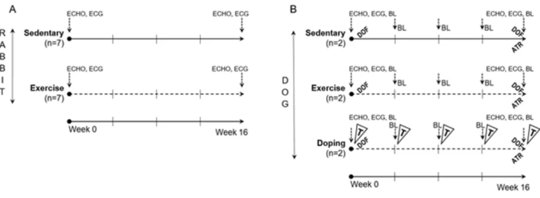
‘Dop’ group were identical to ‘Ex’ group, and therefore the effects of testosterone could not be attributed to diverse training condi- tions. During the 2ndset of experiment blood collection was per- formed at every fourth week from the cephalic or saphenous vein and the levels of serum testosterone, electrolytes, blood chemistry and quantitative blood count parameters were measured. The com- plete experimental protocol is shown inFig. 1.
Running sessions were performed on a self-developed treadmill system, with two separated corridors for the animals and a control panel to modulate speed intensity. ‘Ex’ and ‘Dop’ animals under- went a 16-week-long training session, while ‘Sed’ group did not participate in the training. The protocol started with a 2-week-long warm-up period, thereafter animals were trained for 5 days a week with 20 minutes daily running sessions at speed 2.5-3 km·h−1for 16 weeks (1stset of experiment, rabbits) and with 2×90 minutes at speed 6-10 km·h−1for 16 weeks (2nd set of experiment, dogs).
The training intensity was maintained with the use of 5% to 12%
inclination. The training protocol was tested in preliminary exper- iments and set to the maximum level which could be performed without distress yet.
2.3. Echocardiography
Echocardiography was performed at 0 and 16 weeks of the training protocol. M-mode parasternal long axis view was ap-
plied using 11.5 MHz transducer (GE 10S-RS, GE Healthcare, Chicago, IL, USA), connected to an echocardiographic imaging unit (Vivid S5, GE Healthcare, Chicago, IL, USA). All parameters were analysed by an investigator in a randomised and blinded man- ner. Left ventricular internal diameter during systole (LVIDs) and diastole (LVIDd), thickness of the left ventricular posterior wall (LVPW) and interventricular septum (IVS) were measured in M- mode images. Fractional shortening was calculated as [(LVIDd- LVIDs)/LVIDd]×100.
2.4. Electrocardiography
At 0 and at 16 weeks ECGs were recorded simultaneously with National Instruments data acquisition hardware (PC card, National Instruments, Austin, TX., U.S.A.) and SPEL Advanced Haemosys software (version 3.26, Experimetria Ltd. and Logirex Software Laboratory, Budapest, Hungary).
In anaesthetised rabbits the ECG recording was made with nee- dle electrodes that placed subcutaneously in all four limbs accord- ing to Farkas et al. (Farkas et al,2004). In conscious dogs the ECG was measured using precordial leads. The ECG was digi- tized and stored on a computer for later analysis. RR, PQ, QRS, QT and Tpeak-Tendintervals were measured by manual positioning on screen markers of 40 consecutive sinus beats at the 10thminute after initiation of the recording, then mean values were calculated.
Heart rate was calculated from the RR interval. As QT interval is influenced by the heart rate, baseline data for ventricular heart rates and QT intervals were used to determine the relationship be- tween the RR interval and the QT interval in sinus rhythm accord- ing to (Farkas et al.,2003;Kui et al.,2016). These data were ob- tained from 14in vivorabbits and from 6 dogs. Forty consecutive QT intervals were measured together with the corresponding RR intervals. Simple linear regression revealed a positive correlation between QT and RR intervals in rabbits (QTrabbit = 0.354RR + 51.7) and dogs (QTdog= 0.04RR + 188.5). The equations were re- arranged to allow the calculation of the rate-corrected QT interval in rabbits at an RR interval of 295 ms (i.e. a ventricular rate of 203 beats·min−1) using the formula QTcx= QTx-0.354 (RRx−1-295) and in dogs and at an RR interval 613 ms (i.e. a ventricular rate of 98 beats·min−1) using the formula QTcx= QTx-0.04 (RRx−1-613) (Fig. 2AandFig. 2B). With these equations, plotting QTc−rabbit and QTc−dogagainst the corresponding RR interval produces a re- gression line with a slope of zero (Fig. 2CandFig. 2D), indicating that these corrections remove the influence of heart rate.
2.4.1. Measurement of the beat-to-beat variability and in- stability of the ECG intervals
Beat-to-beat variability and instability (BVI) parameters of the RR, PQ, QRS, QT and Tpeak-Tend intervals (e.g. the ‘root mean square of the successive differences’, the ‘standard deviation of the successive differences’, the ‘short-term variability’, ‘long-term variability’, ‘short-term instability’, ‘long-term instability’, ‘total instability’ and ‘instability’ of the ECG intervals) were derived from 40 consecutive sinus beats as described previously (Farkas et al.,2009;Sarusi et al.,2014;Vincze et al.,2008).
2.5. Dofetilide and atropine challenges in vivo in dogs In conscious dogs, class III antiarrhythmic agent dofetilide per- fusion was performed at 0 and 16 weeks for assessing the QT in-
Figure 1. Experimental protocol in rabbits (A) and dogs (B). Continuous line, Do not participate in training sessions (‘Sed’ groups); Dotted line, Participate in training sessions (‘Ex’ and ‘Dop’ groups); ECHO, Echocardiography; ECG, Electrocardiography; BL, Blood collection;
DOF, Dofetilide challenge, (0.035 mg/kg iv.); ATR, Atropine sulfate challenge, (0.04 mg/kg iv.); T, Testosterone-undecanoate treatment, (14.3 mg/kg im.).
Figure 2. Correlation between individual values of the QT and RR intervals in rabbit (A) and in dog (B) heartsin vivo. Correlation be- tween individual values of the ‘rate-corrected’ QT intervals and the RR intervals in rabbit (C) and in dog (D) heartsin vivo. Rabbit panels contain 560 baseline data points obtained from 14 rabbitsin vivo.
Dog panels contain 480 baseline data points obtained from 6 dogs in vivo. QTc, heart rate-corrected QT interval.
terval prolonging potential and testing the proarrhythmic sensitiv- ity before and after the long-term training protocol. Drug-free resting ECG was recorded for 20 minutes. Resting ECG values were determined at the 19th minute after initiation of the record- ing (‘Baseline’; 1 minute before dofetilide treatment). It was fol- lowed by a bolus administration of intravenous dofetilide (a se- lective inhibitor of the rapidly activating inward rectifying com- ponent of net delayed rectifier K+current, ‘IKr’) at a dosage of 0.035 mg per kg. ECG intervals were measured 15 minutes after dofetilide administration, in the 40th experimental minute (‘Dof’
period). Dofetilide was dissolved in dimethyl sulphoxide (DMSO).
Dofetilide and DMSO were obtained from Sigma-Aldrich, Inc.
(Vienna, Austria).
In conscious dogs at 16th week heart rate response was tested by intravenous administration of the parasympatholythic agent at- ropine sulfate (Egis Pharmaceuticals PLC, Budapest, Hungary).
After recording resting ECGs for 20 minutes (‘Baseline’ period), 0.04 mg per kg atropine, dissolved in saline, was administered.
The mean values of 40 consecutive RR intervals were measured at drug-free control period (T1) and 2, 5, 10, 20 minutes after at- ropine treatment (T2-T5).
3. Statistics
IBM SPSS Statistics V25 software package was used for statis- tical analysis. Continuous data were expressed as mean ± standard error of the mean (S.E.M.). Student's t-test was applied to estimate whether there is a statistically significant difference between the means in independent groups. Data were considered statistically significant whenp< 0.05.
4. Results
4.1. Structural and functional echocardiographic parame- ters
The 16-week endurance-training program resulted in signifi- cantly greater internal end-diastolic diameter of the left ventricle (LVIDd) in the ‘Ex’ rabbit group, and in an increasing tendency in LVIDd in ‘Ex’ dogs. A moderate internal end-systolic diame- ter of the left ventricle (LVIDs) increment was also seen in ‘Ex’
rabbits and ‘Ex’ dogs (Table. 1). Interestingly, the thickness of the left ventricular posterior wall (LVPW) and the interventricular septum (IVS) did not differ significantly between the groups nei- ther in rabbit nor in dog models (Table. 1). The left ventricular muscle began to dilate and enlarge as a result of endurance ex- ercise training, without an increase in ventricular wall thickness.
The ejection fraction (EF) and the fractional shortening (FS) did not indicate any difference between the groups. The complete list of measured echocardiographic parameters is shown inTable. 1.
4.2. Mean ECG depolarization parameters and RR beat-to- beat variability parameters
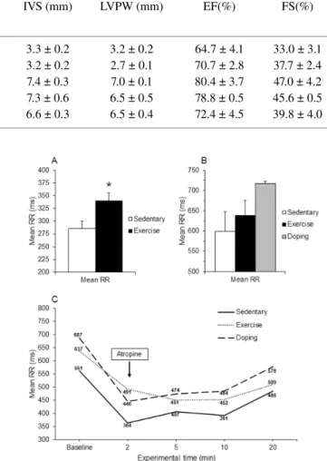
The ECG showed lengthened mean RR intervals in all ex- ercised groups of both species, which corresponded to training
Table 1. Echocardiographic cardiac dimensions and performance in rabbits (above) and dogs (below) after 16 weeks endurance exercise.
LVIDd and LVIDs, Left ventricular internal diameter in diastole and systole; IVS, Interventricular septum, LVPW, Left ventricular posterior wall; EF, Ejection fraction; FS, Fractional shortening. All values are means ± SEM.∗p< 0.05 vs. ‘Sedentary’.
Exmained Groups
LVIDd (mm) LVIDs (mm) IVS (mm) LVPW (mm) EF(%) FS(%)
RABBIT (N=7) Sedentary 13.6 ± 0.7 9.2 ± 0.7 3.3 ± 0.2 3.2 ± 0.2 64.7 ± 4.1 33.0 ± 3.1 Exercise 17.5 ± 0.6∗ 10.6 ± 0.6 3.2 ± 0.2 2.7 ± 0.1 70.7 ± 2.8 37.7 ± 2.4 DOG (N=2)
Sedentary 24.0 ± 3.0 12.5 ± 0.5 7.4 ± 0.3 7.0 ± 0.1 80.4 ± 3.7 47.0 ± 4.2 Exercise 27.2 ± 0.2 14.9 ± 0.1 7.3 ± 0.6 6.5 ± 0.5 78.8 ± 0.5 45.6 ± 0.5
Doping 22.5 ± 1.5 13.5 ± 0.5 6.6 ± 0.3 6.5 ± 0.4 72.4 ± 4.5 39.8 ± 4.0
bradycardia, indicating increased vagal tone (Fig. 3AandFig. 3B).
There was no difference in depolarizing PQ and QRS intervals af- ter the completion of the training protocol (Rabbit mean-PQ ‘Ex’
vs. ‘Sed’: 74.0 ± 6.5 vs. 67.9 ± 2.0 ms; Dog mean-PQ ‘Ex’ vs.
‘Dop’ vs. ‘Sed’: 90.8±8.0 vs. 95.2±3.2 vs. 91.2±3.5 ms;
Rabbit mean-QRS ‘Ex’ vs. ‘Sed’: 49.8±4.2 vs. 56.5±5.2 ms;
Dog mean-QRS ‘Ex’ vs. ‘Dop’ vs. ’Sed’: 60.3±3.6 vs. 56.5± 0.8 vs. 50.8±9.3 ms).
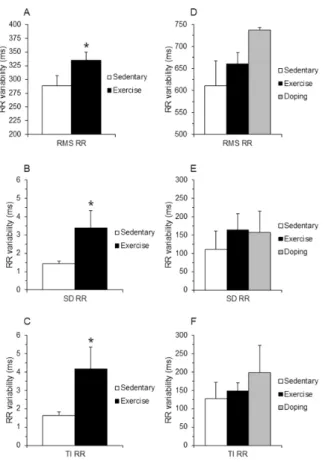
After 16 weeks the beat-to-beat root mean square (RMS), stan- dard deviation (SD) and total instability (TI) of the ‘Ex’ RR inter- vals significantly increased compared to ‘Sed’ in rabbits (Fig. 4A- C). Similar heart rate variability increasing tendency was found in
‘Ex’ and ‘Dop’ dogs (Fig. 4D-F), although significant difference was not found.
4.2.1. Atropine challenge in dogs
Repeated measurements on resting RR intervals during drug- free period (as a first stage before atropine challenge) confirmed the training-induced bradycardia in both ‘Ex’ and ‘Dop’ dogs (Fig. 3C). However, continuous atropine infusion could not retract the lengthened the RR values of ‘Ex’ and ‘Dop’ groups to the level of RR intervals of ‘Sed’ group indicating a decreased atropine re- sponse after long-term exercise (Fig. 3C).
4.3. Mean and beat-to-beat variability ECG QT parameters of ECG intervals
The mean and the beat-to-beat root mean square (RMS) of the QT intervals were mildly longer at the end of the training protocol in the exercised animals in both sets of experience (rabbit mean- QT ‘Ex’ vs. ‘Sed’: 192.6 ± 6.5 vs. 166.7±10.9 ms; RMS-QT:
192.7±6.5 vs. 166.8±10.9 ms; dog mean-QT ‘Ex’ vs. ‘Dop’ vs.
‘Sed’: 219.7±9 vs. 218.2±16.5 vs. 204±2.3 ms; RMS-QT:
219.7±9 vs. 224.8±9.8 vs. 204.0±2.3 ms), although this can be the result of the longer RR intervals due to bradycardia. Ac- cordingly, corrected QT intervals (QTc) did not differ between the groups after the long-term physical exercise (Fig. 5AandFig. 5B).
Some beat-to-beat QT variability values were increased in ‘Ex’
rabbits (e.g. short-term variability (STV): ‘Ex’ vs. ‘Sed’: 4.4 ± 0.4 vs. 4.0 ± 0.5 ms; long-term instability (LTI): 5.5±1.1 vs. 4.1± 0.4 ms; instability (I): 9.6±1.5 vs. 7.6±0.9 ms), although these parameters did not reach statistical significance. No difference and tendencies were found in QT BVI parameters in dogs (STV: ‘Ex’
vs. ‘Dop’ vs. ‘Sed’: 3.6±0.2 vs. 2.8±0.4 vs. 4.5±0.7 ms;
LTI: 4.7±1 vs. 2.6±0.8 vs. 5.3±1.9 ms; I: 7.7±1.5 vs. 5.3±
Figure 3. The meanin vivoRR intervals in rabbits (A) and dogs (B) and the mean RR intervals in dogs after parasympatholytic atropine challenge (C) at 16thweek. All values are means ± SEM.∗p< 0.05 vs. ’Sedentary’.
2.4 vs. 8.7±3.2 ms).
4.3.1. Repolarization sensitivity to proarrhythmic agent dofetilide in conscious dogs
To examine the repolarization sensitivity of athlete’s hearts, IKr inhibitor was administered to the dogs after the training protocol.
As it was expected, dofetilide markedly increased the QTcinter- val in each group (Fig. 5C), however, QTcprolongation was more pronounced in ‘Ex’ and ‘Dop’ hearts compared to ‘Sed’ hearts (Fig. 5C). There was no meaningful difference in QTcinterval be- tween the exercised (‘Ex’) and doping (‘Dop’) groups (Fig. 5C).
Despite the marked QTcinterval lengthening effect, no signif- icant difference was found in the beat-to-beat variability and in- stability parameters of the repolarization after dofetilide treatment (e.g. STV: ‘Ex’ vs. ‘Dop’ vs. ‘Sed’: 4.7±0.6 vs. 4.4±1.0 vs.
4.6±1.0 ms; LTI: 4.8±0.7 vs. 3.5±0.9 vs. 3.6±0.3 ms; I: 9.9
±1.9 vs. 6.4±0.7 vs. 7.7±1.3 ms).
During dofetilide perfusion a low number of ventricular pre-
Figure 4. Heart rate variability parameters in rabbits (A-C) and dogs (D-F) at 16thweek. Values were derived from 40 consecutive ven- tricular complexes during sinus rhythm. RMS, root mean square; SD, standard deviation; TI, total instability. All values are means ± SEM.
∗p< 0.05 vs. ‘Sedentary’.
mature beats occurred, but no significant difference was found be- tween the groups. More complex arrhythmias (e.g. Torsades de Pointes) did not develop during dofetilide perfusion in any groups (data not shown).
4.4. Testosterone levels and laboratory parameters in dogs Testosterone levels were higher in ‘Dop’ dog blood samples over the 16-week long training period than the testosterone-free dogs (‘Dop’ vs. ‘Ex’ vs. ‘Sed’ in male: 19.4±3.2 vs. 10.2±1.4 vs. 11.2±1.4 nmol/l,p< 0.05; in female: 18±4.1 vs. < 0.43 vs.
< 0.43 nmol/l,p< 0.05).
No significant difference was revealed between the groups in any other laboratory parameters (data not shown). Thus, long-term physical exercise did not influence any assessed internal organ lab- oratory parameters.
5. Discussion
Long-term endurance exercise was performed on two non- rodent species that might share similar cardiac electrophysiologi- cal and autonomic neural properties with humans. Cardiac mor- phological and functional adaptation signs, including echocar- diographic findings of left ventricular enlargement and increased parasympathetic tone leading to decreased resting heart rate and increased heart rate variability were found.
Figure 5. The heart rate corrected QT intervals in rabbits (A) and in dogs (B) and the heart rate corrected QT interval changes of dogs after dofetilide challenge (‘Dof’ period) at 16th week (C). Values were derived from 40 consecutive ventricular complexes during si- nus rhythm. QTc, heart rate-corrected QT interval; Baseline, drug-free values; Dof, values after 15 minutes dofetilide challenge. All values are means ± SEM.
Moderate heart rate sensitivity to the parasympatholytic agent atropine and a tendency of higher sensitivity to the QTclengthen- ing dofetilide using a self-calculated QTcequation was found in dogs duringin vivoproarrhythmic challenge. These trends might indicate delayed ventricular repolarization that predisposes to ma- lignant ventricular tachycardia, e.g. Torsades de Pointes.
5.1. Animal models of the human athlete’s heart
In animal models a variety of cardiac morphological-functional processes can be studiedin vivoand evenin vitroat an organ, cel- lular and molecular level, however the choice of a model needs to be considered carefully, since it vitally affects experimental out- comes.
Small rodents are widely used since they are easier to han- dle and have shorter gestation period which allow larger sample size (Milani-Nejad and Janssen,2014) with relatively low finan- cial cost. There are number of well-established rodent training models in the literature, e.g. swimming-induced cardiac hyper- trophy model verified by echocardiography and hystomorphome- try (Radovits et al.,2013) and long-term endurance training mod- els (Benito et al., 2011;Chu et al.,2000). Nevertheless, there are numerous limitations of translating cardiac remodeling signs and mechanisms from rodents to humans, e.g. rodent hearts must contract and relax more rapidly in order to maintain cardiac out- put at very high heart rates (Janssen and Periasamy,2007). Fur- thermore, rodent action potentials have a rapid repolarization and lack a prominent plateau phase compared to human cardiomy- ocytes (Nerbonne,2004). Larger animals, such as rabbit and dog models would characterize the human heart more accurately in
terms of oxygen uptake kinetics, cardiac mechanics, repolariza- tion, excitation-contraction coupling, collateral coronary circula- tion and subcellular architecture. Considering the ion channel ki- netic properties, the IKswhich is presumably affected in cardiac remodeling during chronic exercise, best resembles to those mea- sured in human hearts in dog (Liu and Antzelevitch,1995) and rabbit ventricles (Lengyel et al.,2001). Carroll et al. used rabbits to determine the effects of exercise training during the develop- ment of obesity. They applied a 12-week-long treadmill protocol at 1.2 km·h−1 of maximum speed and 50-60 minute daily run- ning sessions. Exercise trained rabbits had slower resting heart rates in both lean and obese animals (Carroll and Kyser,2002).
Such et al. found lower resting heart rates and longer ventricu- lar effective refractory periods at all the pacing cycle lengths dur- ingin vitroelectrical stimuli compared with the control group in a chronic motor-driven treadmill study (Such et al.,2008). Approxi- mately 10-week endurance training programme based on treadmill running is required to get a significant cardiac response to exer- cise training (Hexeberg et al,1995). Non-rodents mammals would need even more training for longer time to show the properties of athlete’s heart (Wyatt et al.,1974). In our model, after a long pre- liminary period, 16-week-long training was used in both species at speed 2.5-3 km·h−1in rabbits and at speed 6-10 km·h−1in dogs, which induced lower resting heart rates, made the participating animals physically tired and sometime exhausted. As the New Zealand White rabbit is a physically inactive species, this work- load is thought to be convenient to mimic regular, high-intensity human training activity. A greater similarity of rabbit myocardium to humans make them a closer representative of the human heart.
The cost of acquiring and housing rabbits is still significantly much lower than for dogs.
Canine heart rate, and heart weight, excitation-contraction cou- pling, action potential duration and expression patterns of var- ious ion channels are more comparable to humans (Szel et al., 2011). Moreover, canine can increase its heart rate approximately 96–136% during maximal exercise which is close-lower than the 140–170% increase in humans (Haidet et al.,1989;Musch et al., 1987;Stratton et al.,1994). These characteristics of the canine myocardium serve as a very good model of the human heart. Nev- ertheless, the housing and maintenance is considerably more ex- pensive than small rodents and rabbits, the number of animals that can be used is limited, therefore, this issue is often to be taken into consideration in case of long-term studies.
5.2. Cardiac morphological changes after endurance exer- cise training
Physical exercise in elite athletes induces adaptation of the car- diovascular system according to the type and intensity of sport ac- tivity (Morganroth et al.,1975). Endurance trained athletes (e.g.
long distance running, cross-country skiing) usually have evidence of cardiac enlargement (increased LV chamber dimensions), with or without obvious cardiac hypertrophy (increased LV wall thick- ness) (D'Andrea et al.,2010;Mitchell et al.,2005;Pelliccia et al., 2010;Toufan et al.,2012). The present study showed increased cardiac left ventricular end-diastolic diameters in the exercised an- imals. Interestingly, neither interventricular septum, nor posterior wall thickness altered. Our echocardiographic results correspond
to long-term endurance exercise, indicating structural cardiac re- sponse to an increased volume load. Since the duration of long- term exercise has also an impact on the physiological remodel- ing of myocardial structure and function in athletes (Weiner et al., 2015), it may be possible that after several years of continuous training the morphological signs of wall thickness and ventricu- lar hypertrophy could be seen in the model. Further studies (i.e.
considerably longer training sessions) are required to verify this hypothesis.
5.3. Increased parasympathetic activity
Endurance training results in enhanced parasympathetic activ- ity in young athletes (Jensen-Urstad et al., 1997;Macor et al., 1996), which contributes to a higher prevalence of sinus brady- cardia in resting conditions and a slower increase in heart rate at any degree of submaximal oxygen uptake (Uusitalo et al.,1996) Analysis of the beat-to-beat variability of the RR intervals permits insight in this regulation mechanism in a non-invasive way. In the present study a training-induced bradycardia was found which was accompanied by an increase in HRV values in both species during drug-free resting conditions. These findings are related to a higher parasympathetic activity due to the long-term endurance training. This was supported after atropine blockade when the heart rate increased in the exercised dogs. Likewise, some stud- ies that used heart rate variability methods, demonstrated an in- crease in parasympathetic activity (Goldsmith et al.,1992), and its disappearance after vagolytic maneuver. However, atropine chal- lenge could not decrease the heart rate of exercised dogs to that level of the sedentary ones. Some studies have proposed that al- terations in the intrinsic properties of the sinus node, the so-called
‘non-autonomic component’ was responsible for rest bradycardia of athletes (Boyett et al.,2013;Katona et al.,1982). Since we have not investigated this point at the cellular level in our study, intrin- sic adaptations in the conduction system (e.g. as a result of some downregulation of If channel) cannot be ruled out. Considering that only moderate heart rate increment was found after atropine infusion in the exercised dog group, it is possible that long-term endurance training may also induce intrinsic adaptations in the si- nus node.
5.4. Repolarization-related changes and susceptibility for cardiac arrhythmias
Several studies showed that cardiac remodelling, e.g. left ven- tricular hypertrophy and dilatation, induced by different patho- physiologic processes (Janse,2004;Volders et al.,1998) prolonged repolarization, increased electrical inhomogeneity and arrhythmia propensity. However, there are less data about cardiac repolar- ization in exercise-induced physiological cardiac remodeling, es- pecially in animal models. Findings from early research pointed out that repolarization BVI parameters are increased in elite soc- cer players (Lengyel et al.,2011), raising the possibility to an in- creased propensity for ventricular arrhythmias. In our study, some of the BVI QT values tended to increase in the exercised rabbits.
It was hypothesised previously that endurance-exercised animals have some degree of repolarization impairment, even without clin- ical sign or measurable QT interval lengthening under baseline as- sessment. This might be unmasked by potassium channel inhibi- tion (Lengyel et al.,2007). In this study, the selective IKrchannel
blocker dofetilide tended to lengthen the QTcintervals equally in the exercised dogsin vivoas compared with the sedentary group.
This finding might indicate an augmented repolarization prolonga- tion after long-term exercise in these models that could be in con- nection with higher risk of the repolarisation-related proarrhyth- mic side effects.
At a low concentration of dofetilide, which was applied in this study for the identification of mild repolarization changes, only some ventricular premature beats occurred equally in all groups.
Consequently, the exercised dogs were not more susceptible to ar- rhythmias than the controls.
Our study did not find enough hard evidence in any investi- gated species about repolarization-related electrical remodeling.
However, a modest repolarization alteration cannot be excluded in athletes, since the training induced repolarization changes are probably mild (alike in the human athlete’s heart). Even if these effects are found to be marginal, they may be added up with other potentially harmful factors (e.g. non-steroid agents, H1 antihis- tamines, dietary constituents or doping) resulting in a dangerous increase of repolarization inhomogeneity forming a significant ar- rhythmia substrate in the athlete’s heart (Varro and Baczko,2010).
5.5. Doping
Anabolic androgenic steroid abuse has been shown to change lipoprotein metabolism leading to premature atherosclerosis, hy- pertension and myocardial infarction resulting in cardiomyopathy and ventricular arrhythmias (Dhar et al.,2005). However, it is un- known if testosterone or other anabolic steroid can trigger cardiac electrophysiological changes and augment proarrhythmic poten- tial. Our results showed that testosterone in conjunction with a standardized program of endurance exercise did not increase car- diac muscle mass and did not influence other structural and re- polarization parameters in dogs. Undoubtedly, some athletes and bodybuilders may use higher doses of anabolic steroids than that was used in our study. Moreover, athletes often take multiple forms of doping agents simultaneously. It may be also possible that steroid doping has different kind of effects on different type of sports. To clarify these effects of steroid doping in this model, further investigation is needed.
6. Conclusion
Long-term intensive exercise caused left ventricular cavity en- largement and changes in heart rate and in HRV parameters indi- cating an increased vagal tone in both species. These findings are in accordance with human cardiac remodeling in elite endurance athletes. Lower heart rate by itself may favour prolonged repolar- ization and inhomogeneity, furthermore, higher sensitivity to the IKr inhibitor dofetilide may explain higher risk of life-threating arrhythmias. These experimental models might be useful for the further investigations of the cardiovascular effects of the long-term physical exercise in humans.
7. Limitation
Low ‘n’ numbers were used in the dog studies attributed to the long training protocol and to the restrictions in access to large an- imals (i.e. dogs), which did not make possible the proper compar- ison of the groups. Similarly, the verification of mild repolarisa-
tion changes seen in top athletes, a relatively large sample size is required which is really challenging to produce with non-rodent animals. The application of higher sample size in further studies is warranted to thoroughly prove our hypotheses.
Acknowledgments
This work was supported by Hungarian Scientific Research Fund [OTKA K 119992], the UNKP-17- 4 and UNKP-18-4-SZTE- 95 new National Excellence Program of The Ministry of Hu- man Capacities, GINOP-2.3.2-15-2016-00047, the EFOP-3.6.2- 16-2017-00006 and János Bolyai Research Scholarship of the Hun- garian Academy of Sciences (N.N), the NKFIH PD-125402 (N.N) and by FK-129117 (N.N) projects.
Conflict of interest
The authors declare that there is no conflict of interest.
References
Benito B, Gay-Jordi G, Serrano-Mollar A, et al. Cardiac arrhythmo- genic remodeling in a rat model of long-term intensive exercise training. Circulation. 2011;123:13-22.
Boyett MR, D'Souza A, Zhang H, et al. Viewpoint: is the resting bradycardia in athletes the result of remodeling of the sinoa- trial node rather than high vagal tone? J Appl Physiol (1985).
2013;114:1351-1355.
Carroll JF, and Kyser CK. Exercise training in obesity lowers blood pressure independent of weight change. Med Sci Sports Exerc.
2002;34:596-601.
Chu TF, Huang TY, Jen CJ, et al. Effects of chronic exercise on calcium signaling in rat vascular endothelium. Am J Physiol Heart Circ Physiol. 2000;279:H1441-1446.
Corrado D, Basso C, Rizzoli G, et al. Does sports activity enhance the risk of sudden death in adolescents and young adults?J Am Coll Cardiol. 2003;42:1959-1963.
D'Andrea A, Cocchia R, Riegler L, et al. Left ventricular myocardial velocities and deformation indexes in top-level athletes. J Am Soc Echocardiogr. 2010;23:1281-1288.
Dhar R, Stout CW, Link MS, et al. Cardiovascular toxicities of performance-enhancing substances in sports. Mayo Clin Proc.
2005;80:1307-1315.
Farkas A, Batey AJ, Coker SJ. How to measure electrocardiographic QT interval in the anaesthetized rabbit. J Pharmacol Toxicol Meth- ods. 2004;50:175-185.
Farkas A, Curtis MJ. Does QT widening in the langendorff-perfused rat heart represent the effect of repolarization delay or conduction slowing?J Cardiovasc Pharmacol. 2003;42:612-621.
Farkas AS, Makra P, Csik N, et al. The role of the Na+/Ca2+ ex- changer, I(Na) and I(CaL) in the genesis of dofetilide-induced tor- sades de pointes in isolated, AV-blocked rabbit hearts. Br J Phar- macol. 2009;156:920-932.
Farkas AS, Nattel S. Minimizing repolarization-related proarrhyth- mic risk in drug development and clinical practice. Drugs.
2010;70:573-603.
Goldsmith RL, Bigger JT Jr, Steinman RC, Fleiss JL. Comparison of 24- hour parasympathetic activity in endurance-trained and untrained young men. J Am Coll Cardiol. 1992;20:552-558.
Haidet GC, Musch TI, Friedman DB, et al. Cardiovascular ef- fects of dobutamine during exercise in dogs. Am J Physiol.
1989;257:H954-H960.
Hexeberg E, Westby J, Hessevik I, et al. Effects of endurance training on left ventricular performance: a study in anaesthetized rabbits.
Acta Physiol Scand. 1995;154:479-488.
Janse MJ. Electrophysiological changes in heart failure and their re-
lationship to arrhythmogenesis. Cardiovasc Res. 2004;61:208- 217.
Janssen PM, Periasamy M. Determinants of frequency-dependent con- traction and relaxation of mammalian myocardium. J Mol Cell Cardiol. 2007;43:523-531.
Jensen-Urstad K, Saltin B, Ericson M, et al. Pronounced resting brady- cardia in male elite runners is associated with high heart rate vari- ability. Scand J Med Sci Sports. 1997;7:274-278.
Katona PG, McLean M, Dighton DH, et al. Sympathetic and parasym- pathetic cardiac control in athletes and nonathletes at rest. J Appl Physiol Respir Environ Exerc Physiol. 1982;52:1652-1657.
Kui P, Orosz S, Takacs H, et al. New in vitro model for proarrhythmia safety screening: IKs inhibition potentiates the QTc prolonging effect of IKrinhibitors in isolated guinea pig hearts. J Pharmacol Toxicol Methods. 2016;80:26-34.
Lengyel C, Iost N, Virag L, et al. Pharmacological block of the slow component of the outward delayed rectifier current (I(Ks)) fails to lengthen rabbit ventricular muscle QT(c) and action potential du- ration. Br J Pharmacol. 2001;132:101-110.
Lengyel C, Orosz A, Hegyi P, et al. Increased short-term variability of the QT interval in professional soccer players: possible impli- cations for arrhythmia prediction. PLoS One. 2011;6:1-10.
Lengyel C, Varro A, Tabori K, et al. Combined pharmacological block of I(Kr) and I(Ks) increases short-term QT interval variability and provokes torsades de pointes. Br J Pharmacol. 2007;151:941- 951.
Liu DW, Antzelevitch C. Characteristics of the delayed rectifier cur- rent (IKr and IKs) in canine ventricular epicardial, midmyocardial, and endocardial myocytes. A weaker IKs contributes to the longer action potential of the M cell. Circ Res. 1995;76:351-365.
Macor F, Fagard R, Amery A. Power spectral analysis of RR interval and blood pressure short-term variability at rest and during dy- namic exercise: comparison between cyclists and controls. Int J Sports Med. 1996;17:175-181.
Marijon E, Tafflet M, Celermajer DS, et al. Sports-related sudden death in the general population. Circulation. 2011;124:672- 681.
Maron BJ, Doerer JJ, Haas TS, et al. Sudden deaths in young com- petitive athletes: analysis of 1866 deaths in the United States, 1980-2006. Circulation. 2011;119:1085-1092.
Maron BJ, Shirani J, Poliac LC, et al. Sudden death in young compet- itive athletes. Clinical, demographic, and pathological profiles.
Jama. 1996;276:199-204.
Milani-Nejad N, Janssen PM. Small and large animal models in car- diac contraction research: advantages and disadvantages. Phar- macol Ther. 2014;141:235-249.
Mitchell JH, Haskell W, Snell P, et al. Task Force 8: classification of sports. J Am Coll Cardiol. 2005;45:1364-1367.
Morganroth J, Maron BJ, Henry WL, et al. Comparative left ventric- ular dimensions in trained athletes. Ann Intern Med. 1975;82, 521-524.
Musch TI, Friedman DB, Pitetti KH, et al. Regional distribution of blood flow of dogs during graded dynamic exercise. J Appl Phys- iol (1985). 1987;63:2269-2277.
Nerbonne JM. Studying cardiac arrhythmias in the mouse--a reason- able model for probing mechanisms?Trends Cardiovasc Med.
2004;14:83-93.
Pelliccia A, Di Paolo FM, De Blasiis E, et al. Prevalence and clinical significance of aortic root dilation in highly trained competitive athletes. Circulation. 2010;122:698-706.
Radovits T, Olah A, Lux A, et al. Rat model of exercise-induced car- diac hypertrophy: hemodynamic characterization using left ven- tricular pressure-volume analysis. Am J Physiol Heart Circ Physiol.
2013;305:H124-H134.
Sarusi A, Rarosi F, Szucs M, et al. Absolute beat-to-beat variability and instability parameters of ECG intervals: biomarkers for pre- dicting ischaemia-induced ventricular fibrillation. Br J Pharmacol.
2014;171:1772-1782.
Stratton JR, Levy WC, Cerqueira MD, et al. Cardiovascular responses to exercise. Effects of aging and exercise training in healthy men.
Circulation. 1994;89:1648-1655.
Such L, Alberola AM, Such-Miquel L, et al. Effects of chronic exercise on myocardial refractoriness: a study on isolated rabbit heart.
Acta Physiol (Oxf). 2008;193:331-339.
Szel T, Koncz I, Jost N, et al. Class I/B antiarrhythmic property of ranolazine, a novel antianginal agent, in dog and human cardiac preparations. Eur J Pharmacol. 2011;662:31-39.
Toufan M, Kazemi B, Akbarzadeh F, et al. Assessment of electrocar- diography, echocardiography, and heart rate variability in dy- namic and static type athletes. Int J Gen Med. 2012;5:655-660.
Uusitalo AL, Tahvanainen KU, Uusitalo AJ, et al. Non-invasive evalua- tion of sympathovagal balance in athletes by time and frequency domain analyses of heart rate and blood pressure variability. Clin Physiol. 1996;16:575-588.
Varro A, Baczko I. Possible mechanisms of sudden cardiac death in top athletes: a basic cardiac electrophysiological point of view.
Pflugers Arch. 2010;460:31-40.
Vincze D, Farkas AS, Rudas L, et al. Relevance of anaesthesia for dofetilide-induced torsades de pointes in alpha1-adrenoceptor- stimulated rabbits. Br J Pharmacol. 2008;153:75-89.
Volders PG, Sipido KR, Vos MA, et al. Cellular basis of biventricular hypertrophy and arrhythmogenesis in dogs with chronic complete atrioventricular block and acquired torsade de pointes. Circula- tion. 1998;98:1136-1147.
Weiner RB, DeLuca JR, Wang F, et al. Exercise-Induced Left Ventricular Remodeling Among Competitive Athletes: A Phasic Phenomenon.
Circ Cardiovasc Imaging. 2015;8:1-9.
Wyatt HL, Mitchell JH. Influences of physical training on the heart of dogs. Circ Res. 1974;35:883-889.