EXAMINATION OF ENDURANCE AND POWER ATHLETES WITH MODERN ECHOCARDIOGRAPHIC (SPECKLE
TRACKING)
Abstract of the PhD thesis
Dr. Szauder Ipoly
Doctoral School of Sport Sciences University of Physical Education
Consultant: Gábor Pavlik professor emeritus, DSc
Official reviewers: György Ábrahám professor, PhD
István Hartyánszky assistant professor, PhD
Head of the Final Examination Committee:
József Tihanyi professor, DSc Members of the Final Examination Committee:
Csaba Nyakas professor emeritus, DSc Elek Dinya professor, PhD
János Takács cardiologist chief physician, PhD Budapest
2015
INTRODUCTION
The role of heart fitness in preserving health
One of the most important aims of sports and also of sports medicine is to preserve human health thereby achieving better quality life and longer life span. It should be regarded as a model of “health medicine”, which instead of focusing on the already existing illness, strives to prevent it, together with all its benefits to the individual and to the society. Well chosen, regular, dynamic stress tailored to the individual (to the illness) will have a positive effect both on the process and on the complications of cardiovascular diseases and hypertension. In recent decades regular exercise became an organic part of healthy lifestyle, its two most common forms being running and body building. Masses join running races abroad (e.g.
Vivcitta, the New York Marathon) but there are tens of thousands of fans pursuing this sport in Hungary too (e.g. Budapest Marathon). The proliferation of fitness centres is a universally known phenomenon.
The effect of endurance and power sports on the heart
Endurance performance mainly depends on the functioning and fitness of the heart; high quality endurance performance is not possible without having trained the heart in endurance tasks. Different cardiovascular adaptation is required by endurance sports (in other words dynamic, isotonic, aerobic trainings) for example long distance running and swimming, and by power sports like weight lifting, power lifting and body building (in other words static, isometric, anaerobe trainings). There are few sports which contain purely dynamic (endurance) or purely static (strength) exercises. In the case of endurance sports the maximal oxygen uptake, cardiac output and systolic blood pressure is increased with the decrease of the peripheral vascular resistance. With power sports oxygen uptake and cardiac output increases slightly while there is a significant rise in the systolic blood pressure, peripheral vascular resistance and heart beat rate (Csajági 2014, Major, Szauder 2015).
Definition of the athlete’s heart
The athlete’s heart is considered to be an athlete’s heart demonstrating all the cardio-vascular adaptation signs which result from long term training. Morphological fitness signs are:
concentric or eccentric left ventricular hypertrophy, rare right ventricular hypertrophy, richer coronary supply. Signs of functional fitness are: systole and diastole can be measured with different non-invasive methods. Signs of regulated fitness: change in pulse rate and cardiac output due to the change in vegetative regulation. The fitness signs of the heart depend on several factors one of the basics of these being the nature of the movement the given sport requires.
Short overview of the literature on athlete’s heart
In order to demonstrate the different periods I used the historic ages of mankind.
Stone Age: physical examination
Henschen studied cardiomegaly on cross country skiers in 1898 and regarded it as the sign of physiological adaptation. Darling, who studied the rowers of Harvard University in 1899, and White, who studied the runners of the Boston Marathon in 1918 were the first to describe the explicit resting sinus bradycardia of the radial pulse.
Antiquity: the beginnings of instrumental examination of morphology: the rtg examination
Before the appearance of echocardiography until the 1970s only X-ray was able to provide direct imaging information about the size and functioning of the heart. Functional data supplied by an X-ray examination were indirect and considerably inaccurate.
Middle Ages: recognition of cardiac functions begins: the beginning of non-invasive era:
the ECG examination and the mechanocardiography
In the 1970s and 1980s beside ECG, mechanocardiography also played an important role.
This is the recording of the carotis pulse wave by a pressure sensor working on the piezo- electronic principle, which was evaluated through the simultaneously recorded ECG and heart beat sound. The determination of systolic and diastolic time intervals indirectly transmits the stroke volume, the aorta pressure and the end diastolic pressure of the left ventricle. Hungary was among the first to describe the use of the first and second temporal derivative of the carotis pulse wave both in the case of healthy people and people suffering from heart disease.
Hungary was the first to calculate the measured data by computer analysis (Herpai 1978, Simonyi 1977, 1980, 1983, Szauder 1978, 1982).The first and second temporal derivative of the carotis pulse on our recording is very similar to the “wave intensity” wave observed by echocardiography. This is because the derivatives are more distinct due to the temporal
representation thereby making it possible to analyse more data about the carotis stress wave, especially its initial phase, so they can convey more information indirectly about the flow better than the carotis pulse wave curve. There is an especially remarkable similarity between the first temporal derivative of the carotis and the “wave intensity” recording (Szauder 2014).
The dawn of the Modern Age: echocardiography appears
In the beginning the M-method and the two dimensional procedure provided data about the left ventricular hypertrophy. It is a classic assumption that power sports mostly cause concentric, while endurance sports cause eccentric left ventricular hypertrophy. There is only a slight difference between the cardiac adaptations observed in dynamic and static sports. The increase of the left ventricular sinus and the muscle mass is also determined by the body surface and the body mass. Thickening of the wall appears in all sports, but an increase in the internal diameter is more explicit in endurance sports. Adaptation of the heart in the case of long distance runners is twofold: it adopts both to the volume and pressure load as a result of which the fit heart reacts with both the increase of the internal diameter and the thickening of the left ventricular wall. With power sports the training of the heart is slighter, with less increase in the internal diameter of the left ventricle than in endurance sports, but thickening of the wall can be of varying degree. These changes can be explained on one hand by the pressure load developed as a blood pressure reaction which exceeds that of the endurance sports and on the other hand by the volume load generated by the smaller degree of cardiac output. However, there is no clear load pressure in the case of power athletes either, because it combines with volume pressure to a certain extent (Szauder, Csajági Major 2015).
Beside the morphological data the systolic function of the left ventricle is generally measured with the ejection fraction and with the shortening of the circumferential fibre. Several studies have proven that these do not differ greatly from that of non-athletes. This leads to the conclusion that in case of athletes there is no relationship between the geometry of the heart and the left ventricular systolic function.
The present: the Doppler echocardiography. The beginning: the E/A value
In determining the diastolic function the usability of the quotient of E and A waves of the mitral inflow i.e. the E/A value is characterised by limitations and dependences such as the mitral diastolic gradient, the preload of the left ventricle, the afterload of the left ventricle and the heart frequency. In the case of athletes a slightly increased diastolic function is generally
observed, while with hypertonic patients the increase in the left ventricular muscle mass can be accompanied by abnormal diastolic filling (Szauder 2014).
Recent past: the tissue Doppler
Measurements taken by the myocardial Doppler show the speed of the longitudinal, rotational, radial and translational movement of the heart and the origins of these movements.
The E /E’ value is the index of relaxation, which is preload dependent in normal individuals, preload independent in patients suffering from heart disease, the lateral is bigger than the septal, it is in inverse proportion to age, it is abnormally decreased in any type of diastolic dysfunction, it is in inverse proportion to systolic function and shows correlation with capillary wedge pressure and also with left ventricular pressure.
Application of TDI data in sports medicine
With endurance sport athletes E/E’ value was higher than with power athletes and an increase in E’ value and a decrease in A’ value was noticed in comparison to the controls. The increase of the diastolic function in athletes however, also depends on the filling pressure and the heart frequency and it mainly represents the longitudinal movement of the left ventricle (Szauder 2015). The TDI method helps to differentiate between the physiological left ventricular hypertrophy, the fit heart and the pathologic left ventricular hypertrophy.
The present and the future: strain and strain rate imaging (SRI) and the Speckle Tracking Echocardiography (STE)
Strain and strain rate imaging (SRI)
The displacement, the velocity, the strain and the strain rate can be measured. From the perspective of the apex the left ventricle shortens in contraction, this is negative strain. In diastole the strain is positive. In cross section the wall thickens in systole i.e. the strain is positive; in diastole it tapers i.e. the strain is negative. The changes between the endo- and epicardiac layers of the myocardium are represented by the radial strain. If the deformation is shortening, the strain is negative if it is thickening, the strain values are positive. The longitudinal shortening and the transversal thickening can be measured in longitudinal section image, while circumferential shortening and radial thickening are measured in cross section image.
In clinical practice strain and strain rate are used to assess global and segmental left ventricular function. Strain maps the myocardium, and not just the movement of the mitral annulus. Strain and strain rate are in direct proportion to left ventricular functions. It is mainly used for showing ischemia. The disadvantage of TDI is that it is strongly influenced by the insonatic angle of the ultrasound ray in one dimension. It cannot be used in every segment, being noise and object sensitive. These disadvantages are eliminated by the Speckle Tracking Echocardiography.
Speckle Tracking Echocardiography (STE)
The advantage of this is newest, about ten-year old, echocardiographic method is that its mapping does not depend on the angle of the ultrasound ray and it is not influenced by the translational movement of the heart either. Its basis: the different rate dispersion, reflection and interference of the ultrasound in the myocardium shows a special pattern; the spatial and temporal movement and the speed of this speckle can be well represented and measured. So the speckle technique uses the structural inhomogeneity of the myocardium to measure its strain and displacement. Both strain and strain rate can be measured. Its advantage with athletes is that while conventional echocardiography can determine the eccentric or concentric type of the fit heart, STE can provide data about the intrinsic myocardial functions too: the longitudinal, circumferential and radial displacement, strain and strain rate (Szauder 2015).
OBJECTIVES
Few studies compare the cardiac strain caused by the nearly “purely” power sport body building and the nearly “purely” endurance sport marathon running with the latest echocardiographic methods. My objective was to disclose in the most detailed manner the characteristics of the fit heart in the case of marathon runners and body builders representing these two very popular endurance and power sports; to define as precisely and subtly as possible the factors influencing the development of the athlete’s heart with special regards to the latest, only ten year-old, echocardiographic method, the speckle tracking echocardiography. In my assumption the cardiac adaptation of body builders is of different nature, in some aspects may be less positive than that of marathon runners. Clear distinction and better understanding with the help of speckle tracking echocardiography can lead to practical conclusions and can contribute to the creation of a sport-physiologically more favourable training programme for the athletes.
METHODS
Persons, exclusion criteria
The athletes examined were men between 20-30 years of age, 24 marathon-ultra marathon runners and 14 body builders. The control group consisted of 15 non-fit men of sedentary occupation. The athletes had a minimum of 5 years sports history. The marathon runners trained 20,0±1,4 hours per week for 7,8±1,6 years and regularly took part in amateur marathon running races. The body builders took part in 16,8±1,1 hour training weekly for 7,9±1,4 years. The non-athletes were either university students or workers of sedentary occupation, who did not do more than 3 hours of physical activity per week.
Exclusion criteria were: acute or chronic illness, regular medication, use of performance enhancing drugs, smoking, deviation detected by physical examination, laboratory risk factors as abnormal value of serum cholesterol LDL-, HDL cholesterol, triglyceride, serum uric acid or homocysteine, blood sugar level indicating IGT, pathological resting ECG deviation, abnormal value of ankle arm index, thickening of carotis intima detected by Doppler method, pathologic deviation detected with echocardiography, positive ECG under stress (apart from the well-known ECG deviations I considered maximal values over 160/90 Hgmm-t abnormal, due to the suspicion of hypertension).
Screening hypertension based on the new echocardiographic rating of my own development
Blood pressure was determined by 24-hour blood pressure monitor to preclude hypertension with regards to the standing recommendations of the European Society of Hypertension. For hypertension and its echocardiographic detection I used the grading of hypertension severity by multifactorial echocardiography of my own development, which can be useful in recognising undetected hypertension (Szauder 2009, 2011).
Examinations
Basic examinations: recording detailed medical history, measuring body mass and height, cardiological physical examination, recording status: auscultation, measuring blood pressure and taking the pulse. Conventional echocardiographic examinations were carried out in the
morning by an Esaote Biomedica Mylab 25 Gold type echocardiograph with 2,5-3,5 MHz transducer. I took the two dimensional and guided M-mode images from the parasternal longitudinal section and the transmitral flow rate on four cavity image by pulsatile Doppler method. I used the tissue Doppler method to determine the moving speed of the annulus at the septal and lateral stem of the mitral valve. I determined the systolic (PVs) and diastolic (PVd) speed of the pulmonary vein curves by pulsatile Doppler method through a sampling gate inserted into the upper right pulmonary vein. The tricuspid annular plane systolic excursion (TAPSE) was done by M-mode from the tricuspid annulus. We used the average of five cycles for the measurements.
The “conventional” echocardiographic measurements and calculations
I used the Devereux formula to determine the myocardial mass of the left ventricle. I calculated the thickness of the left ventricle wall from the sum of the interventricular septum and the thickness of the back wall, and the rate of the wall thickness and internal diameter of the left ventricle, the muscular quotient. (MQ). Knowing the diameters, first I calculated the end diastolic and end systolic volume, then with the help of the differences in volume the pulse volume and the cardiac output, finally knowing the diameters and the thickness of the wall, the myocardial mass of the left ventricle. Different authors use several different methods to calculate these data. I calculated both the volumes and the left ventricular muscle mass by cubing the transversal diameters of the parasternal image. I related the measurements of the heart to those of the body with formulae in which the power of the numerator and the denominator are equal (Szauder, Csajági Major 2015), and I also provided the myocardial mass related to the cubic metres of the body surface . I calculated the quotient of the early and late diastolic inflow speed (E/A quotient) from the flow rates, and the rate of the septal and lateral early and late diastolic top speed of the mitral annulus from the TDI speed.
Speckle tracking, strain rate measurements and calculations
For speckle tracking analysis in short axis cross-section on the level of mitral valves, on midpapillary level from apex perspective two- three- and four-level apex images were prepared. Appropriate adjustment of the gain, focus, depth and the sector, and the over fifty image frame rate guaranteed the optimal visibility of the left ventricular myocardium. Speckle tracking analysis was prepared by 2D Cardiac Performance Analysis, TomTec Imaging
System Gmbh, (Unterschleissheim Germany) programme. I marked the border of the left ventricular endocardium manually and manual correction was made if the software could not accurately mark it. I measured the segmental systolic strain peak values in the average of two heart cycles. Determining the global longitudinal, circumferential and radial systolic peak values was based on the 16-segment model. I used the Mosteller formula to calculate body circumference, and the Statistical Package for the Social Sciences software version 20 (IBM, Armonk, New York, USA) for statistical analysis. The normal distribution of variables were checked by the Shapiro-Wilk test. I used the Fisher-type post-hoc one-way ANOVA test with normal distribution, and after Kruskal-Wallis ANOVA the Mann-Whitney test in pairs with non-normal distribution. In the tables I show the average values and the standard deviations indicating the significant differences in (p<0,05) value (Szauder 2015).
RESULTS
The age and body measurements of the persons tested
Due to their larger muscle mass body builders showed higher BSA and BMI values.
Morphological data
In the case of body builders the internal diameter of the left ventricle and the absolute and relative value of left ventricular muscle masses were significantly greater than that of non- athletes. The groups did not show difference in MQ. There was no significant difference in the diastolic diameter of the septum and that of the back wall in rel WT and in MQ among the athletes either. The relative left ventricular myocardial mass was no significantly bigger in body builders than in runners. There was no difference in left ventricular hypertrophy between athletes and the control groups. As opposed to the majority of the data recorded in literature neither the endurance nor the power athletes developed significant left ventricular hypertrophy.
Basic heart-circulation data
Body builders had higher heart rate, blood pressure and cardiac output than marathon runners and the control group
Marathon runners had lower resting heart rate, while body builders and non-athletes showed no difference in this respect
Systolic and diastolic blood pressure was significantly lower in marathon runners than in the other two groups
Body builders showed a significantly higher cardiac output than runners and the non- athletes
Left ventricular end diastolic volume was bigger with athletes than with the control group
The relative pulse volume was significantly lower in non-athletes while it showed no difference in the two athlete groups.
There was no difference between the groups in left ventricular ejection function and fractionated fibre shortening
The maximal aortic flow velocity was higher in both athlete groups than in the control but the difference in strength sports was significant.
The functional data of the heart with “conventional” echocardiographic measurements (PW Doppler and TDI data)
Neither the deceleration of mitral E wave nor the isovolumic relaxation time changed significantly. There was no difference in the septal and lateral systolic top speed of the mitral anulus.
Marathon runners
E/A, E/E’, E’/A’ values were not worse, in fact they showed a tendency to improve.
The significantly lower PVd value indicated better diastolic function in the case of runners.
Body builders
Significantly lower E/A quotient
Lateral wall E/E’ significantly lower
Septal E/E’ did not decrease significantly
Septal wall showed significantly lower E/A’ value, which was the result of a decrease in E’ value and an increase in A’ value
Lateral E’/A’ value was significantly higher than in the control group (E’ value increased, A’ value decreased)
Septal A’ was significantly higher
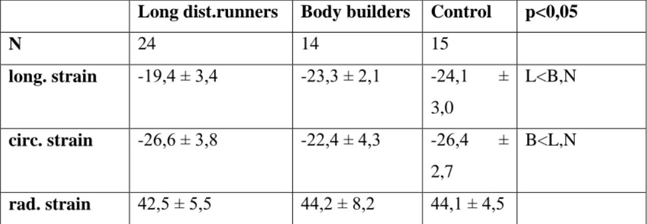
Strain rate and functional data measured by speckle tracking echocardiography
Longitudinal strain
Longitudinal strain decreased in marathon runners.
Its value had good correlation with the body surface and the left ventricular end diastolic volume.
Changes in the circumferential strain
Circumferential strain decreased in body builders.
This is related to the increase of muscle mass and blood pressure.
Values of the radial strain
There was no significant change in the radial strain between the groups.
The radial strain of marathon runners was not significantly lower (1. table).
1. table. The functional data of the heart measured by the new echocardiographic method: the strain rate speckle tracking echocardiography
Long dist.runners Body builders Control p<0,05
N 24 14 15
long. strain -19,4 ± 3,4 -23,3 ± 2,1 -24,1 ± 3,0
L<B,N
circ. strain -26,6 ± 3,8 -22,4 ± 4,3 -26,4 ± 2,7
B<L,N
rad. strain 42,5 ± 5,5 44,2 ± 8,2 44,1 ± 4,5
correlations:
long distance runners: longitudinal strain and EDV: r=0,46; p<0,05 longitudinal strain and BSA: r=0,49; p<0,05
body builders: circumferential strain and muscle mass: r=0,61; p<0,01
DISCUSSION- CONCLUSIONS
Analysis of “conventional” echocardiographic parameters
Results mostly correspond to findings expected in the wake of earlier research: endurance athletes -marathon runners- show better resting hemodynamic regulatory signs, while body builders doing a power sport show slight left ventricular hypertrophy and weaker functional and regulatory results (Szauder, Csajági 2015). In certain aspects the data differ from those of earlier research which is probably due to the nature of the sports under scrutiny. It is quite surprising though that the two groups of athletes did not show significant differences in left ventricular hypertrophy, the most remarkable morphological indicator of fitness. So the results do not correspond to the classic Morganroth-type dichotomy (with endurance athletes the left ventricular hypertrophy is eccentric, while with power athletes it is concentric).
Presumably the pressure load necessary to induce heart hypertrophy is more likely to develop in middle and long distance runners (5-10000 m distance), than in extra-long distance ones, who are amateurs at the same time. It is not surprising that with marathon runners MQ was practically identical with that of the non-athletes since in the case of endurance sports hypertrophy is of concentric type. Body builders do not develop concentric hypertrophy, our data are identical with those of Pelliccia, who having conducted echocardiographic examination on 100 different power athletes, found that there is only slight thickening of the wall, which is still between physiological limits. It is especially easy to understand this in the case of body builders since they do not strive to develop greater static power, or to achieve greater performance but to mould the size and shape of the muscles. Accordingly, they train with less resistance and bigger number of repetitions, than the weight lifters. Consequently it can be assumed that during the training the peripheral resistance does not increase to the extent that would cause concentric-type hypertrophy.
Hemodynamic data
Findings concerning the hemodynamic changes
The fact that left ventricular end diastolic volume was higher with athletes than with non- athletes indicates the cardiac adaptation of the fit heart.
Cardiac output was significantly higher with body builders than with runners and non- athletes, which was caused by the higher pulse volume (endurance athletes vs. non-athletes
p=0,052), rather than the heart rate, in resting vegetative regulation the sympathetic tone increased, i.e. it definitely did not decrease with power athletes. It seems that the ideal resting vegetative tone of the fit body does not develop either in body builders, their resting heart rate and blood pressure was higher than that of the long distance runners and their resting cardiac output was higher than that of both other groups. The resting heart rate is primarily set by the parasympathetic tone. Since ventricular muscles have no parasympathetic innervation cardiac output is primarily the consequence of the resting sympathetic tone and the actual cardiac hypertrophy. Blood pressure is the exact balance of the cardiac output and the peripheral resistance setting. The increase of cardiac output may primarily indicate that in resting vegetative regulation the sympathetic tone increased, i.e. it definitely did not decrease, which can contribute to the less positive diastolic function.
Diastolic function with traditional method
Diastolic function examined by PW and TDI Doppler did not become worse in marathon runners, in fact to an extent it showed an improving trend. The significantly low value of PVd with runners points at a better diastolic function. In the case of body builders the left ventricular hypertrophy was accompanied by the decrease i.e. the relative deterioration of the diastolic function, which was indicated by the significantly lower value of the E/A quotient and by the lower E’/A’ value of the septal wall due to the decrease of the E’ value and the increase of the A’ value. In several studies the analysis of the TDI data of endurance athletes showed an increase of the E’ value and a decrease of the A’ value as compared to the control group. However, the increase of the diastolic function in athletes also depends on the filling pressure and on the heart frequency, and it primarily represents the longitudinal movement of the left ventricle. The lower value of E’/A’ originating from the decrease of the septal E’
value and the increase of A’ value, which I observed with body builders during my examination, shows that the left ventricular hypertrophy did not result in flexible myocardium, but developed the myocardial stiffness growth. At the same time the lateral E’/A’ value was higher (E’ value decreased, A’ value increased) with body builders as compared to that of the control group. This more positive value indicated that fit heart develops to an extent in body builders too, in which case the left ventricular hypertrophy provides a better diastolic function (relaxation) than that of non- athletes.
New findings concerning the diastolic function (changes recorded by PW and tissue Doppler)
Endurance athletes
The diastolic function did not deteriorate, in fact it showed an improving tendency.
The significantly lower value of PVd also indicates better diastolic function.
Based on the diastolic values endurance sport (running) results in better left ventricular mechanics than body building.
Power athletes
Worsening of diastolic function:
Significantly lower E/A quotient.
Lateral E/E’ significantly smaller than that of runners.
Septal E/E’ did not decrease significantly.
The septal lower E’/A’ value, which was due to the decrease of the E’ value and the increase of the A’ value.
As opposed to marathon runners, all three diastolic parameters indicate a negative change showing the rigidity, the growing stiffness of the left ventricular wall.
The left ventricular hypertrophy was accompanied by the decrease of the diastolic function i.e. its relative deterioration resulting in non-expendable, stiffer cardiac muscle increase.
Body builders also developed athlete’s heart to an extent, in which case the left ventricular hypertrophy provides a better diastolic function (relaxation) than that of non-athletes. (The lateral E’/A’ value was significantly higher, the E’ value increased, the A’ decreased as compared to the control group)
Data gained by the strain rate and the speckle tracking echocardiography
There are very few examinations in literature which conduct echocardiographic research on two very different athlete populations complementing the evaluation of ”conventional” data with that of the latest echocardiographic method, the speckle tracking echocardiography.
Longitudinal strain
In my study longitudinal strain in the case of long distance runners had a decreased value, which showed a good correlation with the body surface and with the left ventricular end diastolic volume. This can be explained by the fact that marathon runners’ left ventricular end diastolic volume was relatively higher due to their relatively smaller body surface as compared to the other two groups. Another explanation of the decrease of the longitudinal strain with long distance runners can be that intensive training with volume loads results in volume increase and a bigger ventricle needs less longitudinal shortening for the same resting stroke volume.
We know from the TDI data of endurance athletes that increase of the E’ value and decrease of the A’ value was observed with athletes as compared to the control group. However, the increase of the diastolic function in athletes also depends on the filling pressure and on the heart frequency, and it primarily represents the longitudinal movement of the left ventricle.
On the other hand lower basal strain values with retained ejectional function can refer to a possible functional reserve capacity, which can indicate heart fitness.
The somewhat inconsistent nature of the data in literature can be due to the following: length of training (elite or amateur athletes), the role of different sport physiological effects: body building training is more static than the training of weight lifters, practically the former only train the different muscle groups separately, without any dynamic elements, while the latter do standing-ups, squatting and training of other muscle groups, body muscles. Age: in a metaanalysis of marathon runners with no decrease of GLS, the subjects were 50-65 with no internal control group, their data was compared to the –also inconsistent– data found in literature. Since GLS is age dependent the longitudinal strain shows higher values with time.
Thus GLS is not decreased by training in the elderly, or the age dependent increase can actually cover it up, that is why no GLS decrease develops. Unrecognised cardiovascular diseases: apart from the low sensitivity ECG and medical history there are no objective non- invasive cardiological examinations carried out to exclude hypertension and coronary disease.
Myocardial stiffness may have been caused by other cardiovascular disorders not discerned during the examination. According to Fujimoto left ventricular stiffness gradually increases from young age to middle age and becomes manifested at 50-64. Schwartz observed the paradoxical increase of coronary plaque volume in old marathon runners of 53-65 with coronary CT against a control group.
Changes in the circumferential strain
The decrease of circumferential strain in body builders can be related to the increased muscle mass and higher blood pressure and can also be explained with the age and fitness of the two groups. Generally it is primarily the circumferential strain which is responsible for sustaining the ejectional fraction (EF did not decrease in spite of smaller longitudinal strain with runners), but in a concentrically hypertrophic heart the decrease of the EF will not be observed before long. The correlation of increased left ventricular myocardial mass and the decreased strain values is well known from earlier studies of cases of hypertrophic cardiomyopathy, aorta stenosis and hypertension induced hypertrophy. Based on the circumferential strain values of body builders, and from the correlation of systolic blood pressure and circumferential strain values pathologic remodelling can be suspected in spite of the fact that we excluded arterial hypertension in our examination. Presumably, this type of left ventricular hypertrophy is not physiological and is more negative as in the case of runners. Different left ventricular mechanical patterns can be presumed in different sports, which can be typical and gaining knowledge about them can help to recognise overlapping pathologic conditions. Circumferential strain makes it possible to differentiate between power athletes and endurance athletes based on the different left ventricular mechanics.
Values of the radial strain
The value of the radial strain did not show a great difference in the groups examined. The radial strain increase of the marathon runners was not significantly lower, so this does not make it possible to differentiate between athletes and non-athletes as in the case of longitudinal and circumferential strain.
Conclusions for the practice and for further echocardiographic examinations regarding endurance and strength athletes
Conclusion for the practise: the cardiac fitness of body builders based on the echocardiographic methods and parameters used in the examination does not differ from that of non-athletes. Consequently the static load of body building is not able to develop athlete’s heart, therefore it is advisable to include dynamic load – cardio training – for purposes of prevention. Further development of the in-depth analysis of myocardial dynamics is necessary
to be able to state which strain parameter can be used to differentiate the pathological and the physiological remodelling. At the same time more care should be taken in selecting when examining marathon runners, there should be more thorough screening of the possible risk factors and the already existing but latent cardiovascular diseases. In studies examining older marathon runners it is necessary to perform a coronary CT test to recognise coronary disease and plaques, because these can greatly influence the examination results.
LIST OF OWN PUBLICATIONS
Original publications on the topic of the dissertation
Csajagi E, Szauder I, Major Z, Pavlik G. (2015) Left Ventricular Morphology in Different Periods of the Training Season in Elite Young Swimmers. Pediatr Exerc Sci, 27: 185-191.
Major Zs, Csajagi E, Kneffel Zs, Kovats T, Szauder I, Sidó Z, Pavlik. G (2015)
Comparison of left and right ventricular adaptation in endurance-trained male athletes.
Acta Physiol Hung, 102: 23-33.
Szauder I, Kovacs A, Pavlik G. (2015) Comparison of left ventricular mechanics in runners versus bodybuilders using speckle tracking echocardiography. Cardiovascular Ultrasound, 13:
7.
Other original publications not on the topic of the dissertation
Paszti-Gere E, Barna RF, Kovago Cs, Szauder I, Ujhelyi G, Jakab C, Meggyesházi N, Szekacs A. (2015) Changes in the Distribution of Type II Transmembrane Serine Protease, TMPRSS2 and in Paracellular Permeability in IPEC-J2 Cells Exposed to Oxidative Stress.
Inflammation, 38:775-783.
Simonyi J, Lehoczky J, Herpai Zs, Godry A, Szauder I. (1980) Computer evaluation of human circulation based on non-invasive methods. J Biomed Engng, 2: 177-184.
Szauder I. (1990) Doppler echokardiográfiás módszer alkalmazása a Nitromint aerosol hatásának vizsgálatában koronariabetegeken és bypass műtéten átesetteken. Magyar Belorv Arch, 43:2. 85-88.
Szauder I. (1991) Use of Doppler's echocardiography for the examination of Nitromint spray in coronary disease patient and in patients who underwent coronary bypass surgery. Ther Hung, 39: 142-144.
Szauder I. (1993) Effort angina pectoris kezelése 6 hónapon át izoszorbid dinitrát transzdermális spray-val. Cardiol Hung, 4: 33-39.
Szauder I. (1993) Effort angina pectoris kezelése 6 hónapon át izoszorbid dinitrát transzdermális spray-val. Cardiol Hung, 4: 33-39.
Szauder I. (1995) A Cordaflex spray hatása akut vérnyomás-emelkedéskor a vérnyomásra és a Doppler echokardiográfiával meghatározott szisztolés és diasztolés bal kamra funkcióra. Med Univ, 28: 557-559.
Szauder I. (1995) A hat hónapos Coverex kezelés után létrejött, echokardiográfiával mért bal kamrai szívizomtömeg-változás hypertoniában és korrelációja a vérnyomással. Med Univ, 28:
21-24.
Szauder I. (1997) A Nitromint aerosol vizsgálata terheléses EKG-val effort anginás betegekben. Praxis, 6: 53-55.
Szauder I, Poór F. (1997) Nitromint retard 2,6 mg tabletta hatásának vizsgálata stabil angina pectorisban. Novitates, 3: 23-31.
Szauder I. Kardiológiai-hypertonológiai manuále. Budapest, Medicina, 2004.
Szauder I. (2006) A palpitáció diagnosztikus megközelítése. Orvostovábbképző Szemle, 2:
63-66.
Szauder I. (2009) A hypertoniás szívbetegség és súlyossági besorolása multifaktoriális echokardiográfiás értékelés módszerével. Családorvosi Fórum, 9: 3-7.
Szauder I. (2010) A hypertoniás szívbetegség felismerése és kezelése a háziorvosi gyakorlatban. I. Családorvosi Fórum, 1: 2-7.
Szauder I. (2010) A hypertoniás szívbetegség felismerése és kezelése a háziorvosi gyakorlatban. II. Családorvosi Fórum, 2:2-6.
Szauder I. Kardiológiai-hypertonologiai praktikum. Budapest, Medicina, 2013.
Szauder I, Ujhelyi G. (2012) A hypertonia kronoterápiája - individualizált kezelés a cirkadián vérnyomásprofil ismeretében. Hypertonia és Nephrologia, 16:10-15.
Szauder I. (2014) Eltűnt idők nyomában, avagy mechanokardiográfiás adatok a keringés filozófiájához. Cardiol Hung. 44. 2. 133-134.