920
Histochemical Detection of Enzymes
Franz Duspiva
1. Introduction (Aims of the Method) 2. General
a) Principal techniques b) Requirements c) Sources of error d) Range of application
e) Precautions necessary for the preservation of cell components a) Frozen sections
(3) Fixation of frozen sections y) Paraffin sections
3. Special methods
a) Methods involving precipitation of metallic salts Alkaline phosphatase
Adenosinetriphosphatases Aldolase
Carbonic anhydrase Acid phosphatase Glucose-6-phosphatase Phosphoamidase Lipase
Cholinesterase
^-Glucuronidase
b) Methods employing azonaphthol dyes Hydrolases (esterases, phosphatases) Peptidases
Glycosidases
c) Methods employing indigo dyes Esterases
d) Methods employing indoaniline and azamethine dyes Cytochrome oxidase
e) Methods employing tetrazolium salts Diaphorase
Succinic dehydrogenase
f) Detection of polysaccharide synthesis
Amylo-(1^4)-(l->6)-transglucosidase (branching enzyme) Amylophosphorylase
1. Introduction (Aims of the Method)
For a long time it has been a particular aim of cell physiology to discover the part that the individual components of the cell play in metabolism. In view of the key role of enzymes in metabolism, the investigation of the enzymatic make-up of the cellular constituents is of importance.
At present three methods are used for the study of the intracellular distribution of enzymes.
The biochemist has developed the homogenization method (refer to p. 48). It is used to isolate and purify the cell components. The advantage of this method is that sufficient amounts of cell material are made available so that the usual biochemical methods of ana
lysis can be carried out. However, the structural integrity of the cell is lost. The drastic interference with the ultrastructure of the cell raises the question as to whether the isolated cellular constituents have undergone significant changes both in structural features which are beyond the range of microscopic resolution and in their chemical composition.
According to Briggs and King1* isolated cell nuclei which have been suspended in a medium other than cytoplasm behave like dead structures, i.e. after implantation into denucleated, authentic cells they do not take over the function of the missing nucleus, but are expelled like foreign bodies.
The method of microscopic histochemistry, which has its origins in biology and pathology, seeks to demonstrate qualitatively enzymatic activity under conditions which do the least damage to fine structures of the cell visible with the light and electron microscope. It would be expected that under these conditions the natural distribution of the enzymes would remain practically unchanged up to the moment of locating the enzyme site. Therefore the main aim of enzymatic histochemistry is the localization of enzymes in tissues and cells. A quanti
tative assessment of the enzymatic activity at the site of the enzyme is very difficult.
A middle road is taken by the microdissection technique developed by the Carlsberg Labera- tory in Copenhagen. It is based on the division of a complex biological sample with a knife or microtome into single sections which only consist of one or a few types of cell. After freezing and thawing the sections are suspended in aqueous glycerol. This destroys the struc
tural barriers and allows good contact to be made between the enzyme and substrate. The enzymatic activity is determined quantitatively in the extracts. The minute samples which consist of a few u.1. require the development of special micromethods of enzymatic analysis.
The volume or loss in weight of the section serve as a biological unit and reference standard.
A more precise localization of enzymes in the whole section can be obtained only indirectly by counting specific types of cell in a series of sections and correlating the enzymatic activity with the distribution of the specific types of cells.
Separation of tissue components by dissection can only be successfully carried out on a few objects which are naturally suitable. Several tissues consist of layers of cells (retina, intestinal mucosa), so with suitable orientation of the sample a series of slices can be obtained with a freezing microtome, in which the structural elements of the organ have a different distribution.
The plasmatic components of large unicellular organisms (amoebae, ovae) can be fraction
ated into layers with a centrifuge without affecting the life of the cells. The zones of the
D R. Briggs and Th. J. King in / . Brachet and A. E. Mirsky: The Cell. Academic Press, N e w York, L o n d o n 1959, Vol. I, p. 537.
922 Section C : Measurement of Enzyme Activity
stratified cells can be separated with a glass needle attached to a micromanipulator, and after extraction with suitable media can be analysed enzymatically.
In contrast to the microdissection technique, the microscopic methods of histochemistry 2 - 6>
are applicable to all objects which are found in nature. The continuous refinement of the technique for the preparation of the tissue and the improvement of the chemical methods
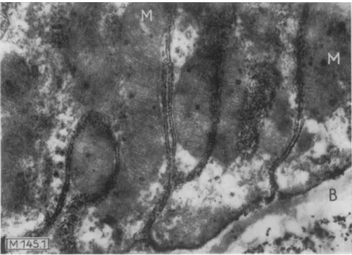
Fig. 1. Electron micrograph o f the basal part o f a tubule epithelial cell from m o u s e kidney with deep indentations of the cell membrane.
Histochemical detection of phosphatase according to a modification o f the method of Gomori. The site of the enzyme is marked with lead phosphate particles which are situated on the cell membrane in a single row, as can be seen in the section on the membranes which have been cut vertically. The representation of the double membrane of the mitochondria (M) indicates that the ultrastructure has been maintained, which is an essential requirement for the precision of the enzyme localization.
(B) Basal membrane. Original magnification: 20000 x, total magnification: 56000 x. (Taken from Mdlbert, Duspiva and v. Deimling1)).
have led in the last 10 years to a significant increase in the precision of the localization. By use of the electron microscope it is now possible in favourable cases to limit the site of an enzyme to an area of the cell which has a dimension of only one or two macromolecules (Fig. 1).
2) G. Gomori: Microscopic Histochemistry, Principles and Practice. Chicago University Press, Chicago 1952.
3) L. Lison: Histochimie et Cytochimie Animales. 2 n d ed., Gautier-Villars, Paris 1953.
4' F. Duspiva in Hoppe-Seyler-Thierfelder: Handbuch der physiologisch- und pathologisch-chemi- schen Analyse. 10th ed., Springer, Berlin-Gottingen-Heidelberg 1955, Vol. 2.
5> A. G. E. Pearse: Histochemistry. 2 n d ed., J. & A . Churchill, L o n d o n 1960.
6> H. W. Deane, R. J. Barrnett and A. M. Seligman in W. Grautnann and K. Neumann: Handbuch der Histochemie. G. Fischer, Stuttgart 1960, Vol. VII, part 1.
7> E. Mdlbert, F. Duspiva and O. v. Deimling, Histochemie 2, 5 [I960].
2 . General a) Principal techniques
As animal tissues are usually not suitable for direct microscopic study, sections must be prepared with a microtome. They are suspended in a medium which will maintain the cell structure. By addition of a buffered solution of the substrate the enzymatic reaction is started. As a rule the primary products of the reaction can diffuse freely; they do not remain in the immediate area of the enzyme molecule, but spread around the enzyme site. A reagent which forms an insoluble compound with one of the reaction products is therefore added with the substrate. The ideal is that the product should be trapped as soon as it is formed and that it should remain in the immediate area of the enzyme site. As the compounds chosen for the histochemical reaction yield insoluble dyes on combination with the products of the reaction, the enzyme site is marked so that it is visible microscopically. The site of the enzyme in the cell can then be determined (the simplest method being visually).
enzyme trapping reaction
Substrate > product > dye The following are the most important methods of quantitative histochemistry:
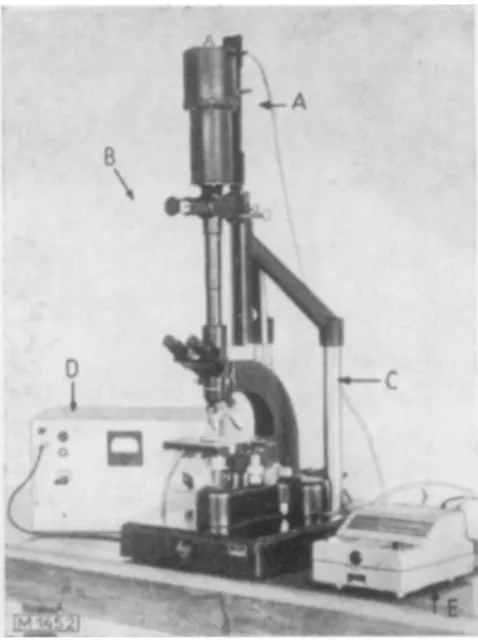
a) Microspectrophotometry
This method is due to Pollister and Ris8K The microspectrophotometer (Fig. 2) is in principle a photomicroscope9). The preparation is illuminated with monochromatic light. The photo- cathode of a photomultiplier is exposed to the image of the enzyme site in the preparation that has been stained histochemically. For the photoelectric determination of the amount of dye accumulated at the enzyme site, the geometry of the object must be taken into account in addition to the optical density. The basic principles of microspectrophotometry have been thoroughly dealt with by Sandritter1®.
P) Interference microscopy
The section is placed in a chamber for continual observation so that the object can be treated several times in succession with incubation medium or rinsing fluid without interrupting the measurements. The precipitate deposited at the enzyme site can be determined quantitatively by measurement of the optical phase differences at suitable successive time intervalsn ). y) Radiochemical methods
The trapping reagent is labelled with a radioactive isotope and has a known specific activity.
By measuring the radiation with a counting tube the amount of precipitate deposited in the tissue section during a histochemical reaction can be determined1 2 1 3). A more precise locali
zation of the precipitate is possible by shielding parts of the preparation or by use of micro- radioautography. The method of counting the silver grains in stripping film allows the quanti
tative evaluation of the radioautogram and a correlation of the radioactivity with individual cells and even with certain cell components.
8) A. W. Pollister and H. Ris, Cold Spring Harbor Sympos. quantitat. Biol. 12, 147 11947].
9) A. Schiihly, A c t a histochem. 9, 156 [1956].
1Q) W. Sandritter in W. Graumann and K. Neumann: Handbuch der Histochemie. G. Fischer, Stuttgart 1958, Vol. I, p. 220.
n ) H. G. Davies, R. Barter and / . F. Danielli, Nature [London] 773, 1234 [1954].
12) T. Barka, S. Szalay, Z. Posalsky and L. Kertesz, Acta Anat. 16, 45 [1952].
13) D. Shugar, H. Sierakowska and A. Szenberg, Acta Biochim. Polonica 5, 27 [1958].
924 Section C : Measurement of Enzyme Activity
b) Requirements
The requirements of the trapping reaction are that 1. it should be a rapid reaction, 2. it should give a non-crystalline, fine-grained precipitate, 3. it should have a high staining power and 4. it should have sufficient substantivity to be firmly bound to the particular region of cell proteins. In this way, mainly by excluding errors due to diffusion, a sharp optical identifi
cation of the enzyme site in the cell can be obtained. Another requirement of histochemical staining methods is that there should be a good correlation between the enzymatic activity at the enzyme site and the amount of dye deposited there.
Holt and O'Sullivan 14> have found that the activity of an enzyme can only be determined by the optical density when the primary products are formed by a zero order reaction, and when the dye formed subsequently follows a first order reaction. To fulfil these requirements the most important condition is a high concentration of substrate in the incubation medium.
With a sufficiently high concentration a steady state soon becomes established in an approxi
mately spherical area of the cell surrounding the enzyme, in which the substrate diffuses in and the reaction products diffuse out. That is to say that in every part of this cell area the substrate concentration depends on the enzyme activity which occurs there.
If the trapping reaction is to approximate to a first order reaction, then the reagent which forms the dye must also be present in high concentration. In practice it is not always possible to fulfil this condition, since high concentrations of these reagents often inhibit the enzymatic reactions. If the dye fulfils the above conditions (2—4), i.e. it produces no gross errors due to diffusion, then the efficiency of the localization of an enzyme depends mainly on the follow
ing factors: 1. the diffusion coefficients of the primary hydrolysis products, 2. the size of the enzyme site and 3. the velocity constant of the trapping reaction. Holt15) has studied this
14) S. J. Holt and D. G. O'Sullivan, Proc. Roy. Soc. [London], Ser. B. 148, 465 [1958].
is) S. J. Holt, J. Histochem. Cytochem. 4, 541 [1956].
Fig. 2. Simple microspectrophotometer according to Schiihly9). A : Photomultiplier
B: Measuring device C: Ortholux microscope D : Powei supply
E: Galvanometer
problem mathematically with a model and has calculated the influence of these factors on the efficiency of the localization. The rate of the trapping reaction is of great importance. With a slow trapping reaction a suitable localization can only be obtained, if the primary product of the enzymatic reaction and the dye have substantive properties.
c) Sources of error
Many of the special methods of enzymatic histochemistry do not satisfy the requirements laid down above. A cloudy or smeared staining of cells or the occurrence of dye clouds during the incubation of the sections in the staining bath always indicate errors due to diffusion.
The cause is not always to be found in the histochemical reaction, since the structural integrity of the tissue section is also important. Because of its unphysiological pH or its content of reactive constituents the incubation medium frequently damages the structure of the tissue.
The granular cell constituents swell in such media and proteins go into solution, with the result that enzymes are lost or are displaced within the cell. To decrease these sources of error the incubation time is always reduced to a minimum. In order to obtain the necessary level of enzyme activity so that this can be done, as little as possible of the enzyme activity should be lost during the preparation of the tissue section. As the sensitivity of enzymes and the extent with which they are bound to other cell proteins varies greatly from tissue to tissue, suitable methods of preparation must be determined in each case by careful preliminary experiments.
d) R a n g e of application
Good histochemical methods of enzyme detection are rare. The biochemist regrets that histo
chemistry is so occupied with hydrolases, especially with phosphatases (whose role in meta
bolism is still practically unknown), instead of being concerned with the key enzymes of metabolism1 6*. The reason for this is that no suitable chromogenic trapping reactions have been found for the products of many enzyme reactions, for example, several of the dehydro
genase reactions which are substrate-specific.
However, it is at present possible to follow an enzymatic reaction histochemically, even though the enzyme is bound to a few tiny granules which are found only in a certain type of cell of a particular tissue. The pH optimum of the enzyme can be measured and the Michaelis constants determined.
e) Precautions necessary for the preservation of cell c o m p o n e n t s
Procedures for the preliminary treatment of tissue which are generally valid cannot be given.
In each case the optimum conditions must be determined in preliminary experiments. The following details are only meant to serve as a guide.
a) Frozen sections
Usually frozen sections which have been cut with a microtome are used. The freezing does not have any effect on the chemical behaviour of the proteins, but on thawing gross changes of the cell structure, including swelling of the mitochondria, are observed. These changes affect the intracellular distribution of the enzymes, since structurally bound proteins are liberated. The unprotected frozen section is therefore not suitable for the histochemical detection 16) G. Siebert, Acta histochem. 2, 122 [1956].
926 Section C : Measurement of Enzyme Activity
of enzymes. To prevent an unintentional or premature thawing of the frozen section, it is recommended that the frozen sections are prepared in a cryostat. This instrument was first used for histochemical purposes by Linderstrom-Lang and Mogensen. The more recent designs include a refrigerating unit. The optimum working temperature is —16 to — 18°C, but can be as low as — 25° C. The apparatus gives sections of even thickness (3 \i thick up
wards as serial sections)1 7 _ 2 0 ).
Method: Cool the freshly excised tissue immediately to 0° C. Cool a few u.1. of the tissue in a cold bath (ether-dry ice) to — 70° C and freeze. Cut the frozen tissue in a cryostat. Trans
fer the sections (without thawing) to cold (0 to 2°C) 0.06 M buffer solution (pH 5.0-6.5), which contains 0.01 M ATP (or ADP, AMP) and 7.5% polyvinylpyrrolidone (PVP). The sections thaw in this solution without swelling of the mitochondria. Transfer the sections directly to an incubation medium which contains one of the following compounds: 0.44 — 0.88 M sucrose, 7.5% PVP or 6% dextran.
If the incubation medium has an alkaline reaction (pH 9.5) the cell structure is always damaged. Therefore in studies on enzymes with alkaline pH optima it is recommended that control experiments at less alkaline pH should be set up. The preliminary treatment of sections described above is also adequate for the detection of mitochondrially bound enzymes, such as succinic dehydrogenase and diaphorase2 1>.
P) Fixation of frozen sections
After incubation with the substrate it is expedient to fix the tissue again so as to stop the enzymatic reaction and to harden the cell structure for the production of permanent preparations. Any of the usual fixation methods employed in histology can be used, providing that the method does not solubilize or chemically change the dye deposited in the section.
A preliminary fixation of the tissue binds the enzymes to their sites and prevents the occur
rence of artifacts due to the loss of loosely bound proteins from the tissue section into the incubation medium. These proteins can carry with them the enzyme to be studied, even if the enzyme is bound to the cell structure (desmoenzyme). Preliminary fixation increases the sharpness of the localization, but unfortunately it usually also causes a partial loss of acti
vity. Numerous hydrolases and the DPN-diaphorase survive fixation of the tissue before the enzymatic reaction when it is carried out with special precautions. Several tissues with a high water content, for example, in the early stages of mammalian foetal development, cannot be cut in the unfixed state to give serial sections in cryostats. These tissues must be fixed first. However, preliminary fixation is also recommended for tissue which is easy to cut, if the enzyme under study can stand the necessary lengthy contact with the fixative. Whilst frozen sections are completely fixed in one of the following fixatives after 30 min., the fixa
tion time required for tissue blocks (volume: several u.1.) can be up to 24 hours.
A mild, but usually insufficient preliminary fixation is obtained by desiccation of the frozen sections in a cryostat at —15 to — 25° C. A better procedure is to fix with one of the following formaldehyde mixtures:
17) K. Linderstrom-Lang and K. R. Mogensen, C. R. Lab. Carlsberg. Ser. chim. 23, 27 [1938].
J8) R. Taugner and U. Wagenmann, Arch. exp. Pathol. Pharmakol. 234, 336 [1958].
19) p. B. Diezel, Mikroskopie 13, 196 [1958].
20) E. Reinholz, V. Belloch-Zimmermann and Ch. Wirth, Experientia 16, 286 [I960].
20 D. G. Scarpelli and A. G. E. Pearse, J. Histochem. Cytochem. 6, 369 [1958].
I . " Chloralformalin" according to Baker, Hew and Fishman 2 2 ).
Mix 20 ml. 40 % formaldehyde solution with 80 ml. distilled water containing 100 mg. chloral hydrate. Adjust the pH to 7.0 with 1 N KOH.
II. "Calcium-Formol Mixture" according to Baker23).
Dissolve 1 g. anhydrous calcium chloride in 100 ml. 4 % formaldehyde solution and add 1 g.
granular calcium carbonate. After thorough shaking, allow the insoluble material to settle and decant off the supernatant.
It is recommended that these solutions should include 0.88 M sucrose, 7.5% PVP or 6%
dextran to preserve the fine structure of the cells.
Both solutions are suitable for the fixation of tissue containing alkaline and acid phosphatase, esterases, lipases and ^-glucuronidases.
Place blocks of freshly excised tissue (not larger than 10x5 x 2 mm.3) in the cold (0—2°C) fixative and incubate for 24—48 hours. To store the fixed tissue samples until ready for further treatment according t o2 4 ), dry the outside of the pieces of tissue and transfer to ice-cold 0.88 M sucrose solution containing 9 g. gum arabic/1000 ml. The tissues can remain in this solution up to 24 hours. They should now be very easy to cut and the fine structure should be well preserved (only suitable with stable enzymes).
The same osmotic protective measures should be observed for the enzymatic reaction as have been described for the treatment of unfixed tissue, since the fixation does not bring about an absolute binding of the enzymes.
"Chloralformalin" or "calcium-formol mixture" are used for the fixation of the tissue after the enzymatic reaction. The "calcium-formol mixture" is most suitable for the preservation of the lipid-rich structure of the cytoplasm. According to Berg25* sections prepared in a cryostat can be fixed at — 10°C in a mixture of 50 ml. acetone, 9 ml. 40% formaldehyde and
1.02 g. MgCl2-6H20 diluted to 100 ml. with distilled water. The sections are rinsed with 40% acetone.
y) Paraffin sections
Embedding in paraffin requires careful dehydration of the tissue sample; the activity of the enzyme and its location must be maintained. The dehydration can also bring about fixation of the tissue if the water is removed with organic solvents (acetone, ethanol).
I. The oldest method for dehydration is the acetone method of Gomori:
Immediately after excision, cool small pieces of tissue to 0°C and place for 12—24 hours in ice-cold, anhydrous acetone. Change the acetone several times. Remove the last traces of water from the tissue samples with absolute ethanol. Embed the tissue by way of celloidin into paraffin (not above 53°C) according to one of the usual histological methods.
Although most tissues are poorly fixed with this method, it has found wide application and very important results have been obtained with it.
22) J. R. Baker, H. Hew and W. H. Fishman, J. Histochem. Cytochem. 6, 244 [1958].
23) j . R. Baker, Quart. J. Micr. Sci. 87, 441 [1946].
24) S. J. Holt and F. J. Withers, Proc. Roy. Soc. [London], Ser. B., 148, 520 [1958].
25) G. G. Berg, J. Histochem. Cytochem. 6, 397 [1958].
928 Section C : Measurement of Enzyme Activity
II. Dehydration by freeze-drying:
Lyophilization2 6) of the tissue sample in high vacuum followed by embedding in paraffin gives excellent results. With correct handling, sections are obtained in which the fine structure is well maintained. However, freeze-drying does not fix cell proteins sufficiently, so that losses and displacement of the cell proteins occur on wetting the section after removal of the paraf
fin. The unfixed paraffin section must therefore in most cases be fixed again.
Method: Cool the tissue which has been removed immediately after the death of the animal to 0°C and cut it into small slices about 1 mm. thick with a razor blade. Introduce a brass cylinder containing isopentane or propane into a Dewar flask containing liquid air or better still liquid nitrogen. To cool the liquid evenly stir with a small metal sieve which is fastened to a long shaft. Allow the sieve to lie at the bottom of the cylinder. Introduce the pieces of tissue into the isopentane by means of a thin wire loop. Meanwhile prepare the freeze- drying flask with some paraffin wax for embedding and cool to — 50° C in a mixture of acetone and carbon dioxide snow. Remove the sieve with the frozen tissue samples from the iso
pentane bath and place in the pre-cooled freeze-drying flask. Connect this to the vacuum.
Maintain the temperature at —30 to — 50°C during the drying*).
On completion of the drying allow the flask containing the tissue sample to warm to room temperature. Melt the paraffin which is at the bottom of the container. Within a few minutes the tissue sinks into the liquid paraffin. Now turn off the vacuum pump and carefully reduce the vacuum by introducing dry air. Remove the liquid paraffin containing the tissue from the container and pour into blocks. Remove the paraffin from the sections before further treatment, but do not use xylene or benzene since these solvents usually cause a loss of enzyme activity. Petroleum ether has proved most successful for the removal of paraffin. The usual practice in histology of transferring the sections through the various concentrations of alcohol must be omitted. After removal of the paraffin allow the sections to dry in the air.
According to Burstone 2 7) the brittleness which is occasionally encountered with freeze-dried sections, and which causes problems during the cutting, can be avoided by double embedding.
Treat the freeze-dried block before the paraffin embedding with celloidin or with a 15%
solution of high molecular weight polyvinylchloride in the purest tetrahydrofuran or with a 20 % solution of an acrylic ester plastic at 0° C. After this impregnation bring the samples to room temperature and embed directly in paraffin.
Sections from which the paraffin has been removed and which have been dried in air cannot be added directly to aqueous buffer solutions. They are transferred through cold 100%, 90%, 75 % and 50 % acetone to the incubation medium, to which several authors add high concen
trations of neutral salts so as to avoid diffusion of the enzyme. G. G. Berg25* recommends that the sections should be fixed before removal of the paraffin. For this, either float the paraffin ribbons on a 0.02 M magnesium acetate solution (pH 6) at 40° C or treat at —10° C with a mixture of 50 ml. acetone, 9 ml. 40% formaldehyde and 1.02 g. MgCl2-6 H2O diluted to 100 ml. with distilled water, rinse in 50% acetone and fix to the slides.
*) The freeze-drying apparatus G 01 from Leybold, Cologne, Germany is suitable. The apparatus is equipped with a rotary pump and a diffusion pump which has a suction capacity of 100 l./sec.
and removes ca. 0.36 g. water/hour at a pressure of 1 0-3 Torr.
26) K. Neumann in W. Gaumann and K. Neumann, Handbuch der Histochemie. G. Fischer, Stuttgart 1958, Vol. I. part. 1.
27) M. S. Burstone, J. Histochem. Cytochem. 6, 322 [1958].
3. Special Methods a) Method involving precipitation of metallic salts
For about 2 decades only a few methods for the detection of oxidases in blood smears and histological sections were known; their value was much disputed. Now about 30 different enzymes can be detected histochemically. The first enzyme to be detected by histochemical means was alkaline phosphatase which was demonstrated simultaneously by Gomori^ and
Takamatsu2). The orthophosphate liberated from glycerophosphate by the phosphatase was precipitated as the calcium salt (hydroxylapatite). In the original method the precipitate was transformed to silver phosphate (according to3)) which became black on exposure to light and was therefore made visible. In 1941 Gomori4) introduced the method of converting the calcium phosphate to cobalt phosphate, which can be transformed to the black sulphide by means of ammonium sulphide (cobalt sulphide method). One of the modifications published by Gomori5) is still used a great deal.
The calcium-cobalt method is unsuitable for enzymes which have their pH optimum in the acid range, since calcium phosphate is soluble below pH 8. Calcium is therefore replaced by lead, which can be used in the weakly acid range up to pH 6 and can be converted to the black sulphide directly with H2S.
Apart from the lead phosphate method there is also one employing iron-hydroxyquinoline
(formation of Berlin blue).
The cobalt sulphide method is used to detect all enzymes having esters as substrates which on hydrolysis produce an anion that can be precipitated with C a2 +. Glucose-6-phosphatase, fructose-1,6-diphosphatase, ATPase, esterases and lipases can be detected by this method.
According t o6) the cobalt sulphide method does not give quantitative results, since the ex
change of Ca for Co phosphate is not complete. On the other hand, the exchange with silver is quantitative 7>. However, the silver precipitate is frequently coarse-grained and gives a less detailed picture. Losses of Ca and Co phosphate occur on rinsing the preparations. For recent suggestions to avoid this error see the individual methods.
Alkaline Phosphatase *) Preliminary Treatment of the Tissue
Fix in cold acetone, doubly embed through celloidin into paraffin according to p. 928.
Reagents
1. Diethylbarbituric acid (veronal), sodium salt
2. (3-Glycerophosphoric acid (glycerol-2-phosphoric acid)
disodium salt-5 H 2 O
3. Calcium chloride, CaCl2-6 H20
4. Magnesium sulphate, A. R., MgS04-7 H2O
*) Original procedure according t o5) .
1) G. Gomori, Proc. Soc. exp. Biol. Med. 42, 23 [1939].
2) H. Takamatsu, Trans. Soc. Path. Japan 29, 492 [1939].
3) J. v. Kossa, Beitr. path. Anat. u. allgem. Path. 29, 163 [1901].
4) G. Gomori, J. cell. comp. Physiol. 17, 71 [1941].
s) G. Gomori, Amer. J. clin. Pathol. 16, 347 [1946].
6) H. Hotter, S. Lovtrup and J. Rubin, Acta chem. scand. 5, 194 [1951].
7) D, Shugar, H. Sierakowska and A. Szenberg, Acta biochim. polonica 5, 27 [1958].
930 Section C: Measurement of Enzyme Activity
5. Cobalt nitrate, A. R., C o ( N 03)2 • 6 H20 6. Ammonia solution, cone., sp. gr. 0.88, A. R.
7. Hydrogen sulphide (from a Kipp's apparatus) 8. Sodium hydroxide, A. R., 0.1 N
9. Cysteine
In addition, solvents and reagents for fixing and counter-staining the section.
Preparation of Solutions
I. Incubation solutions (pH 9.5):
a) Dissolve 0.5 g. Na-p-glycerophosphate and 0.5 g. Na veronal in 25 ml. portions of distilled water. Mix the solutions and add 50 ml. distilled water. Then add 5 ml. of a solution of 2 g. CaCl2-6 H2O/100 ml. and 2 ml. of a solution of 2 g. MgS04- 7 H2O/100 ml. Mix and adjust to pH 9.5 with 0.1 N NaOH.
b) As a), but without the (^-glycerophosphate solution; replace with 25 ml. distilled water.
c) As a), but dissolve 60 mg. cysteine in the mixture.
II. Cobalt nitrate (1 % w/v C o ( N 03)2 • 6 H20 ) :
Dissolve 1 g. Co(N03)2-6 H20 in distilled water and make up to 100 ml.
III. Yellow ammonium sulphide:
Prepare a, ca. 1 % solution by diluting 4 ml. cone, ammonia to 100 ml. with distilled water and gassing with H2S until the solution is yellow, or dilute a commercial prepara
tion of yellow ammonium sulphide.
Procedure
Prepare paraffin sections 5 u, thick. Fix the sections to slides (pre-treated with albumin- glycerol). If necessary, store at 4°C. Remove the paraffin from the sections with petroleum ether and dry in air. They can also be transferred through a series of alcohol concentrations to water.
Pipette into staining jars (glass troughs with grooves):
Experimental (a) 100 ml. incubation solution (la) Controls (b) 100 ml. incubation solution (Ib) (c) 100 ml. incubation solution (Ic) (d) 100 ml. incubation solution (la)
Equilibrate in a water bath at 37° C. Place in the solution in each jar a batch of slides containing the sections. Inactivate tissue for control (d) by immersing in water at 90°C (for 2 to 5 min.).
According to the desired enzyme activity or that determined by preliminary experiment leave for 0.5 to 16 hours in the solution. Rinse with running tap water, immerse for 3 —5 min.
in solution (II) and rinse thoroughly with distilled water. Immerse for 1—2 min. in solution (III). Wash well with distilled water. (If desired counter-stain with 1 % aqueous eosin solu
tion). Dehydrate through the alcohol series, clear in xylene and mount in Canada balsam.
Result
Phosphatase sites are stained brown to black.
N o t e s
If the results are to be evaluated quantitatively, the method must be modified as follows: fixation of the tissue by the acetone technique (p. 928) and preparation of the sections by the paraffin technique (p. 928). D o not fix the tissue sections o n slides, but instead allow to float freely in the incubation medium for the enzymatic reaction.
Replace the veronal buffer (inhibits the activity o f alkaline phosphatase) by veronal-acetate buffer prepared according to Michaelis (0.028 M ; p H 9.5) or tris buffer.
Rinsing with water leads to losses of calcium and cobalt phosphate from the sections. According t o8)
rinse with 40 % acetone instead of tap or distilled water.
The exchange o f calcium phosphate for cobalt phosphate is not quantitative, but for silver phosphate it is rapid and stoichiometric. Treat the sections with 1 % silver nitrate solution instead of cobalt nitrate solution (II) and rinse thoroughly with distilled water (no losses). Expose the preparations to bright daylight until the precipitated silver salts have turned black.
Adenosinetriphosphatases9'1 0)
The ATP-cleaving hydrolases, which remain active in frozen sections, can be divided into three groups according to their behaviour with S H c o m p o u n d s :
1. Phosphatases which are only active in the presence o f S H groups. They can be detected in skeletal and heart muscle fibres and in the mitochondria of the cells of kidney tubules. They are inhibited by /7-chloromercuribenzoate or salyrgan*), but can be reactivated by cysteine or B A L . The specific substrate for this group of enzymes is A T P ; only the terminal phosphate group is hydrolysed. A D P and A M P are not attacked. A member of this group is myosin ATPase.
2. Phosphatases which are inhibited by S H groups. A M P is hydrolysed in addition to A T P (5'- nucleotidase). Cysteine and B A L inhibit. /?-Chloromercuribenzoate or salyrgan have n o effect or activate. Enzymes which hydrolyse phosphomonoesters (alkaline phosphatases) and fructose-1,6- diphosphate belong to this group.
3. Phosphatases which are not acted upon by S H groups. The enzymes of this group occur in the endothelium and in the s m o o t h muscle of the blood vessels. If A D P is used as substrate, enzyme sites containing a true ATPase (enzymes of Group 1) d o not stain.
A modification of the method of Gomori (1941) is used for the detection of ATPases.
Preliminary Treatment of the T i s s u e
Prepare frozen sections 5 (j, thick in a cyrostat**). Take the necessary precautions to maintain the structure o f the cells (see p. 925); also prepare a less alkaline control reaction mixture***).
Reagents
All the reagents for the determination of phosphatase (p. 929) with the exception of (3-glycero- phosphate. In addition:
10. Adenosine triphosphate, ATP
disodium salt, ATP-Na2H2-3 H2O; commercial preparation see, p. 1006.
*) Trade name of Farbwerke Hoechst for a mixture of the N a salt of ocarboxymethylsalicylic acid-(3-hydroxymercuri-3-methoxypropyl)-amide and theophyllin.
**) Fixation with chemical agents inactivates the enzyme; freeze-drying causes no inactivation.
***) Recommended by the author.
8) G. Gomori, J. Lab. clin. Med. 37, 526 [1951].
9) H. A. Padykula and E. Herman, J. Histochem. Cytochem. 3, 161 [1955].
!o) H. A. Padykula and E. Herman, J. Histochem. Cytochem. 3, 170 [1955].
932 Section C : Measurement of Enzyme Activity
11. Adenosine diphosphate, ADP
trisodium salt, A D P- N a 3 ; commercial preparation, see p. 1004.
12. 2,3-Dimercapto-l-propanol, BAL 13. /7-Chloromercuribenzoate, PCMB 14. Salyrgan*)
15. Sodium hydroxide, 1 % w/v
In addition, the solvents and reagents for fixing and counter-staining the sections.
Preparation of Solutions
I. Incubation solutions (ca. 0.012 M ATP; pH 9.4):
a) Containing ATP: mix 20 ml. 0.1 M Na veronal solution (2.06 g. Na veronal/100 ml.) with 0.18 M CaCl2 solution (2 g. CaCl2/100 ml.) and 30 ml. distilled water. Add 1 g. ATP-Na2H2-3 H20 . Adjust to pH 9.4 with 0.1 N NaOH (glass electrode) and dilute to 100 ml. with distilled water.
b) Without substrate: like a), but without ATP.
c) Containing substrate and 2.5 x 10~3 M cysteine: like a), but dissolve 30 mg. cysteine in the final mixture. Adjust the pH as for a).
d) Containing substrate and 5 x 10~3 M BAL: like a), but add 0.1 ml. BAL to the final mixture. Adjust the pH as for a).
e) Containing substrate and 2.5 x l 0 ~3 M PCMB: like a), but instead of the distilled water add 25 ml. PCMB solution (351 mg. PCMB dissolved by the dropwise addition of 0.1 N NaOH and diluted to 100 ml. with distilled water) to the mixture. Adjust the pH as for a).
f) Containing substrate and 4 x 10~4 M salyrgan: like a), but instead of distilled water add 4 ml. salyrgan solution (48 mg. salyrgan dissolved by the dropwise addition of 1% Na2C03 solution and diluted to 10 ml. with distilled water) to the mixture.
Adjust the pH as for a).
g) Containing 0.018 M ADP as substrate and containing 5 x 1 0-3 M BAL or 5 x 10~3 M cysteine: like a), but instead of ATP add 1 g. ADP-Na3 and also add 60 mg. cysteine or 0.1 ml. BAL to the final mixture. Adjust the pH as for a).
II. Cobalt nitrate (1 % w/v C o ( N 03)2 • 6 H20 ) :
Dissolve 1 g. Co(N03)2-6 H20 in distilled water and make up to 100 ml.
III. Yellow ammonium sulphide:
See p. 930.
Procedure
Preliminary remarks: The ATPase sites can only be determined by comparison with controls.
Therefore in addition to mixtures without substrate and after heat inactivation of the enzyme the following are prepared:
1. Experimental reaction mixtures containing ATP
*) For the chemical designation and the manufacturer, see footnote*) on p. 931.
2. Controls a) containing anti-SH compounds (PCMB or salyrgan): ATPase is inhibited, while the other phosphatases remain active.
b) containing SH compounds (BAL or cysteine): ATPase is active, while the other phosphatases are inhibited.
c) containing ADP as substrate and with the addition of BAL or cysteine:
ATPase does not react, while other phosphatases of Group 2 (p. 931) are inhibited. Phosphatases of the endothelium and smooth muscle which are not acted upon by SH groups are active (Group 3, p. 931).
The rest of the procedure is as for alkaline phosphatase (see p. 930).
Method:
Pipette into staining jars:
Experimental (a) 100 ml. incubation solution (la) Controls (b) 100 ml. incubation solution (Ie) or (If)
(c) 100 ml. incubation solution (Ic) or (Id) (d) 100 ml. incubation solution (Ig) (e) 100 ml. incubation solution (Ib) (f) 100 ml. incubation solution (la).
Equilibrate in a water bath at 37° C and proceed as described on p. 930.
Result
ATPase sites are stained brown to black.
Aldolase1 1)
Aldolase cleaves fructose-1,6-diphosphate to give glyceraldehyde-3-phosphate and dihydroxyacetone phosphate. The triose phosphates precipitate in the presence of magnesia mixture. Their further degradation by way of the Embden-Meyerhof pathway is inhibited by iodoacetate. The precipitated phosphate is converted to the cobalt salt which is transformed to the black sulphide with ( N H ^ S . The transformation of triosephosphate to cobalt phosphate depends on the spontaneous liberation of orthophosphate from triosephosphate at room temperature in alkaline medium. This reaction does not proceed sufficiently rapidly to exclude diffusion. The authors use tissue fixed in 8 0 % alcohol, which after thorough rinsing to remove the alcohol is frozen prior to the preparation of frozen sections.
The method has not been developed further; little is known of its reliability.
Acid Phosphatase 12> 13>
This method was developed by Gomori in 1941 and further modified by him in 1950. The enzyme is more sensitive than alkaline phosphatase. Frozen sections give more uniform results than paraffin sections.
Preliminary Treatment of the Tissue
Preliminary fixation of the tissue samples in cold formalin (see p. 927). Preparation of the frozen tissue sections in a cryostat.
I D R. J. L. Allen and G. H. Bourne, J. exp. Biology 20, 61 [1943], 12) G. Gomori, Ar-h. Path. 32, 189 [19411.
13) G. Gomori, Stain Tech. 25, 81 [1950].
934 Section C : Measurement of Enzyme Activity
Reagents
1. Sodium acetate-3 H2O, A. R.
2. Acetic acid, A. R., I N
3. Lead nitrate, A. R., P b ( N 03)2
4. p-Glycerophosphoric acid (glycerol-2-phosphoric acid) disodium salt - 5 H2O.
5. Ammonia solution, cone, sp. gr. 0.880, A. R.
6. Hydrogen sulphide (from a Kipp's apparatus) 7. Sodium fluoride
In addition, solvents for the fixing the sections.
Preparation of Solutions
I. Incubation solutions (pH 5.0):
a) Mix 10 ml. 1 M acetate buffer pH 5.0 (mix 70.5 ml. of a solution of 136.1 g. Na acetate/1000 ml. with 29.5 ml. 1 N acetic acid), 3 ml. 5% lead nitrate solution (5 g.
Pb(NO3)2/100 ml.), 20 ml. distilled water and 10 ml. 2% Na-(3-glycerophosphate (2 g./lOO ml.). Check the pH.
b) Like a), but 10 ml. distilled water instead of the ^-glycerophosphate solution.
c) Like a), but add 2 mg. NaF to the final mixture.
II. Acetic acid (2% w/v):
Dilute 1 volume 1 N acetic acid with 2 volumes distilled water.
III. Yellow ammonium sulphide:
Prepare a ca. 1 % solution by diluting 4 ml. cone, ammonia to 100 ml. with distilled water and gas with H2S until the solution is yellow.
Procedure
Prepare frozen tissues 5 —15 u. thick (see p. 926) and fix to slides (pre-treated with gelatine) Prepare controls without substrate and with fluoride to inhibit the acid phosphatase.
Pipette into staining jars:
Experimental (a) 40 ml. incubation solution (la) Controls (b) 40 ml. incubation solution (I b) (c) 40 ml. incubation solution (Ic)
Equilibrate in a water bath at 37 °C. Place in the solution in each jar a slide containing a section.
According to the enzyme activity (estimated or previously determined) allow to stand for 0.5 — 16 hours in the incubation solution. Rinse for a short time under running tap water.
Stop the enzyme reaction by immersing the sections for ca. 2 min. in solution (II), rinse again and immerse in solution (III) for 1—2 min. Rinse thoroughly with water. (Counter-stain as desired.) Mount in glycerol-gelatine.
Result
Phosphatase sites are brown to blach. With good fixation fine granular localization (lysosomes) is obtained.
Glucose-6-phosphatase1 4' 15>
Frozen sections (6—10 u. thick) prepared with a cryostat are incubated for 6 — 10 min. in a medium containing lead nitrate and K or N a glucose-6-phosphate buffered at p H 6.7 as substrate. T h e incubation medium consists of 1 part of a 1 % potassium glucose-6-phosphate solution (pH 6.7) and 2 parts of a 0 . 2 ^ P b ( N O 3 ) 2 solution. A s a control the sections are inactivated by heat or the incuba
tion medium is replaced by Lugol's iodine. A s the substrate is also hydrolysed by unspecific alkaline phosphatases, the glucose-6-phosphatase enzyme sites can only be determined in a comparative study using several substrates.
Phosphoamidase
According to Gomori1^ the staining pattern in the histochemical reaction with />-chloroanilido- phosphoamide at p H 5.6 (in a mixture corresponding to that used for the detection of acid phospha
tase, p. 933) differs from that given by acid phosphatase and indicates the existence of a special enzyme.
However, E. Pearsel7) could not find any difference in the staining reaction using this substrate as compared that obtained with the usual assay for the detection of acid phosphatase. Meyer and Wein
mann1^ have checked the method and studied the distribution of the enzyme which catalyses the hydrolysis of /?-chloroanilidophosphoamide in rat tissues 1 9>. The reaction catalysed by the enzyme in living cells is unknown. A phosphoamide b o n d occurs in creatine phosphate and arginine phosphate.
Hotter and Si-Oh 1 9 a> have suggested that the enzyme is identical with phosphoprotein phosphatase.
The highest activity is found in epithelium and nerve tissue. The enzyme has a well-defined distri
bution pattern in tissue and is found particularly in tissues which are insulin insensitive.
Preliminary Treatment of the Tissue
Fix the freshly excised tissue in ice-cold absolute acetone. Prepare the paraffin sections as for the detection of alkaline phosphatases (p. 929).
Reagents
1. Sodium hydroxide 2. Maleic acid
3. Lead nitrate, P b ( N 03)2 4. /?-Chloroanilidophosphoamide
prepared according t o2 0 ).
5. Sodium hydroxide, 1 N 6. Sodium chloride
7. Ammonia solution, cone, sp. gr. 0.880 8. Hydrogen sulphide (from a Kipp's apparatus) Preparation of Solutions
I. Substrate solution:
Dissolve 2.08 g. /?-chloroanilidophosphoamide in 14 to 15 ml. 1 N NaOH and dilute to 100 ml. with distilled water.
14) A. D. Chiquoine, J. Histochem. Cytochem. /, 429 [1953].
i-s> A. D. Chiquoine, J. Histochem. Cytochem. 3, 471 [1955].
if) G. Gomori, Proc. Soc. exp. Biol. Med. 68, 354 [1948].
•7) E. Pearse, Histochemistry Theoretical and Applied. London [1954].
•8) J. Meyer and J. P. Weinmann, J. Histochem. Cytochem. 3, 134 [1955].
19> J. Meyer and J. P. Weinmann, J. Histochem. Cytochem. 5, 354 [1957].
19a) H. Holier and Li Si-Oh, C. R. Trav. Lab. Carlsberg Ser. Chim. 27, 393 [1950].
20) Si-Oh. Li, Acta chem. scand. 4, 610 [1950]; P. Otto, Ber. dtsch. chem. Ges. 28, 616 [1895].
936 Section C: Measurement of Enzyme Activity
II. Incubation solution (pH 5.6):
To 900 ml. maleate buffer (62 ml. 1 N NaOH + 5.8 g. maleic acid/1000 ml.) add 24.6 ml.
0.1 M P b ( N 03)2 solution (3.3 g. (Pb(NO3)2/100 ml.) and 45 ml. substrate solution (I).
Dissolve 2.05 g. NaCl in this mixture. Adjust to pH 5.6 with ca. 16.5 ml. 1 N NaOH.
III. Yellow ammonium sulphide:
See p. 930.
Procedure
Prepare paraffin sections 5 \L thick (see p. 927) and fix the sections to slides. Inactivate control sections with 1 N H N 03. Remove the paraffin from all sections with petroleum ether. Pour incubation solution (II) into flat glass dishes and lay the slides horizontally in the incubation solution with the surface to which the section is fixed underneath and about 7 mm. from the bottom of the incubation vessel. Incubate for 1.5 to 4 hours at 40—42° C. Rinse the sections under running tap water, treat with dilute ammonium sulphide solution (III) and proceed as described in the section "Acid phosphatase" (p. 933).
Result
Phosphoamidase sites are brown to black.
Lipase21 >
Only water soluble esters, which on hydrolysis give products having insoluble calcium salts, are suitable for the detection of lipase. This condition is fulfilled by the " T w e e n s " which are prepared by the Atlas Powder Comp. for technical purposes and are fatty acid esters of polyglycols and poly- sorbitol. True lipases hydrolyse the esters of both saturated fatty acids and unsaturated fatty acids, while typical esterases only attack the esters of saturated fatty a c i d s2 2) .
Preliminary Treatment of the Tissue
Fix in cold acetone and then embed in paraffin or prepare frozen sections with a cryostat.
Reagents 1. Substrates
a) Tween 80 (oleic acid ester of a polysorbitol) as substrate for true lipase b) Tween 60 (stearic acid ester of a polysorbitol) as substrate for esterase 2. Tris-hydroxymethyl-aminomethane, tris
3. Calcium chloride, CaCl2-6 H20 4. Lead nitrate, P b ( N 03)2
5. Ammonia solution, cone, sp. gr. 0.880 6. Hydrogen sulphide (from a Kipp's apparatus) Preparation of Solutions
I. Substrate solution:
a) Dissolve 5 g. Tween 80 in 100 ml. distilled water.
b) Dissolve 5 g. Tween 60 in 100 ml. distilled water.
21) G. Gomori, Proc. Soc. exp. Biol. Med. 58, 362 11945].
22) G. Gomori, Proc. Soc. exp. Biol. Med. 72, 697 [1949].
II. Tris buffer (0.5 M; pH 7.2-7.4):
Dissolve 6.1 g. tris-hydroxymethyl-aminomethane in 20 ml. distilled water, adjust to pH 7.2-7.4 with 41.5 ml. 1 N H Q and dilute to 100 ml. with distilled water.
III. Calcium chloride (10% w/v):
Dissolve 10 g. CaCl2-6 H20 in 100 ml. distilled water.
IV. Incubation solution:
Mix 5 ml. tris buffer (solution II) with 2 ml. CaCl2 solution (III), add 2 ml. substrate solution (I) and 40 ml. distilled water.
V. Lead nitrate (1 - 2 % w/v):
Dissolve 1 - 2 g. P b ( N 03)2 in 100 ml. distilled water.
VI. Yellow ammonium sulphide:
See p. 930.
Procedure
Incubate the sections in solution (IV) for 3 — 12 hours at 37° C. Wash with distilled water and incubate for 15 min. at 20°C with lead nitrate solution (V). Wash for 5 min. under running tap water and immerse for 1—2 min. in solution (VI). Wash thoroughly with distilled water, mount in glycerol-gelatine or dehydrate through the alcohol series and mount in Canada balsam.
Result
Lipase sites are brown to black.
Cholinesterases2 3)
On hydrolysis in the presence of C u2+ ions thioanalogues of acetylcholine and butyrylcholine form a precipitate of copper thiocholine at the enzyme site, which can be transformed to the black sulphide.
T o avoid errors due to diffusion the reaction is carried out in a medium saturated with copper thio
choline and in the presence of sodium sulphate (to immobilize the enzyme). Acetylcholinesterase hydrolyses acetylthiocholine2 4), but not butyrylthiocholine2 5) which is a g o o d substrate for serum cholinesterase. If the sections are incubated for 30 min. with diisopropylfluorophosphate solution (final concentration 1 0 ~7 M), the unspecific (serum) cholinesterase is inhibited 100%, while acetyl
cholinesterase is inhibited less than 5 % 26).
Preliminary Treatment of the Tissue
Prepare frozen sections from freshly excised, unfixed tissue. Freeze-drying does not inhibit the acti
vity o f cholinesterases.
Reagents 1. Glycine
2. Copper sulphate, CuS04-5 H20 3. Maleic acid, monosodium salt
23) G. B. Koelle and / . S. Friedenwald, Proc. Soc. exp. Biol. Med. 70, 617 [1949].
24) G. B. Koelle, J . Pharmacol, exp. Therap. 100, 158 [1950].
2-s> G. B. Koelle, J. Pharmacol, exp. Therap. 103, 153 [1951].
2 6> G o o d results are also obtained by the thioacetic acid method of M. Crevier and L. F. Belanger, Science [Washington] 722, 556 [1955].
938 Section C: Measurement of Enzyme Activity
4. Sodium hydroxide, 1 N
5. Sodium sulphate, N a2S 04- 1 0 H20 6. Magnesium chloride, MgCl2 • 6 H20 7. Acetylthiocholine iodide
prepared according t o2 7) ; available from Borden Co., Chem. Div., Philadelphia, Penn., U S A .
8. Butyrylthiocholine iodide
prepared according t o2 7) ; available from Borden Co., Chem. Div., Philadelphia, Penn., U S A .
9. Diisopropylfluorophosphate, DFP*) 10. Propyleneglycol, anhydrous
11. Copper sulphide, CuS
12. Ammonia solution, cone, sp. gr. 0.88 13. Hydrogen sulphide (from a Kipp's apparatus) 14. Formaldehyde, 10% solution
15. Copper thiocholine
Filter incubation solution (which has already been used) (see below, Table 1), incubate for 2 — 3 days at 38° C (hydrolysis of the substrate), centrifuge off the precipitate, wash with water and dry in a desiccator.
Preparation of Solutions I. Copper sulphate-glycine:
Dissolve 3.75 g. glycine and 2.5 g. CuS04-5 H20 in distilled water and make up to 100 ml.
II. Maleate buffer (pH 6.0):
Dissolve 9.6 g. sodium hydrogen maleate in 52.2 ml. 1 N NaOH and dilute to 100 ml.
with distilled water.
III. Sodium sulphate (40% w/v; pH 6.0):
Dissolve 40 g. N a2S 04- 1 0 H20 in 50 ml. distilled water, adjust to pH 6.0 with 0.1 N NaOH and dilute to 100 ml. with distilled water.
IV. Magnesium chloride (1 M):
Dissolve 20.33 g. MgCl2-6 H20 in 100 ml. distilled water.
V. Copper sulphate (0.1 M):
Dissolve 2.5 g. CuS04-5 H20 in 100 ml. distilled water.
VI. Acetylthiocholine iodide:
Stir together 23 mg. acetylthiocholine iodide, 1.2 ml. distilled water and 0.4 ml.
C u S 04 solution (V) and centrifuge. Decant the supernatant.
VII. Butyrylthiocholine iodide:
Stir together 43 mg. butyrylthiocholine iodide, 1.8 ml. distilled water and 0.6 ml.
C u S 04 solution (V) and centrifuge. Decant the supernatant.
*) e.g. from Dr. Th. Schuchardt G m b H , Munich, Germany.
27) G. B. Koelle and / . S. Friedenwcdd, Proc. Soc. exp. Biol. Med. 70, 617 [1949].
VIII. Diisopropylfluorophosphate, D F P : a) Stock solution (0.1 M):
Dissolve 184 mg. diisopropylfluorophosphate in 10 ml. anhydrous propyleneglycol.
b) Dilute solution (10~6 M):
Dilute 0.1 ml. stock solution a) with anhydrous propyleneglycol in the volume ratio 1:105. Do not allow the dilute solution to stand longer than 30 min.
IX. Preservative solution 1:
Mix 4.5 ml. distilled water, 9 ml. N a2S 04 solution (III) and 1.5 ml. dilute DFP solution (VIIIb).
X. Preservative solution 2:
Mix 6 ml. distilled water and 9 ml. Na2SC>4 solution (III).
XI. Preservative solution 3:
Mix 6 ml. distilled water and 10.5 ml. Na2SC>4 solution (III).
XII. Sodium sulphate (20% w/v, saturated with Cu thiocholine):
Dissolve 20 g. Na2SC>4-10H2O in 100 ml. distilled water, add a spatula tip of Cu thiocholine, shake, centrifuge and decant the supernatant fluid.
XIII. Sodium sulphate (10% w/v, saturated with Cu thiocholine):
Dissolve 10 g. Na2SCV 10 H20 in 100 ml. distilled water, add a spatula tip of Cu thio
choline and proceed as for solution XII.
XIV. Copper thiocholine (saturated):
Shake a spatula tip of Cu thiocholine in 100 ml. distilled water, centrifuge and decant the supernatant fluid.
XV. Ammonium sulphide (saturated with CuS):
Prepare a solution of yellow ammonium sulphide according to p. 930. For each 100 ml.
add a spatula tip of CuS, shake, centrifuge and decant the supernatant fluid.
XVI. Formaldehyde (10% w/v, saturated with CuS):
To 100 ml. 10% formaldehyde solution add a spatula tip of CuS, shake, centrifuge and decant the supernatant fluid.
XVII. Incubation solutions:
Incubation
solution For
Dist.
water [ml.] I
[ml.] II [ml.]
Solution N o . I l l IV [ml.] [ml.] VI
[ml.] VII [ml.]
Cu thio
choline A Specific
cholinesterase 2.1 0.6 1.5 9.0 0.6 1.2 — trace B Unspecific
cholinesterase 0.6 0.6 1.5 10.5 0.6 — 1.2 trace
C Control 1.4 0.4 1.0 6.0 0.4 — 0.8 trace
Procedure
Free fresh frozen sections from adhering water (drain). Place the sections for the detection of acetylcholinesterase and the control sections in preservative solution 1 (solution IX) for 30 min. and then transfer to preservative solution 2 (solution X) for 30 sec. Finally place the samples in incubation solution XVII A and the controls in incubation solution XVII C.
940 Section C : Measurement of Enzyme Activity
Place the sections for the detection of serum cholinesterase and the control sections in pre
servative solution 2 (solution X) for 5 min. Then place the samples in incubation solution XVII B and the controls in incubation solution XVII C.
Leave the sections in the incubation solutions for 5—60 min. at 38°C (30min. is usually sufficient). Then wash in 20% N a2S 04 solution (XII) for 5 min. and in 10% N a2S 04 solution (XIII) for 1 min. and place in Cu thiocholine solution (XIV) for 5 min.
For the colour reaction transfer the sections to ammonium sulphide solution (XV) for 20sec, rinse rapidly with distilled water and fix in solution XVI. Dehydrate the sections through a series of alcohol concentrations and clear in xylene and mount in Canada balsam. It is recommended that the preparation is examined with a phase contrast microscope.
Result
Esterase sites are dark-brown.
Carbonic Anhydrase 28>
A method published by Kurata (1953) depends on the enzymatic conversion of soluble metal hydrogen carbonates (Ca, Ni, Co) to insoluble carbonates, which can be made visible by means o f H2S. The method has been criticised on several occasions 29,30). Hausler has described a stabilized incubation solution which contains cobalt ions in the presence of sodium hydrogen carbonate. If CO2 is con
tinually removed from the reaction mixture, the equilibrium of reaction (1) and (2) is displaced to the right. Reaction (2) is accelerated by carbonic anhydrase:
(1) 2 H C 03- CO32- + H2CO3
(2) H2CO3 H20 + C 02
The CC>32~ ions are precipitated by C o2+ ions. R C 0 2-, CI" and NC>3~ ions inhibit the enzyme;
therefore they are to be avoided in the incubation mixture.
Preliminary Treatment of the Tissue
Prepare frozen sections 5 — 15 u. thick in a cryostat from fresh, unfixed tissue. Place the frozen sections in ice-cold acetone for 1 — 1.5 hours. Methanol, ethanol or formaldehyde are less suitable.
Reagents
1. Cobalt sulphate, A. R., CoS04-7 H20 2. Sulphuric acid, A. R., 0.05 M
3. Sodium hydrogen carbonate, A. R., NaHCC>3 4. Sodium sulphate, A. R., N a2S 04- 1 0 H20 5. Ammonia solution, cone, sp. gr. 0.88, A. R.
6. Hydrogen sulphide (from a Kipp's apparatus) Preparation of Solutions
I. Cobalt sulphate (0. 1 M):
Dissolve 197 mg. C0SO4 • 7 H20 in 1 ml. distilled water and add 6 ml. 0.05 M H2S 04.
28) C. Hausler, Histochemie 1, 29 [1958].
29) O. Braun-Falco and B. Rathjens, Experientia 11, 229 [1955].
30) S. B. Fancl, H. J. Levine and H. L. Erwin, J. Histochem. Cytochem. 7, 27 [1959].
II. Sodium sulphate-sodium hydrogen carbonate (0.1 M N a2S 04; ca. 0.24 M NaHC03):
Dissolve 1.61 g. N a2S O41 0 H2O in 50 ml. distilled water and dissolve 1 g. N a H C 03 in this solution. Prepare the solution freshly for each experiment.
III. Incubation solution:
Just before use add 50 ml. solution II to 7 ml. solution I.
IV. Ammonium sulphide:
Prepare a ca. 1 % solution by diluting 4 ml. cone, ammonia to 100 ml. with distilled water and gassing this solution with H2S until yellow.
Procedure
Transfer the sections directly from the fixative into the incubation solution. The sections float on the surface of the solution and spread out well, thus allowing the easy escape of C 02. Temperature of incubation: 18 — 20°C. Length of incubation: 1.5 to 2 hours. Rinse the sections in distilled water, immerse in ca. 1 % aqueous ammonium sulphide solution, rinse again with water, counter-stain with eosin or light green and mount in glycerol- gelatine.
Result
Carbonic anhydrase sites are brown to black.
Controls
Diamox (2-acetylamino-l,3,4-thiadiazole-5-sulphonamide) inhibits carbonic anhydrase. If 4 x 10~3 to 6 x 10~3 M Diamox is added to the incubation mixture, no precipitate of C0CO3 due to enzyme action is formed. The sections can also be heated for a short time at 90° C instead of inhibiting the enzyme with Diamox.
^-Glucuronidase3 1' 32>
A s a result of the enzymatic hydrolysis of 8-hydroxyquinoline glucuronide in the presence of ferric sulphate, a precipitate of ferric hydroxyquinoline forms at the enzyme site and this can be made visible by the formation of Berlin blue.
Preliminary Treatment of the Tissue
Fix the tissue samples in a cold, neutral 1 0 % formaldehyde solution for 4—48 hours. Prepare the sections in a cryostat and wash with cold acetate buffer (0.1 M ; p H 5.2).
Reagents
1. 8-Hydroxyquinoline glucuronide 2. Sodium acetate-3 H20 , A. R.
3. Acetic acid, A. R., I N
4. Ferric sulphate, A. R., Fe2(S04)3 5. Sodium oxalate, A. R.
6. Oxalic acid, A. R.
3D J. S. Friedenwald and B. Becker, J. cell. comp. Physiol. 31, 303 [1948].
32) J. F. Burton and A.G.E. Pearse, Brit. J. exp. Path. 33, 87 [1952].
942 Section C: Measurement of Enzyme Activity
7. Potassium ferricyanide, A. R., K4[Fe(CN)6] • 3 H20 8. Hydrochloric acid, A. R., I N
9. Neutral red 10. Phenol, A. R.
11. Fuchsin hydrochloride 12. Calcium saccharate Preparation of Solutions
I. Acetate buffer (1 M; pH 5.2):
Dissolve 6.48 g. Na acetate (anhydrous) in 50 ml. distilled water, adjust to pH 5.2 (glass electrode) with 21.0 ml. 1 N acetic acid and dilute to 100 ml. with distilled water.
II. Acetate buffer (0.1 M; pH 5.2):
Dilute 10 ml. solution I to 100 ml. with distilled water.
III. Incubation solution:
a) Dissolve 321 mg. 8-hydroxyquinoline glucuronide in 100 ml. 0.1 M acetate buffer (solution II).
b) Dissolve 1.2 g. Fe2(S04)3 in 100 ml. distilled water. Mix 13 ml. solution a) with 9 ml.
solution b) and add 1 ml. 1 M acetate buffer (solution I). Incubate the mixture for 2 hours at 37° C, centrifuge and store the supernatant at 4°C in the dark.
IV. Oxalate buffer (0.25 M; pH 4.4):
Dissolve 2.87 g. Na oxalate and 0.47 g. oxalic acid in distilled water and make up to 100 ml.
V. Ferricyanide (1 % w/v):
Dissolve 1 g. K4[Fe(CN)6]-3 H20 in 100 ml. distilled water.
VI. Staining mixture:
a) Dissolve 0.5 g. neutral red in 50 ml. distilled water.
b) Dissolve 0.5 g. phenol in 10 ml. distilled water.
c) Dissolve 0.1 g. fuchsin hydrochloride in 1 ml. ethanol. Mix solutions b) and c).
then mix 1 volume of the mixture with 15 volumes solution a).
Procedure
Allow frozen sections to float freely in the incubation solution (III) for 5 to 24 hours at 37° C. Rinse the sections quickly with distilled water and fix to slides which have been treated with albumin-glycerol. Rinse briefly in distilled water and place the slides with the sections in oxalate buffer (solution IV) for 15 min. Rinse with distilled water, place in a mixture of equal volumes ferricyanide solution (V) and 1 N HC1 for 15 min. and rinse with distilled water. Place in the staining mixture (solution VI) for 1 min., rinse briefly with distilled water and dehydrate through a series of alcohol concentrations, clear with xylene and mount in Canada balsam.
Place the controls in an incubation mixture to which calcium saccharate to a final concentra
tion of 5 x 10~3 M has been added. All other steps as for the sample.
Result
Glucuronidase sites are blue, cell nuclei are red.