Váradi
Ludovic Martin, Katalin Monostory, Sándor Paku, Balázs Sarkadi, Gergely Szakács and András Viola Pomozi, Olivier Le Saux, Christopher Brampton, Ailea Apana, Attila Iliás, Flóra Szeri,
ABCC6 Is a Basolateral Plasma Membrane Protein
Print ISSN: 0009-7330. Online ISSN: 1524-4571
Copyright © 2013 American Heart Association, Inc. All rights reserved.
is published by the American Heart Association, 7272 Greenville Avenue, Dallas, TX 75231 Circulation Research
doi: 10.1161/CIRCRESAHA.111.300194
2013;112:e148-e151; originally published online April 26, 2013;
Circ Res.
http://circres.ahajournals.org/content/112/11/e148
World Wide Web at:
The online version of this article, along with updated information and services, is located on the
http://circres.ahajournals.org/content/suppl/2013/04/26/CIRCRESAHA.111.300194.DC1.html
Data Supplement (unedited) at:
http://circres.ahajournals.org//subscriptions/
is online at:
Circulation Research Information about subscribing to
Subscriptions:
http://www.lww.com/reprints
Information about reprints can be found online at:
Reprints:
document.
Permissions and Rights Question and Answer about this process is available in the
located, click Request Permissions in the middle column of the Web page under Services. Further information Editorial Office. Once the online version of the published article for which permission is being requested is
can be obtained via RightsLink, a service of the Copyright Clearance Center, not the Circulation Research
in
Requests for permissions to reproduce figures, tables, or portions of articles originally published Permissions:
at SEMMELWEIS UNIV OF MED (B13/62 on February 3, 2014 http://circres.ahajournals.org/
Downloaded from http://circres.ahajournals.org/ at SEMMELWEIS UNIV OF MED (B13/62 on February 3, 2014 Downloaded from http://circres.ahajournals.org/ at SEMMELWEIS UNIV OF MED (B13/62 on February 3, 2014 Downloaded from http://circres.ahajournals.org/ at SEMMELWEIS UNIV OF MED (B13/62 on February 3, 2014 Downloaded from http://circres.ahajournals.org/ at SEMMELWEIS UNIV OF MED (B13/62 on February 3, 2014 Downloaded from
A
lthough the physiological function of the ABCC6 protein is currently unknown, overwhelming experimental and genetic evidence supports its involvement in the pathogenesis of soft tissue calcification.1 ABCC6 is the gene mutated in pseudoxanthoma elasticum and generalized arterial calcifica- tion of infancy.2,3 Because ABCC6 is mostly expressed in the liver, whereas aberrant calcification occurs in the periphery, pseudoxanthoma elasticum is considered a metabolic dis- ease.4,5 Based on compelling evidence, ABCC6 is believed to be responsible for the sinusoidal efflux of a metabolite from the hepatocyte toward the bloodstream. In a recent article in Circulation Research, Martin et al6 have challenged this para- digm, suggesting that ABCC6 is localized in the mitochon- dria-associated membrane (MAM) and not in the plasma membrane (PM). Because MAM localization is inconsistent with published localization data7–10 and the presumed role of ABCC6, we conducted a series of experiments to confirm the cellular localization of the protein. In contrast to Martin et al,6 we find that under physiological conditions ABCC6 indeed localizes to the basolateral membrane of hepatocytes and is not associated with MAM.See Response, p e152
Methods
C57BL/6J mice (Jackson Laboratories) and Abcc6−/− mice11 were used. The experiments were approved by the Animal Care and Use Committee of the University of Hawaii and that of the Research Center for Natural Sciences, Hungarian Academy of Sciences. Human frozen liver sections were analyzed as approved by the Ethics Committee of Semmelweis University of Budapest. Negative controls were per- formed on samples obtained from Abcc6−/− mice (Online Figure I).
Further experimental details and the list of antibodies are shown in the online-only Data Supplement.
Results
Our aim was to use immunohistochemistry because this technique evaluates proteins in their native tissue environment.
Given the sharp discrepancy between the findings of Martin et al6 and previously published work, we designed the experiments with special care. Immunohistochemistry was performed independently in 2 laboratories, with various monoclonal (M6II-7, M6II-24, M6II-68) and polyclonal (S-20) anti-Abcc6/ABCC6 antibodies and colocalization markers. Our
© 2013 American Heart Association, Inc.
Circulation Research is available at http://circres.ahajournals.org DOI: 10.1161/CIRCRESAHA.111.300194
Rationale:
ABCC6 plays a crucial role in ectopic calcification; mutations of the gene cause pseudoxanthoma elasticum and general arterial calcification of infancy. To elucidate the role of ABCC6 in cellular physiology and disease, it is crucial to establish the exact subcellular localization of the native ABCC6 protein.Objective:
In a recent article in Circulation Research, ABCC6 was reported to localize to the mitochondria- associated membrane and not the plasma membrane. As the suggested mitochondrial localization is inconsistent with published data and the presumed role of ABCC6, we performed experiments to determine the cellular localization of ABCC6 in its physiological environment.Methods and Results:
We performed immunofluorescent labeling of frozen mouse and human liver sections, as well as primary hepatocytes. We used several different antibodies recognizing human and mouse ABCC6. Our results unequivocally show that ABCC6 is in the basolateral membrane of hepatocytes and is not associated with the mitochondria, mitochondria-associated membrane, or the endoplasmic reticulum.Conclusions:
Our findings support the model that ABCC6 is in the basolateral membrane, mediating the sinusoidal efflux of a metabolite from the hepatocytes to systemic circulation. (Circ Res. 2013;112:e148-e151.)Key Words: arterial calcification, generalized, of infancy ■ hepatocytes ■ pseudoxanthoma elasticum
■ soft tissue calcification
Original received September 29, 2012; revision received January 11, 2013; accepted January 14, 2013. In March 2013, the average time from submission to first decision for all original research papers submitted to Circulation Research was 14.5 days.
From the Institute of Enzymology, RCNS, Hungarian Academy of Sciences, Budapest, Hungary (V.P., A.I., F.S., G.S., A.V.); Department of Cell and Molecular Biology, John A. Burns School of Medicine, University of Hawaii, Honolulu, HI (O.L.S., C.B., A.A.); PXE Consultation Center (INSERM1083 CNRS 6214), L’UNAM University, Angers, France (L.M.); Institute of Molecular Pharmacology, RCNS and Membrane Research Group, Hungarian Academy of Sciences, Budapest, Hungary (K.M., B.S.); and Tumor Progression Research Group, Joint Research Organization of the Hungarian Academy of Sciences and Semmelweis University, Budapest, Hungary (S.P.).
*These authors contributed equally.
The online-only Data Supplement is available with this article at http://circres.ahajournals.org/lookup/suppl/doi:10.1161/CIRCRESAHA.
111.300194/-/DC1.
Correspondence to András Váradi, Institute of Enzymology, RCNS, Hungarian Academy of Sciences, Karolina ut 29, Budapest, 1113 Hungary. E-mail varadi@enzim.hu
ABCC6 Is a Basolateral Plasma Membrane Protein
Viola Pomozi,* Olivier Le Saux,* Christopher Brampton, Ailea Apana, Attila Iliás, Flóra Szeri,
Ludovic Martin, Katalin Monostory, Sándor Paku, Balázs Sarkadi, Gergely Szakács, András Váradi
Pomozi et al ABCC6 Is a Plasma Membrane Protein e149
results show unambiguously that in frozen sections of mouse and human livers, Abcc6/ABCC6 is colocalized with the PM markers cadherin (Figure 1A and 1D) and catenin (not shown), as well as the basolateral membrane marker Na,K-ATPase (Figure 1B).
To further determine the subcellular location of ABCC6, we visualized the bile salt export pump (Bsep/Abcb11) that is ex- pressed in the apical (canalicular) membrane of hepatocytes.
Staining with the anti-Abcb11 and the anti-Abcc6 antibodies clearly delineated the canalicular and the sinusoidal com- partments, respectively (Figure 1C). Immunohistochemical
studies performed on frozen human liver sections gave identi- cal results (Figure 1E). Furthermore, cross-sectional analysis of the confocal images indicated that Abcc6 is uniquely local- ized in the PM (Figure 1G).
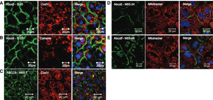
In line with the lack of apparent intracellular staining in frozen liver sections, we were unable to detect colocalization between Abcc6/ABCC6 and the mitochondrial marker cytochrome C oxidase (complex IV) (CoxIV) (Figure 2A and 2C) or the endoplasmic reticulum/MAM-specific anticalnexin antibody (Figure 2B). To corroborate our findings, we have also analyzed cultured primary mouse hepatocytes from wild-type and Abcc6−/− mice. Cells were treated with MitoTracker Red before fixing and immunostaining with 2 different monoclonal antibodies recognizing Abcc6. Consistent with the results obtained with frozen tissue sections, Abcc6 was exclusively found in the PM of primary hepatocytes (Figure 2D and 2E), showing colocalization with catenin (not shown) but not with MitoTracker Red (Figure 2D and 2E).
Nonstandard Abbreviations and Acronyms MAM mitochondria-associated membrane PM plasma membrane
Figure 1. Colocalization of the mouse Abcc6 and human ABCC6 proteins with various plasma membrane markers in mouse and human liver samples. A, The monoclonal antibody M6II-68 revealed the plasma membrane localization of Abcc6 (green), which clearly colocalized (yellow) with the plasma membrane marker cadherin (red). B, The combined use of the polyclonal antibody S-20 specific to the mouse Abcc6 (red) and an antibody recognizing the basolateral plasma membrane marker Na,K-ATPase (green) also showed significant overlap with large areas of colocalization (yellow). C, The monoclonal antibody recognizing Abcb11 (TU236) delineates the canalicular (apical) membranes of hepatocytes (red), whereas the anti-Abcc6 M6II-68 labels the basolateral plasma membrane (green).
Minimal colocalization is visible. D, Immunofluorescence staining of frozen human liver sections with the monoclonal antibody recognizing human ABCC6 (M6II-7, green) or the plasma membrane marker cadherin (red) shows evidence of colocalization. E, The anti-ABCC6 antibody M6II-7 and the anti-ABCB11 monoclonal antibody TU236 reveal plasma membrane staining but at distinct plasma membrane compartments (ABCC6, green, basolateral; ABCB11, red, canalicular or apical). F, The strong punctuate staining (yellow) on D and E represents nonspecific fluorescent signals often encountered in human liver cells as evidenced by the negative control staining performed without primary antibodies. G, Z-stack projections obtained of frozen mouse liver sections and stained with the M6II-68 antibody. All images were collected by confocal microscopy, except for those of B that were obtained by a standard fluorescent microscope. Nuclei were counterstained with 4',6-diamidino-2-phenylindole (blue).
at SEMMELWEIS UNIV OF MED (B13/62 on February 3, 2014 http://circres.ahajournals.org/
Downloaded from
Discussion
To elucidate the physiological role of ABCC6 in ectopic calci- fication, the exact subcellular localization of the native protein must be firmly established. We have performed experiments to reveal the localization of the ABCC6 protein in its natural tissue environment. Sharply contrasting the findings of Martin et al6 and consistent with previously published literature re- garding the rat,8 mouse,7,9 and human ABCC6 proteins,10,12 we unequivocally show that the ACBC6 protein is expressed in the basolateral PM (Figure 1). Furthermore, we detected no significant intracellular localization for Abcc6/ABCC6 using the same markers used by Martin et al6 for labeling mitochon- dria, the endoplasmic reticulum, and MAM organelles. The same conclusion could be drawn in a second experimental setup using primary mouse hepatocytes (Figure 2).
Contrary to the methods used by Martin et al,6 our methods avoided multistep fractionation of homogenized hepatic tissue samples. It is worth noting that cell disruption and fractionation experiments have led to inaccurate conclusions concerning the (intra)cellular localization of membrane-embedded proteins.13 Martin et al6 found no evidence of PM expression of Abcc6 by cell surface biotinylation of primary hepatocytes. Based on the overwhelming evidence of our experiments, we believe that the lack of biotinylation should be explained by the unavailability of surface biotinylation targets. Indeed, our topology analysis sug- gests that the extracellular loops of ABCC6 are relatively short and contain only 2 potentially reactive lysine residues.
The precise biosynthetic pathway and the mechanisms reg- ulating the PM targeting of ABCC6 are still largely unknown,
yet a recent study has suggested a role for endoplasmic re- ticulum chaperones in cellular trafficking.5 The present study has reestablished that ABCC6 is mainly localized in the PM, which is probably where the physiological action of the pro- tein is exerted.
Acknowledgment
Thanks are due to Tamás Arányi for the numerous valuable discussions.
Sources of Funding
Financial support for this work was provided to O. Le Saux by the Hawaii Community Foundation grant 11ADVC-49234, the American Heart Association grant 11GRNT5840005, and National Institutes of Health (NIH) grants R21HL087289 and RO1HL108249. A. Váradi was funded by NIH R01AR055225, and PXE International and Hungarian research grants OTKA NK 81204 and OTKA K 104227.
G. Szakács is supported by the Momentum grant of the Hungarian Academy of Sciences.
Disclosures
None.
References
1. Váradi A, Szabó Z, Pomozi V, de Boussac H, Fülöp K, Arányi T.
ABCC6 as a target in pseudoxanthoma elasticum. Curr Drug Targets.
2011;12:671–682.
2. Le Saux O, Urban Z, Tschuch C, Csiszar K, Bacchelli B, Quaglino D, Pasquali-Ronchetti I, Pope FM, Richards A, Terry S, Bercovitch L, de Paepe A, Boyd CD. Mutations in a gene encoding an ABC transporter cause pseudoxanthoma elasticum. Nat Genet. 2000;25:223–227.
Figure 2. Absence of colocalization of mouse Abcc6 and human ABCC6 with mitochondrial or endoplasmic reticulum (ER)/
mitochondria-associated membrane (MAM) markers. A, Immunofluorescence staining with the polyclonal S-20 antibody recognizing Abcc6 (green) and a monoclonal antibody reacting with the mitochondrial marker CoxIV (red) show no colocalization of Abcc6 and mitochondria in frozen mouse liver sections. B, No colocalization is visible between the S-20 (green) and an antibody recognizing calnexin, an ER/MAM marker (red). C, Staining with the anti-ABCC6 monoclonal antibody M6II-7 (green) and the mitochondrial marker CoxIV (red) reveals the lack of colocalization of ABCC6 and mitochondria in human frozen liver sections. The yellow punctuate pattern is a result of nonspecific staining (as shown in Figure 1F). D and E, There is no colocalization between Abcc6 and mitochondria in cultured mouse primary hepatocytes stained with MitoTracker (red) and M6II-24 (D, green) or M6II-68 (E, green). Note that the green staining within the nuclei represents nonspecific signals (for control experiments, see Online Figure IC and ID). All images were collected by confocal microscopy, except for those of A and B that were obtained with a standard fluorescent microscope. Nuclei were counterstained with 4',6-diamidino-2-phenylindole (blue).
Pomozi et al ABCC6 Is a Plasma Membrane Protein e151
3. Le Boulanger G, Labrèze C, Croué A, Schurgers LJ, Chassaing N, Wittkampf T, Rutsch F, Martin L. An unusual severe vascular case of pseu- doxanthoma elasticum presenting as generalized arterial calcification of infancy. Am J Med Genet A. 2010;152A:118–123.
4. Jiang Q, Oldenburg R, Otsuru S, Grand-Pierre AE, Horwitz EM, Uitto J.
Parabiotic heterogenetic pairing of Abcc6-/-/Rag1-/- mice and their wild- type counterparts halts ectopic mineralization in a murine model of pseu- doxanthoma elasticum. . 2010;176:1855–1862.
5. Le Saux O, Martin L, Aherrahrou Z, Leftheriotis G, Váradi A, Brampton CN. The molecular and physiological roles of ABCC6: more than meets the eye 2012. Front Genet. 2012;3:289. doi: 10.3389/fgene.2012.00289.
6. Martin LJ, Lau E, Singh H, et al. ABCC6 localizes to the mitochondria- associated membrane. Cir Res. 2012;111:516–520.
7. Le Saux O, Fülöp K, Yamaguchi Y, Iliás A, Szabó Z, Brampton CN, Pomozi V, Huszár K, Arányi T, Váradi A. Expression and in vivo rescue of human ABCC6 disease-causing mutants in mouse liver. PLoS One. 2011;6:e24738.
8. Madon J, Hagenbuch B, Landmann L, Meier PJ, Stieger B. Transport func- tion and hepatocellular localization of mrp6 in rat liver. Mol Pharmacol.
2000;57:634–641.
9. Beck K, Hayashi K, Nishiguchi B, Le Saux O, Hayashi M, Boyd CD. The distribution of Abcc6 in normal mouse tissues suggests multiple functions for this ABC transporter. J Histochem Cytochem. 2003;51:887–902.
10. Scheffer GL, Hu X, Pijnenborg AC, Wijnholds J, Bergen AA, Scheper RJ. MRP6 (ABCC6) detection in normal human tissues and tumors. Lab Invest. 2002;82:515–518.
11. Gorgels TG, Hu X, Scheffer GL, van der Wal AC, Toonstra J, de Jong PT, van Kuppevelt TH, Levelt CN, de Wolf A, Loves WJ, Scheper RJ, Peek R, Bergen AA. Disruption of Abcc6 in the mouse: novel insight in the pathogenesis of pseudoxanthoma elasticum. Hum Mol Genet.
2005;14:1763–1773.
12. Sinkó E, Iliás A, Ujhelly O, Homolya L, Scheffer GL, Bergen AA, Sarkadi B, Váradi A. Subcellular localization and N-glycosylation of human ABCC6, expressed in MDCKII cells. Biochem Biophys Res Commun.
2003;308:263–269.
13. Kiss K, Brozik A, Kucsma N, Toth A, Gera M, Berry L, Vallentin A, Vial H, Vidal M, Szakacs G. Shifting the paradigm: the putative mitochondrial protein ABCB6 resides in the lysosomes of cells and in the plasma mem- brane of erythrocytes. PLoS One. 2012;7:e37378.
at SEMMELWEIS UNIV OF MED (B13/62 on February 3, 2014 http://circres.ahajournals.org/
Downloaded from
Antibodies
The primary antibodies to mouse Abcc6 (sc-5787), β-catenin (Sc-7199), and NA,K- ATPAse (sc-28880) were purchased from Santa Cruz Biotechnology Inc. (Santa Cruz, CA). The anticadherin (Ab16505) and anticalnexin (10286) antibodies were purchased from Abcam (Cambridge, MA). AntiCoxIV (3E11) rabbit mAb was from Cell Signaling Technology, Danvers, MA, USA. The Rat M6II-7 and M6II-68 monoclonal antibodies were generous gifts from George Scheffer (University Medical Center Amsterdam), while the polyclonal antibody TU236 was kindly provided by Daniel F. Ortiz (Tufts University, Medford, Ma)., The appropriate secondary antibodies were Alexa Fluor 488 and Alexa Fluor 594 (Invitrogen, Carlsbad, CA).
Immunochemistry of frozen liver sections
For immunohistochemical staining of mouse liver samples C57BL/6 mice were used, and Abcc6-/- mouse strain was used as negative control to prove the specificity of anti- Abcc6 antibodies. Mice were terminated by cervical dislocation, multiple liver lobes were quickly harvested, placed in Optimum Cutting Temperature (OCT) compound and stored at -800C.
Human frozen normal liver sections were prepared and stained at the 1st Pathology Department, Semmelweis University as approved by the Ethical Committee of Semmelweis University of Budapest. Immunofluorescent staining of liver tissues was performed using 8 µm- frozen sections. Samples were fixed with ice-cold methanol for 5 min and rinsed 3 times for 5 min in DPBS (PBS + 1 mM CaCl2 + 0.5 mM MgCl2). The samples were then blocked for 30 min in DBPS with 2mg/ml BSA, 1% fish gelatin, 0.15 % Triton X-100, 5% of the appropriate serum). Mouse liver sections were stained with a rat monoclonal anti-Abcc6 antibody (M6II-68) as described [2], or the goat polyclonal anti-Abcc6 antibody (S-20) to specifically detect Abcc6.
The human ABCC6 protein was detected on the human liver samples using anti-ABCC6 monoclonal antibody M6II-7 [1]. As colocalization markers, the primary antibodies against cadherin, β- catenin, Na,KATPase or Abcb11/Bsep were used. Images were obtained with an Axioscope 2 fluorescent microscope (Zeiss, Thornwood, NY) and a Zeiss LSM 710 confocal laser scanning microscope.
Isolation and culture of mouse hepatocytes
Experiments were carried out by using hepatocytes prepared from female C57BL/6 mice (weighing 20 g). Liver cells were isolated by the published method [2]. Hepatocytes having viability better than 90%, as determined by trypan blue exclusion, were used in the experiments. Cells were plated in collagen-precoated dishes in serum containing medium described [3]. After attachment of cells medium was changed and renewed every 24h in the absence of serum. Treatment with MitoTracker Red CMXRos (Life Technologies,Grand Island, NY, USA) was perfored at 200 nM concentration for 30 minutes at 370C and subsequent fixation with 4% paraformaldehide, immunocytochemistry and confocalimaging was carried out after 72 hours of cell culturing.
Supplemental Material References
1. Le Saux O, Fülöp K, Yamaguchi Y, Iliás A, Szabó Z, Brampton CN, Pomozi V, Huszár
K, Arányi T, Váradi A Expression and in vivo rescue of human abcc6 disease-causing mutants in mouse liver. 2011 PLoS One 2011;6:e24738
2. Bayliss KM, Skett P Isolation and culture of human hepatocytes, in: G.E. Jones, (Ed.),
Human Cell Culture Protocols, Humana Press, Totowa, 1996. pp.369-390
3. Ferrini JB, Ourlin JC, Pichard L, Fabre G, Maurel P Human hepatocyte culture, in: I.R.
Phillips, E.A. Shephard (Ed.), Cytochrome P450 Protocols, Humana Press, Totowa, 1998. pp. 341-352
Supplemental Figure I.
Controls for antibody specificity.