Angiotensin type 1A receptor regulates β-arrestin binding of the β2-adrenergic 1
receptor via heterodimerization 2
3
András D. Tóth1, Pál Gyombolai1,2, Bence Szalai1,2, Péter Várnai1, Gábor Turu1,2, 4
László Hunyady1,2 5
6
1Department of Physiology, Faculty of Medicine, Semmelweis University, P. O. Box 7
2, H-1428 Budapest, Hungary 8
2MTA-SE Laboratory of Molecular Physiology, Hungarian Academy of Sciences and 9
Semmelweis University, Budapest, Hungary 10
11 12
Abstract 13
Heterodimerization between angiotensin type 1A receptor (AT1R) and β2-adrenergic 14
receptor (β2AR) has been shown to modulate G protein-mediated effects of these 15
receptors. Activation of G protein-coupled receptors (GPCRs) leads to β-arrestin 16
binding, desensitization, internalization and G protein-independent signaling of 17
GPCRs. Our aim was to study the effect of heterodimerization on β-arrestin 18
coupling. We found that β-arrestin binding of β2AR is affected by activation of AT1Rs.
19
Costimulation with angiotensin II and isoproterenol markedly enhanced the 20
interaction between β2AR and β-arrestins, by prolonging the lifespan of β2AR-induced 21
β-arrestin2 clusters at the plasma membrane. While candesartan, a conventional 22
AT1R antagonist, had no effect on the β-arrestin2 binding to β2AR, TRV120023, a β- 23
arrestin biased agonist, enhanced the interaction.
24
These findings reveal a new crosstalk mechanism between AT1R and β2AR, and 25
suggest that enhanced β-arrestin2 binding to β2AR can contribute to the 26
pharmacological effects of biased AT1R agonists.
27 28 29
Highlights:
30
Heterodimerization between AT1R and β2AR enhances β-arrestin coupling of β2AR.
31
Heterodimerization increases the lifespan of β-arrestin2 clusters after β2AR 32
stimulation.
33
Biased AT1R ligands alter the function of heterodimerized β2AR.
34
*Manuscript
Click here to view linked References
35
Keywords: GPCR, heterodimerization, arrestin, BRET, biased signaling 36
37
Abbreviations: G protein-coupled receptors (GPCR), Angiotensin type 1A receptor 38
(AT1R), β2-adrenergic receptors (β2AR), serotonin 2C receptor (5HT2CR), angiotensin 39
II (AngII), isoproterenol (ISO), Renilla luciferase (Rluc), Super Renilla luciferase 40
(Sluc) 41
42
1. Introduction 43
44
G protein-coupled receptors (GPCRs) are the largest plasma membrane receptor 45
superfamily, and according to estimations ~40% of the marketed drugs target GPCRs 46
(Whalen, Rajagopal and Lefkowitz, 2011). Although the monomeric form of GPCRs is 47
functional, a large number of evidence has accumulated demonstrating that they are 48
also capable to form higher order complexes (Milligan, 2013). A very intriguing finding 49
is that dimerization or oligomerization can greatly influence the signaling properties of 50
GPCRs (Ferre, Casado, Devi et al., 2014). It has been reported that GPCR 51
dimerization can result in altered ligand binding, receptor conformation or effector 52
functions (Smith and Milligan, 2010,Szidonya, Cserzo and Hunyady, 2008).
53
Heterodimerization between GPCRs widens the number of the possible physiological 54
receptor crosstalk mechanisms, and helps fine tune receptor functions (Ferre, Baler, 55
Bouvier et al., 2009,Jonas, Rivero-Muller, Huhtaniemi et al., 2013,Rivero-Muller, 56
Jonas, Hanyaloglu et al., 2013). On the other hand, receptor dimerization can also 57
cause unexpected drug interactions.
58
Angiotensin type 1A receptor (AT1R) and β-adrenergic receptors (βAR) play crucial 59
role in the regulation of heart function and vascular tone under physiological and 60
pathophysiological conditions, therefore they are pivotal drug targets in 61
cardiovascular diseases, including heart failure or hypertension (Whalen et al., 2011).
62
Moreover, they were shown to form dimeric complexes and the blockade of either 63
protomer with an antagonist can result in simultaneous hindering of the other 64
protomer’s G protein activation (Barki-Harrington et al., 2003).
65
In addition to G proteins, β-arrestin molecules are also considered to be effector 66
proteins of GPCRs. β-arrestins govern GPCR desensitization, endocytosis and also 67
participate in G protein-independent signaling pathways (Shenoy and Lefkowitz, 68
2011). β-arrestins regulate β2AR function via several mechanisms. β-arrestin2 69
induces desensitization and internalization of β2AR, and these effects have been 70
linked to tachyphylaxis of β2-adrenergic agonists (Deshpande, Theriot, Penn et al., 71
2008). This phenomenon greatly limits the use of β2-agonist drugs in the treatment of 72
bronchial asthma. β-arrestins also mediate signaling of β2AR. β-arrestin2 initiates the 73
activation of MAPK cascade independently of G protein activation (Shenoy, Drake, 74
Nelson et al., 2006), and β-arrestins promote cardiomyocyte contraction (Carr, 75
Schilling, Song et al., 2016). Chronic activation of β2-adrenergic receptor by 76
catecholamines leads to DNA damage via β-arrestin1 (Hara, Kovacs, Whalen et al., 77
2011). β-arrestin1 facilitates the MDM2 promoted ubiquitination and degradation of 78
p53. In the absence of β-arrestin1 this effect of β2AR is greatly abrogated. These 79
examples show the central role of β-arrestins in the function of β2AR.
80
Activation of G proteins by AT1R is considered to evoke deleterious effects in several 81
pathophysiological conditions. However, stimulation of the G protein-independent, β- 82
arrestin-mediated signaling pathways through AT1R has been shown to have 83
beneficial outcomes (Hunyady and Catt, 2006,Whalen et al., 2011). The clinically 84
used conventional AT1R antagonist drugs antagonize both pathways, so the desired 85
β-arrestin-mediated favorable effects are also blocked. Thus, it is proposed, that 86
ligands which are able to antagonize the G protein activation of a GPCR, but still able 87
to induce the β-arrestin dependent signaling, could be prosperous drugs in many 88
pathological circumstances(Whalen et al., 2011). Such β-arrestin biased agonist 89
ligands have been already discovered for AT1R. The first such ligand was 90
[Sar1,Ile4,Ile8]-AngII, however its clinical use was seriously hindered because of its 91
poor receptor affinity (Wei, Ahn, Shenoy et al., 2003). Since then, new peptides with 92
higher affinity, like TRV120023 or TRV120027, have been developed, which offered 93
the possibility of the clinical application (Rajagopal, Ahn, Rominger et al., 94
2011,Szakadati, Toth, Olah et al., 2015,Violin, Crombie, Soergel et al., 2014).
95
In this study, we investigated the consequences of angiotensin type 1A 96
receptor-β2-adrenergic receptor (β2AR) heterodimerization on β-arrestin binding 97
using a bioluminescence resonance energy transfer (BRET)-based approach. We 98
found that dimerization alters the β-arrestin binding of β2AR. The physiological AT1R 99
agonist angiotensin II or the β-arrestin biased AT1R ligand TRV120023, but not the 100
unbiased AT1R inverse agonist candesartan could potentiate β-arrestin coupling to 101
the β2AR. These findings reveal a possible new physiological crosstalk mechanism 102
between AT1R and β2AR.
103 104
2. Materials and methods 105
2.1. Materials 106
The AT1R, AT1R-DRY/AAY (Gaborik, Jagadeesh, Zhang et al., 2003), AT1R-∆319 107
(Hunyady, Bor, Balla et al., 1994), β2AR, β2AR-Sluc, untagged 5HT2CR-VGV (I156V, 108
N158G, I160V), 5HT2CR-VGV-Sluc, PM-mRFP (mRFP fused to plasma membrane 109
target sequence of Lyn) (Toth, Toth, Gulyas et al., 2012), AT1R-Rluc (Szakadati et 110
al., 2015), AT1R-Venus, β-arrestin1-Venus, β-arrestin2-Venus (Gyombolai, Boros, 111
Hunyady et al., 2013), β-arrestin2-Rluc, β2AR-Venus (Turu, Szidonya, Gaborik et al., 112
2006), Cameleon D3 Ca2+-BRET sensor (Gulyas, Toth, Toth et al., 2015), EPAC 113
cAMP-BRET sensor (Erdelyi, Balla, Patocs et al., 2014) and L10-Venus (Venus fused 114
to plasma membrane target sequence of Lck) (Toth, Gulyas, Toth et al., 2016) 115
constructs were previously described. 5HT2CR-Venus was generated by replacing the 116
Super Renilla luciferase (Sluc) tag to monomeric Venus (Venus) in the 5HT2CR-Sluc 117
construct. To create the Cerulean tagged β2AR construct, the Sluc tag of β2AR-Sluc 118
was replaced with Cerulean. To generate the YFP-β-arrestin2 construct the cDNA of 119
rat β-arrestin2 was cloned into pEYFP-N1 vector between AgeI and KpnI restriction 120
sites. The plasmids encoding HA epitope-tagged wild type and K44A mutant 121
dynamin-2A were kindly provided by Dr. K. Nakayama (Tsukuba Science City, 122
Ibaraki, Japan).
123
Cell culture reagents were from Invitrogen (Carlsbad, CA). Cell culture dishes and 124
white 96-well plates for BRET measurements were obtained from Greiner 125
(Kremsmunster, Austria). TRV120023 (Sar-Arg-Val-Tyr-Lys-His-Pro-Ala-OH) was 126
synthetized by Proteogenix (Schiltigheim, France). Coelenterazine h was purchased 127
from Regis Technologies (Morton Grove, IL). Unless otherwise stated, all other 128
chemicals and reagents were from Sigma (St. Louis, MO).
129 130
2.2. Cell culture and transfections 131
HEK 293T and COS-7 cells were cultured in DMEM supplemented with 100 IU/ml 132
penicillin, 100 µg/ml streptomycin and 10% fetal bovine serum in 5% CO2 at 37 ºC.
133
For the BRET-experiments, the transfection was performed on cell suspension using 134
Lipofectamine 2000 in OptiMEM according to the manufacturer’s instructions, 135
thereafter the cells were plated on polylysine covered white 96-well plates. The 136
measurements were performed 24 or 48 hours after transfection of HEK 293T and 137
COS-7 cells, respectively.
138
CHO cells were cultured in Ham’s F12 supplemented with 100 IU/ml penicillin, 100 139
µg/ml streptomycin and 10% FBS. The day before transfection the cells were plated 140
on 6-well plates, the transfection was achieved using Lipofectamine 2000 according 141
to manufacturer’s protocol.
142
For confocal microscopy experiments, HEK 293T cells were grown on glass 143
coverslips in 6-well plates the day before transfection, and were transfected with 144
plasmids encoding β2AR-Cerulean, AT1R-∆319 and β-arrestin2-Venus (1 µg, 4 µg, 145
and 0.5 µg pro well, respectively) using Lipofectamine 2000. The experiments were 146
performed the day after transfection.
147 148
2.3. Bioluminescence resonance energy transfer (BRET) measurements 149
After a washing step, the medium of HEK 293T or COS-7 cells was changed to 150
modified Kreb’s-Ringer medium containing 120 mM NaCl, 4.7 mM KCl, 1.2 mM 151
CaCl2, 0.7 mM MgSO4, 10 mM glucose 10 mM, pH 7.4 Na-HEPES, unless otherwise 152
stated. 5 µM coelenterazine h, as Renilla luciferase substrate, was added to cells, 153
thereafter luminescence was measured at 480 nm and 530 nm wavelengths by a 154
Thermoscientific Varioskan Flash Reader (Perkin Elmer). BRET ratio was calculated 155
by dividing the emission collected at 530 nm with the emission measured at 480 nm.
156
BRET signal of CHO cells was measured in cell suspension using Mithras LB 940 157
multilabel reader (Berthold Technologies), as earlier described (Gyombolai, Toth, 158
Timar et al., 2014).
159
For the statistical analysis Two-Way-ANOVA tests were performed. An effect was 160
considered statistically significant, when the p value of the interaction between the 161
two treatments was less than 0,05.
162 163
2.4. BRET-titration experiments 164
Increasing amount of donor (Sluc containing) and acceptor (Venus containing) 165
proteins were expressed in HEK 293T cells. Similarly to the conventional BRET 166
experiments, before measurement the medium was changed to modified Kreb's- 167
Ringer medium. Before addition of 5 µM coelenterazine h, Venus fluorescence was 168
measured by excitation at 510 nm and emission collected at 535 nm. After 169
coelenterazine h treatment, luminescence was measured using 480 nm and 530 nm 170
filters and total luminescence was determined without filter. The data analysis in 171
details was earlier described (Szalai, Hoffmann, Prokop et al., 2014). Briefly, 172
measured points were grouped into high/low luminescence group by the median 173
luminescence value for β2AR-Sluc and AT1R-Venus expressing cells. The effect of 174
luminescence on the measured BRET ratio was evaluated by covariance analysis, 175
forcing the regression line through the origin.
176 177
2.5. Confocal laser-scanning microscopy and image analysis 178
The media of the cells were changed to modified Kreb's-Ringer medium. Time-series 179
images were taken every 10 seconds for 190 seconds from the bottom of the cells 180
with a Zeiss LSM 710 confocal laser-scanning microscope using a 63x objective at 181
37 °C. Size of the images was 79.38 x 79.38 m with 1024x1024 resolution.
182
Individual cells were selected and cropped in Fiji ImageJ software and processed for 183
further analysis. -arrestin puncta were identified on images with neural network 184
algorithm using Keras and sklearn libraries in python programming language 185
(https://github.com/fchollet/keras, http://scikit-learn.org/). Stacks of images were 186
sliced into samples with sliding window of 20x20 pixels and each sample was 187
classified as β-arrestin puncta or background. Classifier was trained on examples 188
which were selected from images taken in separate experiments. 2809 negative and 189
664 positive samples were used total which were randomly divided into 2326 training 190
and 1147 cross-validation examples. With a network with two hidden layers, the 191
cross-validation resulted in an average of 98 percent both for precision and recall.
192
After classification of the samples, the original sized binary image was reconstructed 193
and was further processed for tracking with trackpy library (https://github.com/soft- 194
matter/trackpy). Particles present on the tenth image were selected with size of at 195
least 5 pixels and were tracked with memory set to 3. Duration of puncta was 196
determined and the puncta were divided into two subgroups based on their lifespan.
197
The distributions were statistically compared with Fischer’s exact test.
198
For determination of the β-arrestin binding phenotype, images were taken from the 199
middle cross section of the cells 20-40 minutes after stimulation at 37 °C.
200 201
3. Results 202
203
3.1. β2AR and AT1R form heterodimers 204
The existence and the functional relevance of the β2AR-AT1R heterodimer have been 205
reported earlier (Barki-Harrington et al., 2003). To verify the presence of 206
heterodimerization between β2AR and AT1R, we performed BRET-titration 207
experiments in HEK 293T cells. Sluc-tagged β2AR was used as BRET donor and 208
Venus-tagged AT1R as BRET acceptor. In the classical BRET-titration experiments 209
the amount of the donor molecule-encoding plasmid is held constant, while the 210
acceptor-encoding plasmid is gradually increased. Despite of the constant amount of 211
donor-encoding plasmid, the donor molecule expression was strongly dependent on 212
the number of the acceptor molecule in our system, namely increased fluorescence 213
levels led to a drop in the measured luminescence (Suppl. Fig. 1). Formerly we and 214
others have shown that the correct interpretation of the classical approach is 215
seriously hindered when the expression of the BRET donor is not maintained 216
constant (Lan, Liu, Li et al., 2015,Szalai et al., 2014). With the use of computer 217
simulations and in vitro experiments, we recently developed a new approach for the 218
analysis of quantitative BRET data, where the BRET ratio is plotted as the function of 219
the acceptor-labeled receptor expression at various donor receptor expression levels 220
(Szalai et al., 2014). Briefly, we found that in case of non-specific interactions the 221
BRET ratio is only dependent on the number of the acceptor molecules. In case of 222
specific interactions, the BRET ratio is dependent both on the amount of acceptor 223
and donor molecules (for more details see: (Szalai et al., 2014)). In our experiments, 224
we confirmed the specific interaction between β2AR and AT1R, since a linear 225
regression with lower steepness could be fitted on high luminesce points compared 226
to the low luminescence points (Fig. 1A). On the other hand, we detected no specific 227
interaction between Sluc-tagged β2AR and Venus-tagged serotonin 2C receptor 228
(5HT2cR), as the donor expression did not influence the slope of the linear regression 229
(Fig. 1B). This result shows that β2AR forms heterodimer with AT1R, but not with 230
5HT2cR.
231 232
3.2. Activation of AT1R influences the β-arrestin2 binding to β2AR within a 233
heteromer 234
To investigate the crosstalk between the β2AR-AT1R heterodimer, we designed a 235
BRET-based experimental approach. We cotransfected the cells with plasmids 236
encoding Sluc-tagged β2AR, C-terminally Venus-tagged β-arrestin2 and untagged 237
AT1R. Using this experimental setup, we were able to selectively monitor the β- 238
arrestin2 binding of the β2AR and the impact of the AT1R stimulation on the β2AR-β- 239
arrestin2 association (Fig. 2A). Isoproterenol (ISO, 10 μM), a β2AR agonist, induced 240
an increase in the BRET ratio, reflecting the β-arrestin2 binding to the β2AR (Fig. 2B).
241
Angiotensin II (AngII, 100 nM), which exerts its main physiological effects via AT1R, 242
alone induced only a slight increase in the BRET ratio. Strikingly, during 243
simultaneous activation of the two receptors, the association between β-arrestin2 and 244
β2AR was significantly potentiated. Similar results were obtained in COS-7 and CHO 245
cells (Suppl. Fig. 2A and B, in case of CHO cells β2AR was tagged with acceptor and 246
β-arrestin2 with donor). Since the BRET ratio is also dependent on the relative 247
orientation of the donor and acceptor molecules, we tested the interaction with N- 248
terminally YFP-tagged β-arrestin2 (Suppl. Fig. 3). We observed a similar effect 249
indicating that the BRET increase does not originate from conformational changes, 250
but reflects the increased interaction of β2AR and β-arrestin2. The β-arrestin1 binding 251
of β2AR was also examined by BRET, and a very similar response was found (Suppl.
252
Fig. 4). We have also investigated the dose-dependence of the ISO effect on β- 253
arrestin2 binding to β2AR in the presence or absence of AngII (Fig. 2C). We found 254
that 100 nM AngII could increase the ISO-mediated β-arrestin2 binding already at 255
lower ISO concentrations. In addition to the increased maximal response, AngII 256
treatment also caused a left-shift in the β-arrestin2 binding curve (log EC50 (M) -7.48 257
vs -7.17, p<0.05, tested with Student’s t-test), thus at lower ISO concentrations AngII 258
raised the β2AR-β-arrestin2 association more markedly.
259
Since β-arrestin2 also binds to the AT1R, one could assume that, in case of 260
costimulation, β-arrestin2 translocates to the AT1R, and nonspecific BRET is 261
detected between β2AR and membrane-translocated β-arrestin2. To rule out this 262
possibility, we transfected a C-terminally truncated AT1 receptor (AT1R-∆319), which 263
is impaired in the ability of β-arrestin2 binding, because it lacks the major docking site 264
of β-arrestins (Fig. 2D) (Balla, Toth, Soltesz-Katona et al., 2012,Qian, Pipolo and 265
Thomas, 2001). In contrast to the small BRET ratio elevation when wild type AT1R 266
was used, AngII stimulation alone did not lead to any change in basal BRET signal.
267
However, a significant increase in the BRET signal was still present after 268
costimulation of β2AR and AT1R-∆319, indicating that the association between β2AR 269
and β-arrestin2 was enhanced.
270
Next, we checked whether β2AR could also influence the β-arrestin2 binding of the 271
AT1R. In these experiments the AT1R was tagged with Rluc and the β2AR was 272
untagged (Suppl. Fig. 5). Nonetheless, the β2AR stimulation with ISO (10 μM) had no 273
significant effect on the BRET between AT1R-Rluc and β-arrestin2-Venus after AngII 274
treatment. We concluded that the strong β-arrestin2 binding of AT1R cannot be 275
further increased by β2AR stimulation.
276 277
3.3. Signaling pathways originating from AT1R are not essential for the 278
modulation of β2AR signaling 279
To reveal the underlying mechanism of the AT1R induced potentiation of the β2AR β- 280
arrestin2 binding, we used an AT1R mutant that is deficient in G protein activation 281
(AT1R-DRY/AAY) (Gaborik et al., 2003). After stimulation of this mutant with AngII the 282
β-arrestin binding of the β2AR was increased similar to the wild type AT1R (Fig. 3A).
283
However, the kinetics of the potentiation was slower compared to that of the wild type 284
AT1R.
285
Wild type AT1R is coupled to Gq/11 proteins, thus after receptor activation the second 286
messengers inositol trisphosphate (IP3) and diacylglycerol (DAG) are produced by 287
phospholipase C (Hunyady and Catt, 2006). IP3 is responsible for the calcium release 288
from the intracellular stores, while DAG is important in the activation of protein kinase 289
C. However, administration of a specific inhibitor of protein kinase C 290
(Bisindolylmaleimide I /BIM/,2 µM) or calcium depletion of the cells in calcium-free 291
media with calcium chelator EGTA (100 µM) and 200 nM thapsigargin /TG/) could not 292
block the AT1R mediated increase in the β2AR β-arrestin2 binding (Fig. 3B). Calcium 293
depletion abolished the AngII-induced calcium signaling, which is shown using a 294
calcium responsive BRET biosensor (Gulyas et al., 2015) (Suppl. Fig. 6).
295
Coactivation of untagged 5HT2CR, which receptor is coupled to similar signaling 296
pathways as AT1R (Balla et al., 2012), but does not dimerize with β2AR, could not 297
induce the potentiation of β-arrestin binding (Suppl. Fig. 7). These results and the 298
data obtained with the AT1R-DRY/AAY mutant suggest that G protein activation is not 299
necessary for this effect.
300
In the past years it has become evident that AT1R can also signal in the absence of 301
G protein activation. Among others, β-arrestin dependent Src and MAP kinase 302
activation has been described (Fessart, Simaan and Laporte, 2005,Hunyady and 303
Catt, 2006). Src (PP1, 1 µM) and MEK (PD98059, 20 µM) inhibitors did not interfere 304
with the increased BRET ratio during costimulation of the receptors (Fig. 3B). These 305
results, and the fact that the stimulation of the β-arrestin binding deficient AT1R 306
mutant (AT1R-∆319) was capable to increase the β-arrestin2 binding of β2AR, 307
suggest that β-arrestin mediated signaling is not required for the observed 308
phenomenon.
309
β-arrestin2 dissociates from β2AR after its internalization (Oakley, Laporte, Holt et al., 310
1999), therefore an increased β2AR-β-arrestin2 interaction could origin from the 311
inhibition of receptor endocytosis. To block β2AR endocytosis, we overexpressed a 312
dominant negative mutant dynamin2A (dynamin2A-K44A), which has been shown to 313
inhibit agonist induced internalization (Scarselli and Donaldson, 2009,Zhang, 314
Ferguson, Barak et al., 1996). Indeed, the ISO-induced BRET signal was significantly 315
elevated (Suppl. Fig. 8). However, the cotreatment with AngII and ISO still increased 316
the BRET signal under these circumstances, showing that the observed effect cannot 317
be explained by AT1R induced blockade of β2AR internalization.
318
Since the observed effect was independent on activation of the investigated signaling 319
pathways, we concluded that it is mediated by heterodimerization between the β2AR 320
and AT1R.
321 322
3.4. β-arrestin2 binding of β2AR is dependent on the expression of AT1R 323
Since β2AR can be present in both monomeric and dimeric states, only a portion of 324
β2ARs interact with AT1R. Presuming random pairing of the two receptors, the 325
relative number of β2AR-AT1R heterodimers should be elevated by increasing the 326
AT1R-β2AR expression ratio. Therefore, we increased the amount of the AT1R 327
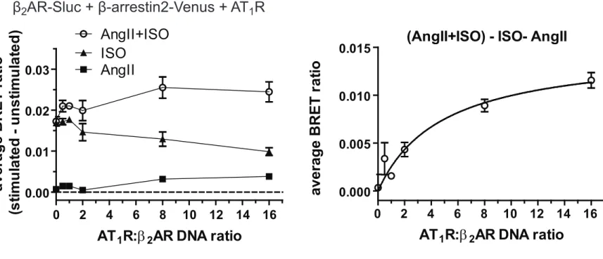
encoding plasmid during the transfection, while keeping the amount of the β2AR-Sluc 328
plasmid constant. As shown in Fig. 4A and B, no AngII effect was detected in 329
absence of AT1R. By increasing the AT1R:β2AR DNA ratio, when compared to ISO 330
stimulation, the costimulation with AngII and ISO caused gradually increased BRET 331
signal. This elevation was not due to higher plasma membrane expression of β2AR- 332
Sluc, since ISO stimulation itself led to slightly decreased BRET signals (Fig. 4A).
333
These results show that the magnitude of β-arrestin translocation in this system 334
depends on the relative expression ratio of AT1R and β2AR, which is consistent with 335
the role of heterodimers.
336 337
3.5. Biased activation of AT1R affects the β-arrestin2 binding of β2AR 338
It has been shown earlier that a conventional AT1R antagonist can simultaneously 339
block the G protein mediated signaling of both AT1R and β2AR (Barki-Harrington et 340
al., 2003). Therefore, we investigated the effects of different AT1R antagonists on the 341
β-arrestin2 binding of the AT1R-β2AR heterodimer. The cotreatment with ISO and the 342
unbiased antagonist candesartan (10 µM) had no effect on the β-arrestin2 343
translocation (Fig. 5A). However, when we costimulated the cells with the β-arrestin 344
biased AT1R agonist TRV120023, we detected an increase in the the β-arrestin2 345
binding of β2AR, similarly to AngII-cotreatment (Fig. 5B), but the kinetics of the 346
potentiation was slower. Other β-arrestin biased agonists (TRV120027 and 347
[Sar1,Ile4,Ile8]-AngII) induced a very similar response (data not shown). These results 348
suggest, in good agreement with the data obtained with the G protein activation- 349
deficient AT1R mutant, that the β-arrestin activating conformation of AT1 receptor 350
enhances the β-arrestin2 binding of β2AR.
351 352
3.6. Coactivation of β2AR and AT1R increases the lifespan of β-arrestin2 353
clusters 354
Upon β2-adrenergic receptor activation, β-arrestin2 translocates to the plasma 355
membrane and forms clusters at the clathrin coated pits via interaction with β2- 356
adaptin (Laporte, Oakley, Holt et al., 2000). To address the mechanism of the 357
increased β-arrestin2 binding, we measured the lifetime and intensity of β-arrestin2 358
puncta of cells expressing β2AR-Cerulean, AT1R-Δ319 and β-arrestin2-Venus by 359
confocal microscopy (Fig. 6A). Images were taken every 10 seconds at the bottom of 360
the cells, and the lifespan of the individual puncta was determined. The lifespan of 361
these β-arrestin2-Venus dots was comparable to those detected in previous studies 362
(Eichel, Jullie and von Zastrow, 2016). AngII treatment did not lead to detectable 363
puncta formation, since AT1R-Δ319 lacks the major binding site for β-arrestin2 (data 364
not shown). However, the longevity of β-arrestin2 puncta was altered upon 365
costimulation with AngII and ISO, compared to ISO stimulation alone. After 366
costimulation, the fraction of puncta with longer lifespan was increased (Fig. 6A and 367
B). On the other hand, we found no difference between the average fluorescence 368
intensity values of the puncta (Suppl. Fig. 9). These results indicate that the detected 369
increase in β-arrestin2 binding is the consequence of the stabilized interaction 370
between β2AR and β-arrestin2. Increased β-arrestin2 localization at the plasma 371
membrane upon costimulation was also found by BRET measurements between 372
plasma membrane targeted Venus and β-arrestin2-Rluc in cells expressing untagged 373
β2AR and AT1R-Δ319 (Fig. 6C). The raise of bystander BRET rises from the 374
enrichment of β-arrestin2 in the juxtamembrane region.
375
β-arrestins dissociate from β2AR before entering the early endosomes, therefore 376
β2AR is classified as a class A receptor (Oakley, Laporte, Holt et al., 2000).
377
Nevertheless, we still could not observe β-arrestin2 at early endosomes after 378
costimulation, therefore this more stable interaction is not strong enough to convert 379
the interaction into a class B endocytic pattern (Suppl. Fig 10).
380 381
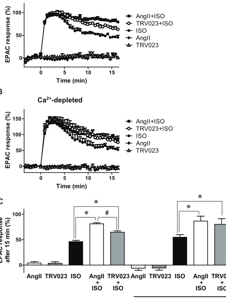
3.7. Simultaneous activation of AT1R prolongs the β2AR mediated cAMP 382
signaling 383
The generally considered main role of arrestins is the termination of G protein 384
signaling (Shenoy and Lefkowitz, 2011).However, several studies have shown that 385
noncanonical cAMP signaling arises from receptor-arrestin-G protein complexes 386
(Feinstein, Wehbi, Ardura et al., 2011,Feinstein, Yui, Webber et al., 2013,Thomsen, 387
Plouffe, Cahill et al., 2016,Wehbi, Stevenson, Feinstein et al., 2013). The magnitude 388
of the noncanonical arrestin-dependent cAMP formation was associated with the 389
stability of the receptor-arrestin interaction (Thomsen et al., 2016). Therefore, we 390
investigated whether the sustained β-arrestin2 binding to β2AR is accompanied by 391
prolonged cAMP signaling. We coexpressed AT1R and a BRET-based cAMP 392
biosensor in HEK 293T cells (Erdelyi et al., 2014), and the cAMP signaling of 393
endogenous β2AR was monitored. Neither AngII nor TRV023 treatment alone could 394
generate cAMP (Fig. 7A and C). Remarkably, cotreatment with AngII or TRV023 395
prolonged the ISO induced cAMP signal. Previously we have shown that calcium 396
dependent pathways can potentiate the cAMP formation (Baukal, Hunyady, Catt et 397
al., 1994). In calcium depleted cells we could still observe the prolonged cAMP 398
signaling upon AngII and TRV023 costimulation, showing that the cAMP signaling 399
was prolonged also via a calcium-independent way (Fig. 7B and C). These results 400
show that AT1R activity influences, in addition to β-arrestin binding, the G protein 401
dependent signaling of β2AR.
402 403
4. Discussion 404
Here we show that the β-arrestin binding of β2AR is regulated by AT1R 405
coactivation. These results are in good agreement with an earlier report, where the 406
authors gave evidence that β2AR and AT1R are working as a functional unit (Barki- 407
Harrington et al., 2003). Nowadays it is widely accepted that receptor dimerization 408
has important impact on the properties of receptor signaling. In elegant studies, using 409
ligand-binding deficient and signaling deficient luteinizing hormone receptors, 410
dimerization was shown to rescue the defective GPCR function both in vitro and in 411
vivo (Jonas, Fanelli, Huhtaniemi et al., 2015,Rivero-Muller, Chou, Ji et al., 2010).
412
Altered G protein activating ability was shown in case of D1-D2 dopamine receptor 413
heterodimer and its possible role was raised in the pathogenesis of major depression 414
(Pei, Li, Wang et al., 2010,Rashid, So, Kong et al., 2007).
415
Several studies have shown that β-arrestin binding can be influenced by receptor 416
heterodimerization. Altered β-arrestin binding was found in case of the V1-V2
417
vasopressin receptor dimer, the µ-δ opioid receptor dimer or the CXC chemokine 418
receptor 2-α1A adrenergic receptor heterodimer (Mustafa, See, Seeber et al., 419
2012,Rozenfeld and Devi, 2007,Terrillon, Barberis and Bouvier, 2004).
420
In our system, the stimulation of the untagged wild type AT1R with AngII alone led to 421
a slight increase of the BRET ratio between the Sluc-tagged β2AR and Venus-labeled 422
β-arrestin2. Since this increase was diminished when we used the β-arrestin binding- 423
deficient AT1R-∆319 mutant, we concluded that this signal reflects the β-arrestin 424
translocation to the untagged AT1R resulting in nonspecific BRET between β- 425
arrestin2 and β2AR. It is worth noting that the AT1R-∆319 mutant was reported to 426
bind β-arrestin2 very weakly (Anborgh, Seachrist, Dale et al., 2000), however it was 427
not detectable under our experimental conditions.
428
A similar system, named BRET heteromer identification technology (BRET-HIT), was 429
earlier introduced as a useful approach for GPCR heteromer detection (See, Seeber, 430
Kocan et al., 2011). This system is based on the close proximity of the heteromer 431
partners. Thus, the β-arrestin translocation to the untagged protomer can result in the 432
elevation of the BRET ratio between the tagged protomer and β-arrestin, because the 433
small distances in the molecular complex allow resonance energy transfer. In case of 434
non-dimerizing receptors this phenomenon cannot occur. However, we and others 435
have shown previously that after stimulation of a GPCR, BRET increase can be 436
detected between β-arrestin2-Rluc and a plasma-membrane targeted Venus, where 437
the interaction was clearly nonspecific (Donthamsetti, Quejada, Javitch et al., 438
2015,Gyombolai et al., 2014). This implicates that the reliability of the BRET-HIT 439
approach is weakened at high receptor expression levels because of the high 440
probability of nonspecific BRET signal. The BRET ratio increase after the 441
costimulation of the Sluc-tagged β2AR and the β-arrestin binding-deficient AT1R- 442
∆319 mutant clearly shows that the β-arrestin2 binding to the β2AR is elevated, and 443
this signal does not originate from a nonspecific interaction. The results obtained with 444
the C-terminally truncated AT1R mutant also suggest that AT1R activation alone 445
cannot induce β-arrestin recruitment to the β2AR. These results show that the AT1R- 446
β2AR heterodimer functions somewhat differently than the AT1R homodimer or the 447
CXC chemokine receptor 2-α1A adrenergic receptor heterodimer, where activation of 448
one protomer alone results in β-arrestin binding to the other protomer (Mustafa et al., 449
2012,Szalai, Barkai, Turu et al., 2012) . 450
Increased BRET signal can originate from increased association or from changes in 451
orientation between the BRET partners. The latter is unlikely to occur here, since we 452
detected similar changes using N- and C-terminally tagged β-arrestin2 variants. In 453
addition, a simple change in orientation could not explain the leftward shift of the 454
dose-response curve of β2AR-β-arrestin2 binding after coactivation of AT1R.
455
Increased association of β2AR with β-arrestin2 after costimulation of the two 456
receptors hypothetically could have three possible mechanisms. After AT1R 457
activation 1) a higher fraction of β2ARs could bind β-arrestin; 2) one β2AR could bind 458
to more β-arrestins concurrently; 3) the interaction between β-arrestin and β2AR 459
could become more stable, and the elevated BRET ratio reflects this new steady- 460
state. The first possibility (more β2ARs recruiting β-arrestin) can be ruled out, since 461
we used saturating agonist concentrations. The possibility of one receptor binding 462
more than one β-arrestin molecule simultaneously is contradicted by the recently 463
solved structure of the β2AR-β-arrestin1 complex (Shukla, Westfield, Xiao et al., 464
2014). In regard to the third possible mechanism, it is well known that the interaction 465
between β2AR and β-arrestin is relatively weak and unstable. This interaction can be 466
strengthened by replacement of the C-terminal of β2AR to the C-terminal of V2
467
vasopressin or AT1 receptor (Anborgh et al., 2000,Oakley et al., 1999). Therefore it is 468
reasonable to assume that the increased stability of the interaction leads to 469
enhanced BRET signal. In fact, the increased stability of the complex was 470
demonstrated in our confocal experiments, which showed that the lifespan of the 471
β2AR-β-arrestin2 clusters at the plasma membrane is increased after costimulation of 472
AT1R and β2AR. The resolution limit of confocal microscopy does not allow us to 473
determine that the clusters whether originate from the plasma membrane only or also 474
from subplasmalemmal vesicles. However, we did not see β-arrestin2 colocalization 475
with early endosomes, the β-arrestin2 binding was not changed to class B 476
phenotype.
477
The crystal structure of β2AR-β-arrestin1 complex shows that there is a large free 478
interface of β-arrestin1 heading toward the plasma membrane (Shukla et al., 2014). It 479
is therefore possible that the protomers bind one β-arrestin molecule concurrently, 480
which would result in a stabilized interaction between β2AR and β-arrestin. However, 481
the exact nature of the increased stability needs to be addressed in further 482
experiments.
483
We found that the allosteric modulation of β-arrestin binding is asymmetric between 484
β2AR and AT1R, as costimulation of β2AR could not increase the β-arrestin binding of 485
AT1R. Based on its β-arrestin binding properties, AT1R belongs to the family of class 486
B receptors, meaning that β-arrestins stably associate with AT1R and cotraffic to 487
early endosomes (Oakley et al., 2000). The stability of this interaction might be 488
already near to its maximum, which suggests that a further increase in the binding is 489
unlikely.
490
We have reported earlier that the conserved DRY motif of the AT1R is crucial for the 491
allosteric interactions in the AT1R homodimer pair (Karip, Turu, Supeki et al., 492
2007,Szalai et al., 2012). Here we found that activation of the DRY/AAY mutant AT1R 493
was still able to increase the β-arrestin binding properties of β2AR. This finding 494
suggests that the DRY motif, in contrast to the AT1R homodimer, is not obligately 495
necessary for the allosteric interaction between AT1R and β2AR.
496
We found that Gq activation is not necessary for the sustained β2AR-β-arrestin 497
interaction, still we cannot rule out that Gq activation could influence it. It was 498
reported that activation of Gαq subunit targets GRK2 to the plasma membrane, which 499
is important in the regulation of the binding between M3 muscarinic acetylcholine 500
receptor and β-arrestin2 (Wolters, Krasel, Brockmann et al., 2015).
501
There is mounting evidence for noncanonical cAMP signaling of several GPCRs, 502
whereas sustained receptor-β-arrestin interaction prolongs the G protein dependent 503
cAMP signaling (Feinstein et al., 2011,Thomsen et al., 2016). We found that 504
coactivation of AT1R with AngII or the biased agonist TRV120023 prolonged the 505
cAMP signaling of β2AR. It must be noted that in addition to prolonged Gs activation 506
via heterodimer formation, the observed alteration of cAMP signal could also arise 507
from the effect of β-arrestin dependent signaling (e.g. adenylyl cyclase activation or 508
cAMP-phosphodiesterase inhibition) or competition between AT1R and β2AR for the 509
desensitization machinery could also explain the observed effect on cAMP signaling.
510
Nonetheless, our results are in good agreement with a previous study, where the 511
authors have shown that the AT1R biased agonist [Sar1,Ile4,Ile8]-AngII potentiated the 512
cAMP dependent gene regulation of β2AR (Christensen, Knudsen, Schneider et al., 513
2011).
514
The direct interaction between β2AR and AT1R has been reported previously (Barki- 515
Harrington et al., 2003). It was shown that β-blocker drugs inhibit G protein coupling 516
of AT1R, and the conventional AT1R antagonist valsartan interferes with the β2AR-G 517
protein coupling. We investigated whether AT1R antagonists have similar effects on 518
the β2AR-β-arrestin interaction. We showed that the conventional AT1R antagonist 519
candesartan had no effect on the β-arrestin binding of β2AR, while the β-arrestin- 520
biased agonist TRV120023 could increase this interaction. These results suggest that 521
β-arrestin-biased AT1R agonists can have very different effects compared to the 522
conventional AT1R antagonists, not only because they activate the β-arrestin 523
dependent signaling of AT1R, but also because they could modulate the AT1R-β2AR 524
heterodimer. It was reported that the β-arrestin-biased AT1R agonist [Sar1,Ile4,Ile8]- 525
AngII has different effect on B2 bradykinin receptor-AT1R heterodimer function 526
compared to the unbiased AT1R antagonist valsartan (Wilson, Lee, Appleton et al., 527
2013). This suggests that β-arrestin-biased AT1R agonists can have unexpected new 528
effects or side effects, postulating a more careful administration of these drugs in 529
patients in the future.In a recent Phase II clinical trial in heart failure TRV120027 has 530
failed to have the expected positive effects (Trevena, 2016). However, our data show 531
that biased agonists of AT1R have effects on the arrestin binding of receptor 532
heterodimers, which may have functional relevance during the treatment of patients 533
with inhibitors of AT1R in other diseases.
534
In summary, we propose a model in which activation of the AT1R stabilizes the β- 535
arrestin binding of β2AR in the heterodimer of AT1R and β2AR (Fig. 8). The unbiased 536
or biased activation of the AT1R affects the dimer partner β2AR directly, which alters 537
the β-arrestin binding to the β2AR.
538 539 540
Acknowledgements 541
L.H. and P.V. were supported by National Research, Development and Innovation 542
Fund (NKFI K116954 and K105006, respectively). The excellent technical assistance 543
of Ilona Oláh and Eszter Halász is greatly appreciated.
544 545
Declaration of interest 546
The authors declare no conflict of interest.
547 548 549 550 551 552
References 553
554
Anborgh, P.H., Seachrist, J.L., Dale, L.B. and Ferguson, S.S., 2000. Receptor/beta-arrestin complex 555
formation and the differential trafficking and resensitization of beta2-adrenergic and 556
angiotensin II type 1A receptors. Mol Endocrinol. 14, 2040-53.
557
Balla, A., Toth, D.J., Soltesz-Katona, E., Szakadati, G., Erdelyi, L.S., Varnai, P. and Hunyady, L., 2012.
558
Mapping of the localization of type 1 angiotensin receptor in membrane microdomains using 559
bioluminescence resonance energy transfer-based sensors. J Biol Chem. 287, 9090-9.
560
Barki-Harrington, L., Luttrell, L.M. and Rockman, H.A., 2003. Dual inhibition of beta-adrenergic and 561
angiotensin II receptors by a single antagonist: a functional role for receptor-receptor 562
interaction in vivo. Circulation. 108, 1611-8.
563
Baukal, A.J., Hunyady, L., Catt, K.J. and Balla, T., 1994. Evidence for participation of calcineurin in 564
potentiation of agonist-stimulated cyclic AMP formation by the calcium-mobilizing hormone, 565
angiotensin II. J Biol Chem. 269, 24546-9.
566
Carr, R., 3rd, Schilling, J., Song, J., Carter, R.L., Du, Y., Yoo, S.M., Traynham, C.J., Koch, W.J., Cheung, 567
J.Y., Tilley, D.G. and Benovic, J.L., 2016. beta-arrestin-biased signaling through the beta2- 568
adrenergic receptor promotes cardiomyocyte contraction. Proc Natl Acad Sci U S A. 113, 569
E4107-16.
570
Christensen, G.L., Knudsen, S., Schneider, M., Aplin, M., Gammeltoft, S., Sheikh, S.P. and Hansen, J.L., 571
2011. AT(1) receptor Galphaq protein-independent signalling transcriptionally activates only 572
a few genes directly, but robustly potentiates gene regulation from the beta2-adrenergic 573
receptor. Mol Cell Endocrinol. 331, 49-56.
574
Deshpande, D.A., Theriot, B.S., Penn, R.B. and Walker, J.K., 2008. Beta-arrestins specifically constrain 575
beta2-adrenergic receptor signaling and function in airway smooth muscle. FASEB J. 22, 576
2134-41.
577
Donthamsetti, P., Quejada, J.R., Javitch, J.A., Gurevich, V.V. and Lambert, N.A., 2015. Using 578
Bioluminescence Resonance Energy Transfer (BRET) to Characterize Agonist-Induced Arrestin 579
Recruitment to Modified and Unmodified G Protein-Coupled Receptors. Curr Protoc 580
Pharmacol. 70, 2 14 1-14.
581
Eichel, K., Jullie, D. and von Zastrow, M., 2016. beta-Arrestin drives MAP kinase signalling from 582
clathrin-coated structures after GPCR dissociation. Nat Cell Biol. 18, 303-10.
583
Erdelyi, L.S., Balla, A., Patocs, A., Toth, M., Varnai, P. and Hunyady, L., 2014. Altered agonist 584
sensitivity of a mutant v2 receptor suggests a novel therapeutic strategy for nephrogenic 585
diabetes insipidus. Mol Endocrinol. 28, 634-43.
586
Feinstein, T.N., Wehbi, V.L., Ardura, J.A., Wheeler, D.S., Ferrandon, S., Gardella, T.J. and Vilardaga, 587
J.P., 2011. Retromer terminates the generation of cAMP by internalized PTH receptors. Nat 588
Chem Biol. 7, 278-84.
589
Feinstein, T.N., Yui, N., Webber, M.J., Wehbi, V.L., Stevenson, H.P., King, J.D., Jr., Hallows, K.R., 590
Brown, D., Bouley, R. and Vilardaga, J.P., 2013. Noncanonical control of vasopressin receptor 591
type 2 signaling by retromer and arrestin. J Biol Chem. 288, 27849-60.
592
Ferre, S., Baler, R., Bouvier, M., Caron, M.G., Devi, L.A., Durroux, T., Fuxe, K., George, S.R., Javitch, 593
J.A., Lohse, M.J., Mackie, K., Milligan, G., Pfleger, K.D., Pin, J.P., Volkow, N.D., Waldhoer, M., 594
Woods, A.S. and Franco, R., 2009. Building a new conceptual framework for receptor 595
heteromers. Nat Chem Biol. 5, 131-4.
596
Ferre, S., Casado, V., Devi, L.A., Filizola, M., Jockers, R., Lohse, M.J., Milligan, G., Pin, J.P. and Guitart, 597
X., 2014. G protein-coupled receptor oligomerization revisited: functional and 598
pharmacological perspectives. Pharmacol Rev. 66, 413-34.
599
Fessart, D., Simaan, M. and Laporte, S.A., 2005. c-Src regulates clathrin adapter protein 2 interaction 600
with beta-arrestin and the angiotensin II type 1 receptor during clathrin- mediated 601
internalization. Mol Endocrinol. 19, 491-503.
602
Gaborik, Z., Jagadeesh, G., Zhang, M., Spat, A., Catt, K.J. and Hunyady, L., 2003. The role of a 603
conserved region of the second intracellular loop in AT1 angiotensin receptor activation and 604
signaling. Endocrinology. 144, 2220-8.
605
Gulyas, G., Toth, J.T., Toth, D.J., Kurucz, I., Hunyady, L., Balla, T. and Varnai, P., 2015. Measurement of 606
inositol 1,4,5-trisphosphate in living cells using an improved set of resonance energy 607
transfer-based biosensors. PLoS One. 10, e0125601.
608
Gyombolai, P., Boros, E., Hunyady, L. and Turu, G., 2013. Differential beta-arrestin2 requirements for 609
constitutive and agonist-induced internalization of the CB1 cannabinoid receptor. Mol Cell 610
Endocrinol. 372, 116-27.
611
Gyombolai, P., Toth, A.D., Timar, D., Turu, G. and Hunyady, L., 2014. Mutations in the 'DRY' motif of 612
the CB1 cannabinoid receptor result in biased receptor variants. J Mol Endocrinol.
613
Hara, M.R., Kovacs, J.J., Whalen, E.J., Rajagopal, S., Strachan, R.T., Grant, W., Towers, A.J., Williams, 614
B., Lam, C.M., Xiao, K., Shenoy, S.K., Gregory, S.G., Ahn, S., Duckett, D.R. and Lefkowitz, R.J., 615
2011. A stress response pathway regulates DNA damage through beta2-adrenoreceptors and 616
beta-arrestin-1. Nature. 477, 349-53.
617
Hunyady, L., Bor, M., Balla, T. and Catt, K.J., 1994. Identification of a cytoplasmic Ser-Thr-Leu motif 618
that determines agonist-induced internalization of the AT1 angiotensin receptor. J Biol Chem.
619
269, 31378-82.
620
Hunyady, L. and Catt, K.J., 2006. Pleiotropic AT1 receptor signaling pathways mediating physiological 621
and pathogenic actions of angiotensin II. Mol Endocrinol. 20, 953-70.
622
Jonas, K.C., Fanelli, F., Huhtaniemi, I.T. and Hanyaloglu, A.C., 2015. Single molecule analysis of 623
functionally asymmetric G protein-coupled receptor (GPCR) oligomers reveals diverse spatial 624
and structural assemblies. J Biol Chem. 290, 3875-92.
625
Jonas, K.C., Rivero-Muller, A., Huhtaniemi, I.T. and Hanyaloglu, A.C., 2013. G protein-coupled 626
receptor transactivation: from molecules to mice. Methods Cell Biol. 117, 433-50.
627
Karip, E., Turu, G., Supeki, K., Szidonya, L. and Hunyady, L., 2007. Cross-inhibition of angiotensin AT1 628
receptors supports the concept of receptor oligomerization. Neurochem Int. 51, 261-7.
629
Lan, T.H., Liu, Q., Li, C., Wu, G., Steyaert, J. and Lambert, N.A., 2015. BRET evidence that beta2 630
adrenergic receptors do not oligomerize in cells. Sci Rep. 5, 10166.
631
Laporte, S.A., Oakley, R.H., Holt, J.A., Barak, L.S. and Caron, M.G., 2000. The interaction of beta- 632
arrestin with the AP-2 adaptor is required for the clustering of beta 2-adrenergic receptor 633
into clathrin-coated pits. J Biol Chem. 275, 23120-6.
634
Milligan, G., 2013. The prevalence, maintenance, and relevance of G protein-coupled receptor 635
oligomerization. Mol Pharmacol. 84, 158-69.
636