Current Drug Targets, 2017, 18, 000-000 1
REVIEW ARTICLE
1389-4501/17 $58.00+.00 © 2017 Bentham Science Publishers
Kynurenine System and Multiple Sclerosis, Pathomechanism and Drug Targets with An Emphasis on Laquinimod
Zsófia Majláth
1, Ádám Annus
1and László Vécsei
1,2,*1University of Szeged, Department of Neurology, Semmelweis u. 6, H-6725, Szeged Hungary; 2MTA-SZTE Neuroscience Research Group of the Hungarian Academy of Sciences and University of Szeged, Semmelweis u. 6, H-6725 Szeged, Hungary
Abstract: Multiple sclerosis is a common chronic, disabling autoimmune neurological disease affecting mainly young adults. In its pathomechanism, neurodegenerative and acute inflammatory characteristics are both involved. Disease-modifying therapies aim to reduce relapse-rate and slow down the deteriora- tion in neurological functions. The currently available therapies fail to exert neuroprotective effects and most of them are associated with potentially toxic side-effects, therefore, ongoing research aims to de- velop novel drug candidates to cover these therapeutic gaps. The kynurenine pathway has been impli- cated in both the physiological processes of the central nervous system and in the pathomechanism of several neurological disorders as well. Alterations of the kynurenine pathway metabolites have been de- tected in multiple sclerosis and a number of potential therapeutic targets related to this metabolic route have been already identified. Laquinimod is a quinoline carboxamide showing structural similarities with kynurenic acid, which proved to have beneficial effects on reduction of brain atrophy and disabil- ity progression. The kynurenine pathway is therefore a promising target for the development of future drugs for the treatment of autoimmune diseases such as multiple sclerosis.
A R T I C L E H I S T O R Y Received: January 04, 2016 Revised: November 19, 2016 Accepted: November 30, 2016 DOI:
10.2174/138945011766616122312 5417
Keywords: Multiple sclerosis, laquinimod, kynurenine system, neuroprotection, neuroinflammation.
INTRODUCTION
Multiple sclerosis (MS) is a chronic, immune-mediated disease of the central nervous system (CNS) causing demye- lination and axonal damage. It is the second most common cause of neurological disability in young adults [1]. The ex- act pathomechanism of MS is unknown, but both genetic and environmental factors are believed to contribute as trigger factors to the development of an autoimmune process. MS is not an inheritable disease, but several gene loci have been identified as risk factors, among them the importance of the major histocompatibility complex (MHC) HLA DR15/DQ6 allele is outstanding [2]. Ebstein-Barr virus infection, ciga- rette smoking and vitamin D deficiency belong to environ- mental factors which have been linked to an increased risk of MS [2]. The diagnosis of MS is mainly clinical, based on the revised version of the McDonald criteria, the main concept of which is the verification of symptom dissemination in time and space [3]. The most prevalent form is relapsing- remitting MS (RRMS), in which recurring episodes of neu- rological symptoms occur. RRMS can later transform into a secondary progressive form, when a slow but continuous
*Address correspondence to this author at the University of Szeged, De- partment of Neurology, Semmelweis u. 6, H-6725, Szeged Hungary;
Tel: +36-62-545348; Fax: +36-62-545597;
E-mail: vecsei.laszlo@med.u-szeged.hu
progression of disability is present without relapses. Approx- imately 10% of the cases have primary progressive MS [4].
For the time being, disease modifying therapy is only availa- ble to the relapsing-remitting form of the disease. The cur- rently available treatments for MS are all anti-inflammatory which are able to reduce the duration and number of relaps- es. However, no proved neuroprotective molecules are avail- able to facilitate remyelination. First line therapies include different forms of beta-interferon and glatiramer acetate, while intravenous second-line therapies include natalizumab, alemtuzumab and mitoxantrone [2]. Until 2010, only paren- terally administered medications were given to patients.
Since then, three oral medications, namely fingolimod, teri- flunamide and dimethyl-fumarate have been approved for the treatment of RRMS. All these drug were shown to have po- tentially toxic side effects, for example cardiotoxicity of mi- toxantrone, hepatotoxicity of natalizumab and teriflunomide, teratogenicity of teriflunomide or the development of pro- gressive multifocal leukoencephalopathy (PML) in the case of natalizumab, fingolimod or dimethyl-fumarate [2, 5]. Be- cause of the less favourable safety profile, these medications remain second line treatments, and a therapeutic challenge is to carefully choose the appropriate patients. Novel drugs are therefore needed with a more favorable safety profile, and also for the treatment of progressive cases of MS. Several other oral drugs are currently under clinical trials and are expected to add to the armamentarium of MS treatment soon.
The kynurenine pathway (KP) is the main metabolic route of the essential amino acid tryptophan (Trp). In this cascade of enzymatic steps, several neuroactive molecules are produced including both neurotoxic and neuroprotective ones. The KP and its metabolites have been implicated not only in the phys- iological functioning of the central nervous system (CNS) but also in the pathomechanism of several neurological disorders and in the immunoregulation as well [6-9]. The enzyme in- doleamine-2,3-dioxygenase (IDO) is of outstanding im- portance in this process as it may take part in the immunoregu- lation through Trp depletion and the production of kynurenines. Alterations in the balance of neurotoxic and neu- roprotective kynurenines have been revealed in all phases of MS, which is supposed to contribute to the pathomechanism and progression of the disease [10]. In recent years, several compounds related to the KP have been developed with the aim to treat MS: synthetic tryptophan analogs, endogenous tryptophan metabolites, structural analogs, IDO inhibitors and kynurenine-3-monooxygenase inhibitors have been investigat- ed [11]. Among these, laquinimod is the most promising drug.
This review aims to give an overview of experimental findings on the role of the KP in the pathomechanism of MS and also of clinical trials evaluating the mode of action, safety, tolera- bility and efficacy of laquinimod.
THE KYNURENINE PATHWAY
Trp is the precursor of several biologically active com- pounds such as serotonin, melatonin, nicotinamide adenine dinucleotide (NAD) and approximately 1% takes part in pro- tein biosynthesis. However, more than 95% of the Trp is metabolized through the KP (Fig. 1) [12]. The first and piv- otal product of Trp degradation through the KP is L- kynurenine (L-KYN), this rate-limiting step can be catalyzed by the enzymes tryptophan-2,3-dioxygenase (TDO) or IDO. TDO and IDO show differences in organ distribution and inducing factors as well. TDO is located almost exclusively in the liver, while IDO is present in most of the other tissues including the different cells of the CNS. L-KYN can be metabolized into different products of the KP, depending on the enzymat- ic machinery present in the different cell types. The main branch of the KP is a cascade of enzymatic steps leading to the production of NAD, this branch gives rise to several neu- rotoxic kynurenines as well. In this arm, L-KYN is trans- formed by kynurenine-3-monooxygenase (KMO) into 3- hydroxy-kynurenine (3HK), which consequently gives rise to 3- hydroxy-anthranilic acid and quinolinic acid (QUIN). Anoth- er possible transformation of L-KYN is the generation of kynurenic acid (KYNA) via the action of kynurenine- aminotransferases (KATs). At present, four different KAT subtypes have been described, each of them possessing dif- ferent biochemical properties [13]. Another side-arm of the KP is the synthesis of anthranilic acid by kynureninase, which is further transformed into 3-hydroxy-anthranilic acid.
The enzymatic machinery of the KP is differently distributed between the cells of the CNS, e.g. astrocytes produce mainly KYNA because they lack KMO, while microglial cells syn- thesize mainly the neurotoxic kynurenines [14, 15].
Peripheral Kynurenine Metabolism
Under physiological conditions, the enzymatic machinery of the KP has a much higher activity at the periphery than in
the CNS, and the hepatic KP is responsible for the metabo- lism of more than 90% of Trp [16, 17]. Physiological con- centrations of kynurenines have been measured in human plasma, but and hepatic concentrations have been also re- ported measured directly form rat liver homogenates. The physiological concentrations of different kynurenines in the rat liver are 2.86±0.86 for 3HK, 0.10±0.01 for 3-hydroxy- anthranilic acid, 0.07±0.02 for KYNA and 0.16±0.06 for anthranilic acid [16]. The effect of intraperitoneal admin- istration of different kynurenines on hepatic KP has been investigated, and the results demonstrated that the activity of TDO is elevated by KYNA and by 3-hydroxy-anthranilic acid, but not by 3HK. Trp in a dose of 50mg/kg was also able to temporary increase TDO activity and the serum levels of kynurenines. Interestingly, the kyureninase inhibitors ben- serazide and carbidopa were both able to enhance TDO ac- tivity and reduce KAT activity in rat liver in this study [16].
However, previous in vitro investigations yielded confusing results, as they reported an inhibition of TDO by benserazide [18].
Neuroactive and Immunoregulatory Functions of Kynurenines
The neurotoxic effect of QUIN is mainly due to its ca- pacity to act as a potent agonist of N-methyl-D-aspartate (NMDA) receptors thereby inducing glutamatergic excitotox- icity but it may also cause lipid peroxidation and oxidative stress [19, 20]. The other important neuroactive metabolite of the KP, KYNA has been suggested to have neuroprotec- tive properties because it is able to counteract glutamatergic excitotoxicity through the antagonism of ionotropic gluta- mate receptors [21]. KYNA acts as a competitive antagonist on NMDA receptors, in lower concentrations it binds to the strychnine-insensitive glycine binding site while at higher concentrations it is able to block the glutamate-binding site as well [22, 23]. On another ionotropic glutamate receptor, the α-amino-3-hydroxy-5-methyl-4-isoxazolepropionic acid (AMPA) receptors KYNA exerts a concentration-dependent dual effect: in lower, nanomolar concentrations it is able to facilitate them, while in a higher concentration range it acts as an inhibitor [24, 25]. Another targets of KYNA are the presynaptic α7 nicotinic acetylcholine receptors, where it evokes inhibition, thereby reducing presynaptic glutamate release [26, 27].
The possible immunoregulatory function of the KP is mainly related to the activation of IDO. IDO can be induced by proinflammatory cytokines such as interferon-γ, interleu- kin-1 or tumor necrosis factor-α. IDO activation results in the elevated synthesis of kynurenines and Trp depletion. Trp depletion inhibits the proliferation of reactive lymphocytes [7]. Moreover, QUIN and 3-hydroxyanthranilic acid also directly lead to the selective apoptosis of TH1 lymphocytes [28]. IDO activation leads to the preferential inhibition of TH1 cells, but Trp depletion and the elevated level of kynurenines also increase the amount of regulatory T cells, which also inhibit TH2 cells. These effects are considered to contribute to the immunoregulation as a negative feedback [7, 29, 30].
THE ROLE OF KYNURENINES IN THE PATHOMECHANISM OF MS
Experimental autoimmune encephalitis (EAE) is the most widely used animal model for MS because it is histologically similar to human MS [31, 32]. The importance of the im- mune modulating effect of IDO in EAE has been confirmed by the fact that inhibition of the IDO led to a significantly decrease of the neuroinflammatory process and a decrease of the exacerbation [33]. Increased levels of the neurotoxic 3- HK and QUIN have been measured in the spinal cords of EAE rats [34, 35]. QUIN in pathological concentrations re- sults in neuronal, astroglial, and oligodendroglial cell death [36-38]. The clinical evidence on the implication of KP in MS was confirmed by the finding that Trp levels are lower in the serum and cerebrospinal fluid (CSF) of MS patients [39- 41]. On the other hand, the neurotoxic L-KYN was found to be elevated in IFN-β treated MS patients compared to un- treated RRMS patients [42]. Levels of pro-inflammatory cytokines such as IFN-γ and TNF-α rise in MS patients, con- sequently resulting in the activation of IDO [43]. IFN-β has been proved to be able to activate IDO in human macro- phage cultures [44]. Importantly, levels of KAT enzymes in the red blood cells and plasma KYNA concentrations have been measured to be significantly elevated of MS patients compared to controls [45]. KYNA levels have been de- scribed to be elevated also in the CSF of MS patients [46].
On the contrary, in postmortem MS brain sections reduced levels of KAT have been detected [47]. Interestingly, during remission, reduced KYNA serum levels while in acute re- lapses elevated concentrations were measured in the CSF of MS patients [48, 49]. The explanation of these results might be the possible preventing role of KYNA in the acute phase while in the progressive phase the reduced KYNA levels reflect a metabolic shift in the KP towards neurotoxicity.
THERAPEUTIC POSSIBILITIES RELATED TO THE KP
The KP produces both and NMDA antagonist and an NMDA agonist compound, e.g. KYNA and QUIN, respec-
tively. An NMDA receptor antagonist has been described to be able to prevent blood-brain barrier breakdown in EAE [50]. KYNA and its pharmacological derivatives might therefore be a possible candidate for future drug develop- ment. Notably, KYNA itself cannot cross the blood-brain barrier, this is possible only by synthetic KYNA analogs [51, 52]. Several synthetic KYNA analogs have been synthesized in recent years, which proved to have better pharmacological properties than KYNA [53, 54]. Another alternative thera- peutic intervention may be to achieve a shift in the KP to- wards the formation of KYNA to reduce the level of neuro- toxic compounds. Specific inhibitors of KMO, kynureninase and 3-hydroxi-anthranilic acid dioxygenase have been de- veloped [55-57]. The systematic use of KMO inhibitors leads to decrease of 3HK and QUIN in parallel with the ele- vation of the level of KYNA [34]. IDO-1 inhibitors result in significantly decreased QUIN concentration and prevention of oligodendrocyte apoptosis [58].
Several Trp metabolites have been shown to have benefi- cial effects in experimental models of MS. A synthetic Trp metabolite (N-[3,4-dimethoxycinnamoyl]-anthranilic acid (3,4-DAA), also known as Tranilas) resulted in decreased proliferation of myelin-specific T cells and inhibition of the proinflammatory cytokines produced by TH1 cells. The same analog, Tranilas in EAE animals significantly de- creased to number and severity of relapses, and also the amount of inflammatory nodes in the brains and spinal cords of the treated animals. These data indicate the immunosup- pressive effect of this molecule [59].
Type-4 metabotropic glutamate receptor (mGlu4) knock- out mice treated with the endogenous Trp metabolite cin- nabarinic acid, which is the partial agonist of the mGluR4, the immune response was shifted toward T reg cell produc- tion [60].
The above mentioned therapeutic possibilities related to the KP are only in very preliminary phase of investigations.
However, another molecule, laquinimod has already been widely investigated and is a potential immunomodulatory Fig. (1). The simplified scheme of the kynurenine pathway.
oral drug for the management of RRMS. Laquinimod is a quinoline carboxamide, which shows structural similarities with the KP metabolites 3-HAA, 3-HKA and KYNA, and it is also able to cross the blood-brain barrier (Fig. 2) [61-63]. Inter- estingly, laquinimod is currently being investigated in Hun- tington’ disease (NCT02215616), where also significant al- terations of the KP have been described [64, 65]. A synthetic KYNA analog proved to have beneficial effects in an animal models of Huntington’s disease, which suggests that KYNA analogs and possibly laquinimod may be a future candidate for drug development in this disease [66].
Fig. (2). Structural similarity of laquinimod and kynurenic acid.
LAQUINIMOD
Laquinimod was first synthetized by Jonsson et al. as a structural variant of roquinimex (Linomide®) [67]. Roquin- imex showed promising results in phase II and III clinical trials in the treatment of MS, however phase III trials were stopped due to severe adverse events that included pericardi- tis, myocardial infarction and serositis [68, 69]. Modifica- tions were made to the quinolone ring and elongation of the amidic methyl group of roquinimex was carried out to achieve the chemical structure of N-ethyl-N-phenyl-5- chloro-1,2-dihydro-4-hydroxy-1-methyl-2-oxo-3-quinoline- carboxamide (laquinimod) [67]. These alterations led to a significant reduction in severity of side effects and also to a marked increase in the efficacy of the drug.
Pharmacokinetics
Laquinimod is a small molecule with a molar mass of 356,803g/mol [67]. 98% of laquinimod is bound to proteins in the plasma [70]. Since it is a small compound, it is able to diffuse freely across the blood-brain barrier (BBB) [71]. Its estimated concentration in the CNS is 13 % of the blood concentration [72]. For the time being, there are no known receptors for active transport of laquinimod. After admin- istration, the plasma concentration reaches maximum level within an hour [73]. It is metabolized in microsomes of liver cells, mainly by CYP450 3A4 isoenzyme of the CYP 450 (cytochrome P450) enzyme family [62]. 10 % of the drug is excreted without metabolism [74]. The half-life of laquini- mod in humans is approximately 80 hours and it does not seem to accumulate in the body after prolonged administra- tion.
Modes of Action
The exact mode of action of laquinimod has yet to be discovered, however several studies investigated its proper- ties and proposed immunomodulatory and neuroprotective rather than immunosuppressive effects.
Among other quinoline-3-carboxamide compounds, laquinimod binds to S100A9 in EAE mice [75]. This protein
is found on the surface of different monocyte populations and is involved in signaling pathways that lead to secretion of pro-inflammatory molecules. Binding of this surface pro- tein caused inhibition of interaction between S100A9 and two other receptors, toll-like receptor 4 (TLR-4) and receptor of advanced glycation end products (RAGE), therefore hin- dering release of inflammatory cytokines (TNFα and IL-1).
Adhesion and migration of leukocytes in mice were in- hibited by laquinimod by decreasing levels of matrix metal- loproteinase 9 (MMP9) and very late antigen-4 (VLA-4) [76, 77].
Brück et al. studied the effects of laquinimod in rats and found decreased migration of T cells into the CNS, and a shift towards production of anti-inflammatory cytokines (TGFβ and IL-4) instead of pro-inflammatory molecules (TNFα and IL-12) [71]. These effects were found to be dose dependent.
Another study investigated the effects of laquinimod on microglia [78]. Decreased density of microglia in the spinal cord of laquinimod treated EAE mice was found, which cor- responded with reduced axonal damage. Furthermore, de- creased secretion of both pro- and anti-inflammatory cyto- kines was observed. Reduced production of TNFα was elic- ited by stimulation of TLR 2 and 4.
In laquinimod treated patients, decreased secretion of chemokines by mature dendritic cells was found after lipo- polysaccharide stimulation [79]. Also, the number of Cd1c+
and plasmacytoid CD303+ dendritic cells was reduced among peripheral blood mononuclear cells.
Gurevich at el. found in a high-throughput in vitro study that laquinimod reduced expression of MHC class II genes and, therefore hindered antigen presentation [80]. Also, al- tered expression of genes involved in the NFκB pathway and T cell activation of B cells was found.
It was reported that in vivo laquinimod treatment of mice resulted in reduction in CD4+ dendritic cells, which regulate differentiation of T-cells [81]. Consequently, reduced fre- quencies of Th1 and Th17 cells along with an increase in regulatory T-cells were found. Also, augmented develop- ment of type II monocytes and dendritic cells, which cause a shift towards anti-inflammatory cytokine production, was observed in the study.
Laquinimod was shown to inhibit T cell secretion of INFγ, IL-17, granulocyte-macrophage colony-stimulating factor (GM-CSF) and TNFα [79, 81]. In contrast, increased production of IL-4 by CD4+ T cells was observed, again pointing to a shift towards Th2 mediated immune response.
An in vitro study found an increase after laquinimod treatment in CD86+ CD25+ and IL10+ CD25+ subpopula- tions of B cells, which are involved in immunoregulatory functions [82]. Furthermore, these cells reduced T cell pro- liferation and also the percentage of INFγ+ T cells, thus im- plying beneficial effects in MS.
Zilkha-Falb and colleagues examined gene expression al- terations in patient participating in the ALLEGRO study, one and six months after initiation of treatment with laquinimod (0,6 mg/day) [83]. They observed significantly decreased TGFβ and NFκB signaling, furthermore reduced expression
of molecules involved in leukocyte activation, adhesion and transmigration (P-selectin, integrin family members:
ITGB1/3/5/6/8 and ITGA8, metalloproteinase family mem- bers: MMP16/24/26/28 and ADAM12/18/22, inflammatory chemokines: CCL19 and CXCR1/2).
The proposed neuroprotective effect of laquinimod might be exerted by increasing the level of brain derived neu- rotrophic factor (BDNF), which is produced not only by neu- rons but also monocytes, B and T cells [84]. BDNF is essen- tial to the development of the CNS via regulating synaptic plasticity, neuronal and axonal growth [85]. A study found in situ overexpression of BDNF in the striatum, lateral septal nucleus, nucleus accumbens and cortex of EAE mice after laquinimod treatment [86]. Also, increased frequency of immunosuppressive Foxp3+ Treg cells among inflammatory cells was observed. These findings were associated with re- duced astrogliosis, axonal and myelin damage.
Thöne et al. studied blood samples of 203 patients with MS treated with 0,6 mg/day dosage of laquinimod and found a significant increase in serum levels of BDNF in 76 % of patients compared to baseline and placebo-treated partici- pants [87]. The same study group examined EAE mice after laquinimod treatment and found significantly reduced num- bers of spleen-derived CD11b+ monocytes.
Another study showed reduced inducible nitric oxide syn- thase (iNOS) activation and therefore decreased levels of NO in the spinal cord of EAE mice [77]. NO is well known to have neurodegenerative effects by causing oxidative stress.
Reducing the levels of this potent toxic agent might be another possible neuroprotective mechanism of laquinimod.
Synaptic alterations due to laquinimod treatment were al- so hypothesized. It was shown that slowed progression of EAE occurred after glutamatergic excitotoxicity decreased and GABAergic transmission was augmented by laquinimod treatment [88].
In summary, based on the findings mentioned above, we conclude that laquinimod exerts its beneficial effects directly in the CNS and via modulating the immune system peripher- ally. It has a widespread immunomodulatory effect.
Laquinimod reduces activation and modulates the function of APCs (dendritic cells, macrophages, microglia in the CNS and even B-cells) thus hindering proliferation of T and B cells, which have a deleterious role in the pathogeneses of MS. In addition, a Th1/Th17 to Th2 shift has been put for- ward. Consequently, reduction in the production of pro- inflammatory cytokines and increase in anti-inflammatory molecules were observed. Migration of leukocytes into the CNS is also reduced by laquinimod. Furthermore, neuropro- tective effects of laquinimod have been proposed. Elevated level of BDNF after laquinimod treatment, decreased activity of iNOS and reduced glutamatergic excitotoxicity has been reported. All these effects lead to reduced demyelination, axononeural damage and cell death (Fig. 1).
CLINICAL STUDIES Phase I Trials
So far 8 phase I studies have been conducted to investi- gate the safety, tolerability and pharmacokinetic properties
of laquinimod in healthy volunteers and MS patients [62, 89]. Doses between 0.1-2.4 mg/day were studied. Laquini- mod was found to be well tolerated.
Phase II Trials
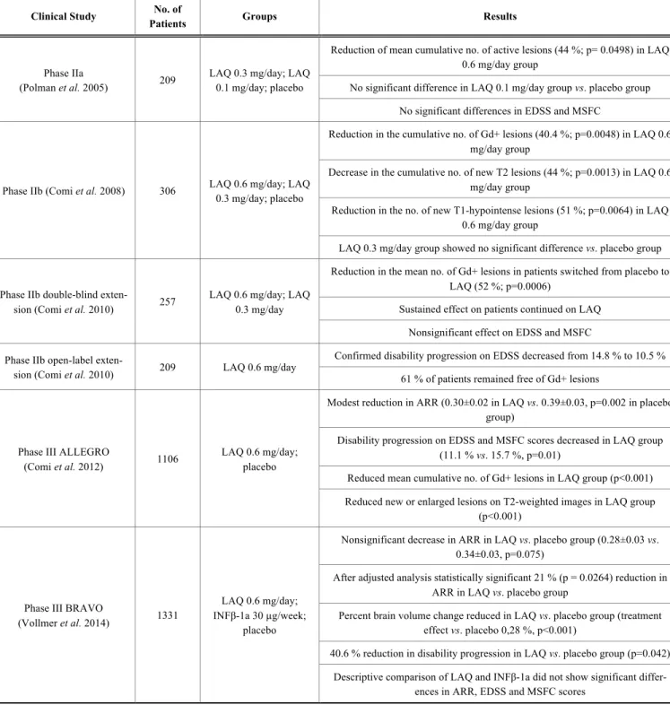
Polman at el. conducted a multicenter, double-blind, ran- domized, placebo controlled and three armed trial [63]. 209 RRMS patients (Expanded Disability Status Scale [EDSS]
no greater than 5.5 and active disease according to MRI cri- teria) were divided into three groups receiving 0.3 mg/day;
0.1 mg/day of laquinimod or placebo for 24 weeks. Primary outcome measure was the mean cumulative number of active lesions on brain MRIs. A significant reduction (44%;
p=0.0498) of the mean cumulative number of active lesions was observed in the 0.3 mg/day laquinimod group compared to the placebo group. A more pronounced effect was found in those patients who had at least one active lesion at base- line MRI (54% reduction; p=0.005). No significant differ- ence was observed in the primary end point in the 0.1 mg/day laquinimod group. Although the study did not aim to assess clinical end points, EDSS and MSFC (Multiple Scle- rosis Functional Composite) were measured, but no signifi- cant differences were found in these scores between groups over the 24 weeks. Laquinimod was found to be well tolerat- ed during the study.
The second phase II study was a multicenter, double- blind, randomized and placebo controlled 36 weeks long trial [90]. 306 RRMS patients (EDSS between 1 and 5, with at least one relapse in the previous year and at least one gado- linium enhancing lesion on brain MRI) were randomly as- signed to 0.3 mg/day; 0.6 mg/day of laquinimod or placebo.
The primary objective was to assess changes in the number of gadolinium enhancing (Gd+) lesions at 24, 28, 32 and 36 weeks. The authors reported 40.4 % (p=0.0048) reduction in the cumulative number of Gd+ lesions in the 0,6 mg/day laquinimod group. Furthermore, 44 % (p=0.0013) decrease in the cumulative number of new T2 lesions and 51 % (p=0.0064) reduction in the number of new T1-hypointense lesions were found in the same group compared to the place- bo arm. Strikingly, the 0.3 mg/day laquinimod group showed no significant difference compared to the placebo group, contradicting the findings of the previous phase II study. It was hypothesized, that the triple dose gadolinium used in the study by Polman et al. resulted in increased sensitivity, and also a possible slower onset of low-dose laquinimod com- pared to the higher dose could explain this observation. One case of Budd-Chiari syndrome occurred in a patient who was heterozygous for factor V Leiden mutation. Herpes simplex and herpes zoster infections were more common in the 0.3 mg/day laquinimod group. Mild arthralgia and transiently elevated liver enzymes were the most common adverse events.
A double-blind, 36 week-long extension of the study was conducted enrolling 257 out of 306 participants [91]. Pa- tients previously on placebo were given either 0.3 mg/day or 0.6 mg/day laquinimod. Main outcome measures were the number of new Gd+ lesions and the number of new hy- pointense T1 lesions. Among the participants who were switched from placebo, a 52 % (p=0.0006) reduction in the mean number of Gd+ lesions was found. The effect on new
hypointense T1-lesions was statistically nonsignificant. Pa- tients who were initially treated with laquinimod continued to have a sustained effect. Beneficial effect on clinical scores (EDSS and MSFC) were also nonsignificant. No deaths or serious adverse events were reported in the study.
An open-label extension of the core and double-blind study conducted by Comi et al. was carried out, in which all 209 participants received 0.6 mg/day of laquinimod, regard- less of what their therapy in the previous trials was [91]. 155 patients reached 24 months of the study. The confirmed dis- ability progression on EDSS decreased from 14.8 % to 10.5
% during the initial 18 months. 61 % of patients remained free of Gd+ lesions on 42 months. The most common ad- verse events were nasopharyngitis, back pain and headache.
Phase III Trials
The ALLEGRO (Assessment of Oral Laquinimod in Pre- venting Multiple Sclerosis) study was a randomized, double- blind, placebo-controlled, phase III clinical trial involving 24 countries [92]. 1106 patients with RRMS (EDSS no greater than 5.5 and a disease duration of at least 6 months) were enrolled. They received 0.6 mg/day laquinimod or placebo for 24 months. The primary end point was the number of confirmed relapses. Secondary clinical end points were disa- bility progression on EDSS and MSFC scores sustained for at least 3 months. Secondary imaging end points were the cumulative number of Gd+ lesions and the cumulative num- ber of new or enlarged lesions on T2-weighted images at 12 and 24 months. The annualized relapse rate showed a statis- tically significant, albeit modest reduction (0.30±0.02 vs.
0.39±0.03, p=0.002) in the laquinimod group. The secondary disability progression end point was significantly decreased in the laquinimod group (11.1% vs. 15.7%, p=0.01). Howev- er, little overall change was observed in the MSFC scores at 24 months. Laquinimod showed beneficial effects on both secondary MRI end points. The mean cumulative number of Gd+ lesions and new or enlarged lesions on T2-weighted images were reduced (p<0.001 in both cases). 112 serious adverse events were reported in 61 patients in the laquinimod group. Appendicitis occurred more often compared to the placebo group (5 vs. 1 respectively). 8 cases of neoplasms occurred in the laquinimod group compared to 6 in the pla- cebo arm. The most frequent adverse events included ab- dominal pain, back pain, cough and elevated levels of ala- nine aminotransferase. This finding was reversible in all cas- es without discontinuation of laquinimod or after 2 months of discontinuation. No cases of liver failure occurred. An open-label extension is currently ongoing with 844 out of the 864 patients who finished 24 months with the study drug.
The BRAVO (Benefit-Risk Assessment of Avonex and Laquinimod) study was the second phase III clinical trial, with the aim to further assess the safety, tolerability and effi- cacy of laquinimod and to descriptively compare its effect with interferon-beta 1a (INFβ-1a, Avonex®) [93]. 18 coun- tries were involved in the randomized, placebo-controlled 24-month trial. 1331 RRMS patients (18-55 years of age;
EDSS no greater than 5.5; at least one relapse in the last 12 months, two relapses in the past 24 months or one relapse in the past 12-24 months and one Gd+ MRI lesion in the previ- ous 12 months) were enrolled, and received 0.6 mg/day
laquinimod, matching placebo or INFβ-1a intramuscular injection 30 µg once a week. 1090 participants completed the trial. The primary endpoint was the annualized relapse rate over the 24 months of the trial. Secondary endpoints were the percent change in normalized brain volume, and disabil- ity progression measured by EDSS and MSFC scores. Ex- ploratory MRI endpoints included the cumulative number of Gd+ lesions and cumulative number of new or enlarging T2 lesions at 12 and 24 months. Laquinimod showed a decrease in annualized relapse rate, which was statistically nonsignifi- cant compared to placebo (0.28±0.03 vs. 0.34±0.03, p=0.075). However, after adjustment for imbalance between groups in baseline MRI disease activity, a statistically signif- icant 21 % (p = 0.0264) reduction in annualized relapse rate was demonstrated. Percent brain volume change was signifi- cantly reduced in the laquinimod group (treatment effect vs.
placebo 0,28 %, p<0.001). In contrast, INFβ-1a treatment did not show beneficial effects on brain volume compared to placebo (treatment effect -0.11 %, P=0.14). The reduction in disability progression in the laquinimod group was 40.6 % (p=0.042) at six months. INFβ-1a showed a nonsignificant reduction in disability worsening (28.3 %, P=0.14) at six months compared to placebo. A nonsignificant reduction in the laquinimod group was observed regarding exploratory MRI endpoints, whereas INFβ-1a treatment showed signifi- cant reductions. Descriptive comparison of laquinimod and INFβ-1a therapy did not show significant differences in an- nualized relapse rate, EDSS and MSFC scores, however laquinimod had a more pronounced beneficial effect on brain volume loss compared to INFβ-1a. One death occurred in the laquinimod group due to sepsis after early termination. The most common adverse events with laquinimod treatment were headache, increased alanine transaminase levels, ab- dominal pain and nausea. No cases of liver failure were re- ported.
The clinical trials with laquinimod (Table 1) showed modest effects on focal inflammatory activity of MS, but demonstrated significant beneficial effects on reduction of brain atrophy and disability progression. Laquinimod was found to be safe and well tolerated. The most common ad- verse event was increase in liver enzymes, however no cases of liver failure were observed in any of the clinical trials.
Ongoing Clinical Trials
The third phase III clinical trial assessing the safety, tol- erability and efficacy of a higher dose of laquinimod is the CONCERTO (The Efficacy and Safety and Tolerability of Laquinimod in Subjects With Relapsing Remitting Multi- ple Sclerosis) trial, which is currently in progress but not recruiting participants. It is a multinational, multicenter, ran- domized, double-blind, parallel-group, placebo-controlled study evaluating two different doses of laquinimod (0.6 mg/day vs. 1.2 mg/day) in patients with RRMS.
Previous clinical trials and animal studies suggest that laquinimod might exert neuroprotective effects. Based on these findings, two ongoing trials investigate the effect of laquinimod in diseases where neurodegeneration plays a prominent role in disease pathomechanism [88].
The ARPEGGIO (A Randomized Placebo-Controlled Tri- al Evaluating Laquinimod in Primary Progressive Multiple
Table 1. Summary of clinical trials with laquinimod.
Clinical Study No. of
Patients Groups Results
Phase IIa
(Polman et al. 2005) 209 LAQ 0.3 mg/day; LAQ 0.1 mg/day; placebo
Reduction of mean cumulative no. of active lesions (44 %; p= 0.0498) in LAQ 0.6 mg/day group
No significant difference in LAQ 0.1 mg/day group vs. placebo group No significant differences in EDSS and MSFC
Phase IIb (Comi et al. 2008) 306 LAQ 0.6 mg/day; LAQ 0.3 mg/day; placebo
Reduction in the cumulative no. of Gd+ lesions (40.4 %; p=0.0048) in LAQ 0.6 mg/day group
Decrease in the cumulative no. of new T2 lesions (44 %; p=0.0013) in LAQ 0.6 mg/day group
Reduction in the no. of new T1-hypointense lesions (51 %; p=0.0064) in LAQ 0.6 mg/day group
LAQ 0.3 mg/day group showed no significant difference vs. placebo group
Phase IIb double-blind exten-
sion (Comi et al. 2010) 257 LAQ 0.6 mg/day; LAQ 0.3 mg/day
Reduction in the mean no. of Gd+ lesions in patients switched from placebo to LAQ (52 %; p=0.0006)
Sustained effect on patients continued on LAQ Nonsignificant effect on EDSS and MSFC Phase IIb open-label exten-
sion (Comi et al. 2010) 209 LAQ 0.6 mg/day Confirmed disability progression on EDSS decreased from 14.8 % to 10.5 % 61 % of patients remained free of Gd+ lesions
Phase III ALLEGRO
(Comi et al. 2012) 1106 LAQ 0.6 mg/day;
placebo
Modest reduction in ARR (0.30±0.02 in LAQ vs. 0.39±0.03, p=0.002 in placebo group)
Disability progression on EDSS and MSFC scores decreased in LAQ group (11.1 % vs. 15.7 %, p=0.01)
Reduced mean cumulative no. of Gd+ lesions in LAQ group (p<0.001) Reduced new or enlarged lesions on T2-weighted images in LAQ group
(p<0.001)
Phase III BRAVO
(Vollmer et al. 2014) 1331
LAQ 0.6 mg/day;
INFβ-1a 30 µg/week;
placebo
Nonsignificant decrease in ARR in LAQ vs. placebo group (0.28±0.03 vs.
0.34±0.03, p=0.075)
After adjusted analysis statistically significant 21 % (p = 0.0264) reduction in ARR in LAQ vs. placebo group
Percent brain volume change reduced in LAQ vs. placebo group (treatment effect vs. placebo 0,28 %, p<0.001)
40.6 % reduction in disability progression in LAQ vs. placebo group (p=0.042) Descriptive comparison of LAQ and INFβ-1a did not show significant differ-
ences in ARR, EDSS and MSFC scores
ALLEGRO: Assessment of Oral Laquinimod in Preventing Multiple Sclerosis, ARR: annualized relapse rate, BRAVO: Benefit-Risk Assessment of Avonex and Laquinimod, EDSS:
Expanded Disability Status Scale, Gd+: gadolinium enhancing, INFβ-1a: interferon beta-1a, LAQ: laquinimod, MSFC: Multiple Sclerosis Functional Composite.
Sclerosis, Gauging Gradations in MRI and Clini- cal Outcomes) trial is a phase II trial aimed at assessing effi- cacy, safety and tolerability of two oral doses of laquinimod (0.6 mg/day or 1.5mg/day) as compared to placebo in prima- ry progressive MS patients.
The third ongoing clinical trial with laquinimod is the phase II LEGATO-HD (Laquinimod Efficacy and Safety in a Global Trial Of Huntington’s Disease) trial, which evaluates the efficacy and safety of three oral doses of laquinimod
(0.5, 1.0 and 1.5 mg/day) in patients with Huntington’s dis- ease.
Despite the positive results of clinical trials, the Commit- tee for Medicinal Products for Human Use (CHMP) rejected approval for laquinimod in 2014 [89]. According to the CHMP, higher occurrence of malignancy was observed after long-term exposure to laquinimod in animal studies. Despite that no treatment related cancer was observed in clinical tri- als, the CHMP stated that long-term cancer risk after
laquinimod treatment could not be ruled out. Also, the CHMP noted that again, based on animal studies, there is a possible teratogenic effect of laquinimod. Since there was only a modest effect of laquinimod in clinical trials, the CHMP concluded, that the potential risk of long-term laquinimod treatment currently outweighs its beneficial ef- fect on reducing disability progression in RRMS patients and rejected its approval in the European Community.
Another setback for laquinimod is that in the CONCER- TO and ARPEGGIO trials 8 cardiovascular (possibly is- chemic) events were reported with higher doses of laquini- mod [90, 91]. Seven adverse events were observed in the CONCERTO trial with 1.2 mg/day laquinimod and one event in the ARPEGGIO trial in the 1.5 mg/day laquinimod group. Consequently, the higher dose arm of these two ongo- ing trials (CONCERTO, ARPEGGIO) and the highest dose of the LEGATO-HD trial were discontinued in early 2016 [92]. No cardiovascular adverse events were reported in the lower dose arm of the studies and all three trials continue with the 0.6 mg/day laquinimod arm and 1 mg/day arm in the LEGATO-HD study.
CONCLUSION
Based on the clinical and experimental studies, laquini- mod might be a suitable medication for combination therapy in MS. Since it is an oral, once-a-day medication, patient compliance, adherence and satisfaction should improve compared to the parenteral medications currently in the first line armamentarium of MS treatment. Influencing the KP might offer a valuable therapeutic option in MS and other autoimmune diseases. KYNA analogs, KP enzyme inhibitors or structural analogs of kynurenines might offer promising candidates for future drug development.
CONFLICT OF INTEREST
The authors confirm that this article content has no con- flict of interest.
ACKNOWLEDGEMENTS
This work was supported by the the MTA-SZTE Neuro- science Research Group of the Hungarian Academy of Sci- ences and the University of Szeged and by the projects enti- tled GINOP-2.3.2-15-2016-00034 and the Hungarian Brain Research Programme (NAP, Grant No. KTIA-13-NAP-A- III/9. and KTIA-13-NAP-A-II/17.).
REFERENCES
[1] Noseworthy JH, Lucchinetti C, Rodriguez M, Weinshenker BG.
Multiple sclerosis. N Engl J Med 2000; 343: 938-52.
[2] Garg N, Smith TW. An update on immunopathogenesis, diagnosis, and treatment of multiple sclerosis. Brain Behav 2015; 5: e00362.
[3] Polman CH, Reingold SC, Banwell B, et al. Diagnostic criteria for multiple sclerosis: 2010 revisions to the McDonald criteria. Ann Neurol 2011; 69: 292-302.
[4] Lublin FD, Reingold SC. Defining the clinical course of multiple sclerosis: results of an international survey. National Multiple Scle- rosis Society (USA) Advisory Committee on Clinical Trials of New Agents in Multiple Sclerosis. Neurology 1996; 46: 907-11.
[5] Bruck W, Gold R, Lund BT, et al. Therapeutic decisions in multi- ple sclerosis: moving beyond efficacy. JAMA Neurol 2013; 70:
1315-24.
[6] Vecsei L, Szalardy L, Fulop F, Toldi J. Kynurenines in the CNS:
recent advances and new questions. Nat Rev Drug Discov 2013;
12: 64-82.
[7] Mandi Y, Vecsei L. The kynurenine system and immunoregulation.
J Neural Transm 2012; 119: 197-209.
[8] Majlath Z, Toldi J, Vecsei L. The potential role of kynurenines in Alzheimer's disease: pathomechanism and therapeutic possibilities by influencing the glutamate receptors. J Neural Transm 2014; 121:
881-9.
[9] Majlath Z, Tajti J, Vecsei L. Kynurenines and other novel thera- peutic strategies in the treatment of dementia. Ther Adv Neurol Disord 2013; 6: 386-97.
[10] Fuvesi J, Rajda C, Bencsik K, Toldi J, Vecsei L. The role of kynurenines in the pathomechanism of amyotrophic lateral sclero- sis and multiple sclerosis: therapeutic implications. J Neural Transm 2012; 119: 225-34.
[11] Rajda C, Majlath Z, Pukoli D, Vecsei L. Kynurenines and Multiple Sclerosis: The Dialogue between the Immune System and the Cen- tral Nervous System. Int J Mol Sci 2015; 16: 18270-82.
Fig. (3). Proposed mechanism of action of laquinimod. CNS: central nervous system; APCs: antigen presenting cells; Th: T helper cells, Treg: regulatory T cells; BDNF: brain derived neurotrophic factor; iNOS: inducible nitric oxide synthase; Glu: glutamate; TNFα: tumor ne- crosis factor α; IL: interleukin; GM-CSF: granulocyte-macrophage colony-stimulating factor; TGFβ: transforming growth factor β.
[12] Leklem JE. Quantitative aspects of tryptophan metabolism in hu- mans and other species: a review. Am J Clin Nutr 1971; 24: 659- 72.
[13] Han Q, Cai T, Tagle DA, Li J. Structure, expression, and function of kynurenine aminotransferases in human and rodent brains. Cell Mol Life Sci 2010; 67: 353-68.
[14] Guillemin GJ, Cullen KM, Lim CK, et al. Characterization of the kynurenine pathway in human neurons. J Neurosci 2007; 27:
12884-92.
[15] Guillemin GJ, Kerr SJ, Smythe GA, et al. Kynurenine pathway metabolism in human astrocytes: a paradox for neuronal protection.
J Neurochem 2001; 78: 842-53.
[16] Badawy AA, Bano S. Tryptophan Metabolism in Rat Liver After Administration of Tryptophan, Kynurenine Metabolites, and Kynureninase Inhibitors. Int J Tryptophan Res 2016; 9: 51-65.
[17] Badawy AA, Dougherty DM. Assessment of the Human Kynurenine Pathway: Comparisons and Clinical Implications of Ethnic and Gender Differences in Plasma Tryptophan, Kynurenine Metabolites, and Enzyme Expressions at Baseline and After Acute Tryptophan Loading and Depletion. Int J Tryptophan Res 2016; 9:
31-49.
[18] Bender DA. Inhibition in vitro of the enzymes of the oxidative pathway of tryptophan metabolism and of nicotinamide nucleotide synthesis by benserazide, carbidopa and isoniazid. Biochem Phar- macol 1980; 29: 707-12.
[19] Perez-De La Cruz V, Carrillo-Mora P, Santamaria A. Quinolinic Acid, an endogenous molecule combining excitotoxicity, oxidative stress and other toxic mechanisms. Int J Tryptophan Res 2012; 5:
1-8.
[20] Stone TW, Perkins MN. Quinolinic acid: a potent endogenous excitant at amino acid receptors in CNS. Eur J Pharmacol 1981; 72:
411-2.
[21] Han Q, Cai T, Tagle DA, Li J. Structure, expression, and function of kynurenine aminotransferases in human and rodent brains. Cell Mol Life Sci 2010; 67: 353-68.
[22] Kessler M, Terramani T, Lynch G, Baudry M. A glycine site asso- ciated with N-methyl-D-aspartic acid receptors: characterization and identification of a new class of antagonists. J Neurochem 1989;
52: 1319-28.
[23] Stone TW. Neuropharmacology of quinolinic and kynurenic acids.
Pharmacol Rev 1993; 45: 309-79.
[24] Prescott C, Weeks AM, Staley KJ, Partin KM. Kynurenic acid has a dual action on AMPA receptor responses. Neurosci Lett 2006;
402: 108-12.
[25] Rozsa E, Robotka H, Vecsei L, Toldi J. The Janus-face kynurenic acid. J Neural Transm 2008; 115: 1087-91.
[26] Hilmas C, Pereira EF, Alkondon M, Rassoulpour A, Schwarcz R, Albuquerque EX. The brain metabolite kynurenic acid inhibits al- pha7 nicotinic receptor activity and increases non-alpha7 nicotinic receptor expression: physiopathological implications. J Neurosci 2001; 21: 7463-73.
[27] Marchi M, Risso F, Viola C, Cavazzani P, Raiteri M. Direct evi- dence that release-stimulating alpha7* nicotinic cholinergic recep- tors are localized on human and rat brain glutamatergic axon termi- nals. J Neurochem 2002; 80: 1071-8.
[28] Fallarino F, Grohmann U, Vacca C, et al. T cell apoptosis by tryp- tophan catabolism. Cell Death Differ 2002; 9: 1069-77.
[29] Belladonna ML, Puccetti P, Orabona C, et al. Immunosuppression via tryptophan catabolism: the role of kynurenine pathway en- zymes. Transplantation 2007; 84: S17-20.
[30] Gonzalez A, Varo N, Alegre E, Diaz A, Melero I. Immunosuppres- sion routed via the kynurenine pathway: a biochemical and patho- physiologic approach. Adv Clin Chem 2008; 45: 155-97.
[31] Paterson PY. Autoimmune diseases of myelin. Prog Clin Biol Res 1980; 49: 19-36.
[32] Gold R, Linington C, Lassmann H. Understanding pathogenesis and therapy of multiple sclerosis via animal models: 70 years of merits and culprits in experimental autoimmune encephalomyelitis research. Brain 2006; 129: 1953-71.
[33] Kwidzinski E, Bunse J, Aktas O, et al. Indolamine 2,3-dioxygenase is expressed in the CNS and down-regulates autoimmune inflam- mation. Faseb J 2005; 19: 1347-9.
[34] Chiarugi A, Cozzi A, Ballerini C, Massacesi L, Moroni F.
Kynurenine 3-mono-oxygenase activity and neurotoxic kynurenine metabolites increase in the spinal cord of rats with experimental al- lergic encephalomyelitis. Neuroscience 2001; 102: 687-95.
[35] Flanagan EM, Erickson JB, Viveros OH, Chang SY, Reinhard JF, Jr. Neurotoxin quinolinic acid is selectively elevated in spinal cords of rats with experimental allergic encephalomyelitis. J Neurochem 1995; 64: 1192-6.
[36] Cammer W. Oligodendrocyte killing by quinolinic acid in vitro.
Brain Res 2001; 896: 157-60.
[37] Guillemin GJ, Wang L, Brew BJ. Quinolinic acid selectively in- duces apoptosis of human astrocytes: potential role in AIDS de- mentia complex. J Neuroinflammation 2005; 2: 16.
[38] Kerr SJ, Armati PJ, Guillemin GJ, Brew BJ. Chronic exposure of human neurons to quinolinic acid results in neuronal changes con- sistent with AIDS dementia complex. Aids 1998; 12: 355-63.
[39] Monaco F, Fumero S, Mondino A, Mutani R. Plasma and cerebro- spinal fluid tryptophan in multiple sclerosis and degenerative dis- eases. J Neurol Neurosurg Psychiatry 1979; 42: 640-1.
[40] Rudzite V, Berzinsh J, Grivane I, Fuchs D, Baier-Bitterlich G, Wachter H. Serum tryptophan, kynurenine, and neopterin in pa- tients with Guillain-Barre-syndrome (GBS) and multiple sclerosis (MS). Adv Exp Med Biol 1996; 398: 183-7.
[41] Sandyk R. Tryptophan availability and the susceptibility to stress in multiple sclerosis: a hypothesis. Int J Neurosci 1996; 86: 47-53.
[42] Sadowska-Bartosz I, Adamczyk-Sowa M, Gajewska A, Bartosz G.
Oxidative modification of blood serum proteins in multiple sclero- sis after interferon or mitoxantrone treatment. J Neuroimmunol 2014; 266: 67-74.
[43] Beck J, Rondot P, Catinot L, Falcoff E, Kirchner H, Wietzerbin J.
Increased production of interferon gamma and tumor necrosis fac- tor precedes clinical manifestation in multiple sclerosis: do cyto- kines trigger off exacerbations? Acta Neurol Scand 1988; 78: 318- 23.
[44] Guillemin GJ, Kerr SJ, Pemberton LA, et al. IFN-beta1b induces kynurenine pathway metabolism in human macrophages: potential implications for multiple sclerosis treatment. J Interferon Cytokine Res 2001; 21: 1097-101.
[45] Hartai Z, Klivenyi P, Janaky T, Penke B, Dux L, Vecsei L.
Kynurenine metabolism in multiple sclerosis. Acta Neurol Scand 2005; 112: 93-6.
[46] Kepplinger B, Baran H, Kainz A, Ferraz-Leite H, Newcombe J, Kalina P. Age-related increase of kynurenic acid in human cerebro- spinal fluid - IgG and beta2-microglobulin changes. Neurosignals 2005; 14: 126-35.
[47] Lim CK, Brew BJ, Sundaram G, Guillemin GJ. Understanding the roles of the kynurenine pathway in multiple sclerosis progression.
Int J Tryptophan Res 2010; 3: 157-67.
[48] Rejdak K, Bartosik-Psujek H, Dobosz B, et al. Decreased level of kynurenic acid in cerebrospinal fluid of relapsing-onset multiple sclerosis patients. Neurosci Lett 2002; 331: 63-5.
[49] Rejdak K, Petzold A, Kocki T, et al. Astrocytic activation in rela- tion to inflammatory markers during clinical exacerbation of re- lapsing-remitting multiple sclerosis. J Neural Transm 2007; 114:
1011-5.
[50] Paul C, Bolton C. Modulation of blood-brain barrier dysfunction and neurological deficits during acute experimental allergic en- cephalomyelitis by the N-methyl-D-aspartate receptor antagonist memantine. J Pharmacol Exp Ther 2002; 302: 50-7.
[51] Stone TW. Kynurenic acid antagonists and kynurenine pathway inhibitors. Expert Opin Investig Drugs 2001; 10: 633-45.
[52] Fuvesi J, Somlai C, Nemeth H, et al. Comparative study on the effects of kynurenic acid and glucosamine-kynurenic acid. Pharma- col Biochem Behav 2004; 77: 95-102.
[53] Nagy K, Plangar I, Tuka B, et al. Synthesis and biological effects of some kynurenic acid analogs. Bioorg Med Chem 2011; 19:
7590-6.
[54] Fulop F, Szatmari I, Toldi J, Vecsei L. Modifications on the car- boxylic function of kynurenic acid. J Neural Transm 2012; 119:
109-14.
[55] Chiarugi A, Carpenedo R, Molina MT, Mattoli L, Pellicciari R, Moroni F. Comparison of the neurochemical and behavioral effects resulting from the inhibition of kynurenine hydroxylase and/or kynureninase. J Neurochem 1995; 65: 1176-83.
[56] Colabroy KL, Zhai H, Li T, et al. The mechanism of inactivation of 3-hydroxyanthranilate-3,4-dioxygenase by 4-chloro-3- hydroxyanthranilate. Biochemistry 2005; 44: 7623-31.
[57] Walsh HA, O'Shea KC, Botting NP. Comparative inhibition by substrate analogues 3-methoxy- and 3-hydroxydesaminokynurenine
and an improved 3 step purification of recombinant human kynureninase. BMC Biochem 2003; 4: 13.
[58] Sundaram G, Brew BJ, Jones SP, Adams S, Lim CK, Guillemin GJ. Quinolinic acid toxicity on oligodendroglial cells: relevance for multiple sclerosis and therapeutic strategies. J Neuroinflammation 2014; 11: 204.
[59] Platten M, Ho PP, Youssef S, et al. Treatment of autoimmune neuroinflammation with a synthetic tryptophan metabolite. Science 2005; 310: 850-5.
[60] Fazio F, Zappulla C, Notartomaso S, et al. Cinnabarinic acid, an endogenous agonist of type-4 metabotropic glutamate receptor, suppresses experimental autoimmune encephalomyelitis in mice.
Neuropharmacology 2014; 81: 237-43.
[61] Bruck W, Wegner C. Insight into the mechanism of laquinimod action. J Neurol Sci 2011; 306: 173-9.
[62] Fernandez O. Oral laquinimod treatment in multiple sclerosis.
Neurologia 2011; 26: 111-7.
[63] Polman C, Barkhof F, Sandberg-Wollheim M, et al. Treatment with laquinimod reduces development of active MRI lesions in re- lapsing MS. Neurology 2005; 64: 987-91.
[64] Wild EJ, Tabrizi SJ. Targets for future clinical trials in Hunting- ton's disease: what's in the pipeline? Mov Disord 2014; 29: 1434- 45.
[65] Zadori D, Klivenyi P, Szalardy L, Fulop F, Toldi J, Vecsei L. Mi- tochondrial disturbances, excitotoxicity, neuroinflammation and kynurenines: novel therapeutic strategies for neurodegenerative disorders. J Neurol Sci 2012; 322: 187-91.
[66] Zadori D, Nyiri G, Szonyi A, et al. Neuroprotective effects of a novel kynurenic acid analogue in a transgenic mouse model of Huntington's disease. J Neural Transm 2011; 118: 865-75.
[67] Jonsson S, Andersson G, Fex T, et al. Synthesis and biological evaluation of new 1,2-dihydro-4-hydroxy-2-oxo-3- quinolinecarboxamides for treatment of autoimmune disorders:
structure-activity relationship. J Med Chem 2004; 47: 2075-88.
[68] Wolinsky JS, Narayana PA, Noseworthy JH, et al. Linomide in relapsing and secondary progressive MS: part II: MRI results. MRI Analysis Center of the University of Texas-Houston, Health Sci- ence Center, and the North American Linomide Investigators. Neu- rology 2000; 54: 1734-41.
[69] Tan IL, Lycklama a Nijeholt GJ, Polman CH, Ader HJ, Barkhof F.
Linomide in the treatment of multiple sclerosis: MRI results from prematurely terminated phase-III trials. Mult Scler 2000; 6: 99-104.
[70] Varrin-Doyer M, Zamvil SS, Schulze-Topphoff U. Laquinimod, an up-and-coming immunomodulatory agent for treatment of multiple sclerosis. Exp Neurol 2014; 262: 66-71.
[71] Bruck W, Wegner C. Insight into the mechanism of laquinimod action. J Neurol Sci 2011; 306: 173-9.
[72] Ali R, Nicholas RS, Muraro PA. Drugs in development for relaps- ing multiple sclerosis. Drugs 2013; 73: 625-50.
[73] Kieseier BC. Defining a role for laquinimod in multiple sclerosis.
Ther Adv Neurol Disord 2014; 7: 195-205.
[74] Thone J, Gold R. Review of laquinimod and its therapeutic poten- tial in multiple sclerosis. Expert Opin Pharmacother 2013; 14:
2545-52.
[75] Bjork P, Bjork A, Vogl T, et al. Identification of human S100A9 as a novel target for treatment of autoimmune disease via binding to quinoline-3-carboxamides. PLOS Biol 2009; 7: e97.
[76] Mishra MK, Wang J, Silva C, Mack M, Yong VW. Kinetics of proinflammatory monocytes in a model of multiple sclerosis and its perturbation by laquinimod. Am J Pathol 2012; 181: 642-51.
[77] Wegner C, Stadelmann C, Pfortner R, et al. Laquinimod interferes with migratory capacity of T cells and reduces IL-17 levels, in-
flammatory demyelination and acute axonal damage in mice with experimental autoimmune encephalomyelitis. J Neuroimmunol 2010; 227: 133-43.
[78] Mishra MK, Wang J, Keough MB, et al. Laquinimod reduces neu- roaxonal injury through inhibiting microglial activation. Ann Clin Transl Neurol 2014; 1: 409-22.
[79] Jolivel V, Luessi F, Masri J, et al. Modulation of dendritic cell properties by laquinimod as a mechanism for modulating multiple sclerosis. Brain 2013; 136: 1048-66.
[80] Gurevich M, Gritzman T, Orbach R, Tuller T, Feldman A, Achiron A. Laquinimod suppress antigen presentation in relapsing-remitting multiple sclerosis: in-vitro high-throughput gene expression study.
J Neuroimmunol 2010; 221: 87-94.
[81] Schulze-Topphoff U, Shetty A, Varrin-Doyer M, et al. Laquini- mod, a quinoline-3-carboxamide, induces type II myeloid cells that modulate central nervous system autoimmunity. PlOS One 2012; 7:
e33797.
[82] Toubi E, Nussbaum S, Staun-Ram E, et al. Laquinimod modulates B cells and their regulatory effects on T cells in multiple sclerosis.
J Neuroimmunol 2012; 251: 45-54.
[83] Zilkha-Falb R, Gurevich M, Hayardeny L, Achiron A. The role of laquinimod in modulation of the immune response in relapsing- remitting multiple sclerosis: Lessons from gene expression signa- tures. J Neuroimmunol 2015; 283: 11-6.
[84] Kerschensteiner M, Gallmeier E, Behrens L, et al. Activated hu- man T cells, B cells, and monocytes produce brain-derived neu- rotrophic factor in vitro and in inflammatory brain lesions: a neuro- protective role of inflammation? J Exp Med 1999; 189: 865-70.
[85] Kalb R. The protean actions of neurotrophins and their receptors on the life and death of neurons. Trends Neurosci 2005; 28: 5-11.
[86] Aharoni R, Saada R, Eilam R, Hayardeny L, Sela M, Arnon R. Oral treatment with laquinimod augments regulatory T-cells and brain- derived neurotrophic factor expression and reduces injury in the CNS of mice with experimental autoimmune encephalomyelitis. J Neuroimmunol 2012; 251: 14-24.
[87] Thone J, Ellrichmann G, Seubert S, et al. Modulation of autoim- mune demyelination by laquinimod via induction of brain-derived neurotrophic factor. Am J Pathol 2012; 180: 267-74.
[88] Ruffini F, Rossi S, Bergamaschi A, et al. Laquinimod prevents inflammation-induced synaptic alterations occurring in experi- mental autoimmune encephalomyelitis. Mult Scler 2013; 19: 1084- 94.
[89] Kolb-Sobieraj C, Gupta S, Weinstock-Guttman B. Laquinimod therapy in multiple sclerosis: a comprehensive review. Neurol Ther 2014; 3: 29-39.
[90] Comi G, Pulizzi A, Rovaris M, et al. Effect of laquinimod on MRI- monitored disease activity in patients with relapsing-remitting mul- tiple sclerosis: a multicentre, randomised, double-blind, placebo- controlled phase IIb study. Lancet 2008; 371: 2085-92.
[91] Comi G, Abramsky O, Arbizu T, et al. Oral laquinimod in patients with relapsing-remitting multiple sclerosis: 36-week double-blind active extension of the multi-centre, randomized, double-blind, parallel-group placebo-controlled study. Mult Scler 2010; 16:
1360-6.
[92] Comi G, Jeffery D, Kappos L, et al. Placebo-controlled trial of oral laquinimod for multiple sclerosis. N Engl J Med 2012; 366: 1000- 9.
[93] Vollmer TL, Sorensen PS, Selmaj K, et al. A randomized placebo- controlled phase III trial of oral laquinimod for multiple sclerosis. J Neurol 2014; 261: 773-83.
PMID: 28017129