REVIEW ARTICLE
1389-5575/20 $65.00+.00 © 2020 Bentham Science Publishers
Medicinal Chemistry of Multiple Sclerosis: Focus on Cladribine
Tamás Biernacki
1, Dániel Sandi
1, Krisztina Bencsik
1and László Vécsei
1,2,*1Department of Neurology, Szent-Györgyi Albert Clinical Center, University of Szeged, Szeged, Hungary; 2MTA-SZTE Neuroscience Research Group, Szeged, Hungary
Abstract: Background: In the recent years, many novel disease-modifying drugs (DMD) have been introduced to the market in the treatment of multiple sclerosis.
Objectives: To provide the reader with an up to date, compact review on the pharmacokinetic proper- ties, mechanism of action, and clinical attributes of one of the most recently approved drugs in the therapy of multiple sclerosis, cladribine.
Conclusion: Cladribine tablets proved to be a highly efficient treatment choice for relapsing-remitting multiple sclerosis (RRMS), especially for patients with high disease activity. It is the first DMD for MS with a complex mechanism of action, by inhibiting the adenosine-deaminase enzyme it increases the intracellular levels of deoxyadenosine triphosphate, which with relative selectivity depletes both T- and B-cells lines simultaneously. However long term follow-up safety and effectiveness data are still missing, and clear treatment protocols are lacking beyond the first two treatment years cladribine should prove to be a valuable addition to the therapeutic palette of RRMS, and potentially for clinically isolated syndrome (CIS) as well.
A R T I C L E H I S T O R Y
Received: August 27, 2018 Revised: April 28, 2019 Accepted: May 19, 2019 DOI:
10.2174/1389557519666191015201755
Keywords: Cladribine, mechanism of action, multiple sclerosis, efficacy, safety, dosing.
1. INTRODUCTION
Multiple Sclerosis (MS) is a chronic inflammatory and neurodegenerative disease of the central nervous system (CNS). There are approximately 2.3 million MS patients worldwide [1]. Among young adults, MS is the most com- mon cause of non-traumatic disability, it is the most common
“rare disease” [2]. Its prevalence shows north to south gradi- ent across the globe, it being the most common in the Scan- dinavian countries (150-200/100 000), roughly half of that in Central Europe (83.7/100 000), and having an even lower prevalence approaching the equator and in southern Baltic states (20-50/100,000) [3-5]. In addition to being the most common "rare-disease", it doesn't just put a heavy strain on one's everyday quality of life – as it was presumed for long -, but it also increases the patients' mortality by 2.57 to 4.10 fold (depending on disease form and severity) [6].
More than 20 years ago the clinical courses of multiple sclerosis have been defined as benign, relapsing-remitting, primary progressive, secondary progressive, and progres- sive-relapsing MS by consensus statement of the internation- al clinical MS community [7]. These disease course types are
*Address correspondence to this author at the Department of Neurology, Faculty of General Medicine, Albert Szent-Györgyi Clinical Centre, Univer- sity of Szeged, Semmelweis u. 6., H-6725, Szeged, Hungary; Tel: +36-62- 545-384; Fax: +36-62-545-597; E-mail: vecsei.laszlo@med.u-szeged.hu
not supported by neuroimaging or other kinds of biomarkers but were solely based on the clinical picture of the patients.
Since then, a substantial amount of knowledge has been gathered regarding the pathophysiology, the nature of and biomarkers in MS. This has invoked a general concern that the "old" classification might not reflect the clinical aspects, the prognosis and the treatment requirement of one's disease with enough accuracy. Hence the classification system has been revised based on the past decades' improved descriptive technologies, biomarkers, imaging technologies and clinical experience [8, 9]. The new classification system re-defined some terms used to describe one's disease. Among the re- defined terms are disease activity (“clinical activity: relapses, acute or sub-acute episodes of new or increasing neurologi- cal dysfunction followed by full or partial recovery, in the absence of fever or infection or evidence of a pseudo- relapse”, “imaging: occurrence of contrast-enhancing T1 hyperintense lesions or new or unequivocally enlarging T2 hyperintense lesions. The term ‘unequivocally’ has been added to highlight the importance of aligning successive scans as closely as possible when gauging enlarging le- sions”), progression (clinical: steadily increasing objectively documented neurological dysfunction/disability without un- equivocal recovery; fluctuations and phases of stability may occur, imaging: no standardized imaging measures of disease progression are currently established; however, measures,
such as an increasing number and volume of T1 hypointense lesions, brain volume loss and changes in magnetization transfer imaging and diffusion tensor imaging are being ex- plored), disease worsening ("is defined as a documented in- crease in neurological dysfunction/disability as a result of relapses or progressive disease or both") and confirmed pro- gression or worsening ("is an increase in neurological dys- function confirmed throughout a defined time interval (e.g.
3, 6 or 12 months); this distinction is particularly relevant in the clinical trial setting [8, 9]. Given that neurological dys- function may improve (especially in relapsing-remitting MS) even if initially confirmed, it is recommended that the term
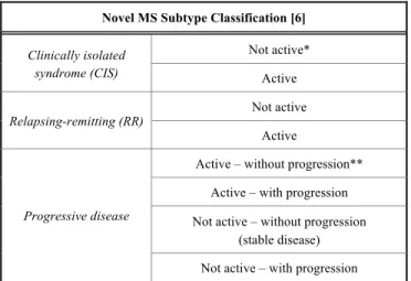
‘sustained' be abandoned unless the disease has been meas- ured over "a long period of time" [8, 9]. Based on the new classifications system a person's disease can be classified as a clinically isolated syndrome (CIS) with or without activity, relapsing-remitting with or without activity, progressive – active with or without progression, inactive with or without progression [10]. Radiologically isolated syndrome (RIS) was omitted and is not considered to be part of the MS spec- trum disease anymore [10].
Table 1. Novel classification of MS subtypes. Activity deter- mined by clinical relapses assessed at least annually and/or MRI activity (contrast-enhancing lesions;
new and unequivocally enlarging T2 lesions).
Novel MS Subtype Classification [6]
Clinically isolated syndrome (CIS)
Not active*
Active
Relapsing-remitting (RR) Not active Active
Progressive disease
Active – without progression**
Active – with progression Not active – without progression
(stable disease) Not active – with progression
**Progression measured by clinical evaluation, assessed at least annually. If assess- ments are not available, activity and progression are “indeterminate” [6].
1.1. Pathomechanism
Although a great number of studies have been dedicated to revealing the exact cellular and molecular pathomecha- nism of multiple sclerosis, the picture behind it is still un- clear in many aspects. What is the first step in developing the disease? In spite of the fact that most findings agree that MS is primarily an immunological disease where secondary neu- rodegeneration is involved, there is some convincing evi- dence pointing towards the opposite direction i.e. the under- lying process may be neurodegeneration itself with the sec- ondary involvement of the immune system [11, 12]. Yet, as the therapies used for slowing the progression and lowering the relapse rate are primarily immunotherapies – and those therapies are effective in halting the disease – we try to con-
centrate on the immunological aspect of MS in this review [13].
1.1.1. The Role of T-Cells
MS is primarily considered to be initialized by the activa- tion and migration of autoreactive Th-1 and Th-17 cells into the CNS, mainly targeting MBP, MOG and HspB5 antigens [14-16]. The arrival of these autoreactive cells initiates a cascade which by recruiting and activating both the cellular and the humoral pathways of the immune response gradually leads to demyelination and to subsequent neurodegeneration.
Evidence was supplied for this theory by many studies: be- ginning with the fact, that autoreactive T-cells were found in all 4 types of MS lesions throughout pathological examina- tions as described by Lucchinetti et al. [17]. Through in vivo animal studies, other evidence was found: EAE-prone mice were found to produce a large amount of IFN-γ, one of the primary inflammatory cytokines produced by Th-1 cells, which neutralizes IL-12, an anti-inflammatory cytokine, whereas Th-2 responses and the inhibition of IL-4 cytokine release is prominent in animals resistant to EAE [18]. It was also shown, that the T-cells of MS patients produce far more IFN-γ than the T-cells of healthy controls, suggesting an important role in CNS inflammation [19]. Furthermore, the dysregulation of T-regulatory and Th-17 cells has been found: these cell types have a similar development pathway and some studies showed that the development of Th-17 cells is up-regulated in MS as compared to other subtypes [20, 21]. These cells were also shown to be the most damag- ing to the blood-brain barrier (BBB) [15]. Other studies con- cluded that the other regulatory subsets of T-cells – under specific circumstances – may become proinflammatory T-17 cells [22]. All these factors lead to myelin damage and axo- nopathy, first initializing, then sustaining the inflammation in the CNS.
The role of CD8+ T-cells is less understood, but seems nonetheless important. Classically, these cells were named
"T-killer" cells due to their cytotoxic potential. Yet, more recent evidence points to the regulatory capabilities of CD8+
T-cells, as a factor in MS [23]. It seems that specific types of these cells are involved in the protection against inflammato- ry processes as they suppress the activation of CD4+ path- ways, thus the down-regulation of these cells play part in the autoimmune process [23].
1.1.2. The Role of B-Cells
Several facts show the direct involvement of the humoral pathway, thus B-cells in the pathology of MS, the presence of oligoclonal gammopathy (OGP) being the most obvious [24]. Many studies concluded, however, that this activation is secondary and unspecified, and it plays an important role in the de facto degradation of myelin and damage of the ax- ons [24-26]. However, other, newer evaluations found, that some peripheral nervous system-specific antibodies are ca- pable of activating CNS-specific T-cells, thus playing an important part in the initialization of the inflammatory pro- cess while still in the periphery [24]. Also, another aspect of B-cells seems to be highly important in the immuno- pathomechanism: B-cells are effective and specific antigen-
presenting agents through major histocompatibility complex II (MHC-II) and B-cell receptor (BCR) molecules, playing a pivotal role in activating and promoting the differentiation of T-cells toward the proinflammatory path [27-29]. In recent studies, the presence of both myelin-recognizing B- and T- cells successfully induced EAE [30, 31]. The other important factor is the role of the regulating B-cell population and the production of cytokines. Several studies indicate, that the chronic activation of B-regulatory cells producing mainly proinflammatory cytokines (e.g. IL-6), thus leading to the shift toward proinflammatory T-cell populations is a major correspondent in CNS inflammation [24]. For instance, the genetically modified, B-cell restricted IL-6 deficient mice population showed a far less severe course of EAE and Th- 17 cell numbers [32, 33]. Also, the levels of secreted IL-6 and other proinflammatory cytokine levels are definitely higher in the B-cells derived from MS patients than from healthy controls [33]. On the other hand, studies showed that the activation of anti-inflammatory cytokine-producing B- cells relates to a less severe course [24, 34].
As described above, the pathomechanism of MS is a very complex process involving most cellular parts of the immune system, however, this process is still unclear in many as- pects. Yet, it is important to see, that these diverse immuno- logical dysfunctions all play important parts in the initiation and sustaining of the CNS inflammation, thus therapies tar- geting these processes can-, and have been proven beneficial.
1.2. Therapy of MS
Currently, MS is an incurable condition: current treat- ments focus on slowing the disease progression by reducing the inflammation in the CNS, lowering the relapse-rate and slowing the emergence of disability. There are several un- met needs, considering MS therapy, aside from a definitive cure. It is a heavy burden for both the patients and the medi- cal service providers, that current disease-modifying thera- pies (DMT) - except for alemtuzumab - should be used and monitored continuously for decades. Because of constant immunosuppression, patients are more prone to opportunistic infections. The efficacy of the injectable DMTs (interferon-ß and glatiramer-acetate) is lower than the newer therapies, they often cause local side effects, thus patients often cannot toler- ate the long-term use. On the other hand, the higher effectivity of monoclonal antibodies (natalizumab, alemtuzumab, ocreli- zumab, daclizumab), which are not taken daily, can lead to serious side effects (progressive multifocal leukoencephalo- pathy (PML) with natalizumab, daclizumab, which was withdrawn from the market in early 2018 due to severe, po- tentially lethal side effects, listeriosis following treatment with alemtuzumab) [35-37]. The effectivity of per os drugs is similar to the injectable DMTs, but they require regular blood test control, some should be taken more than once a day and in case of missed doses or stopping the treatment, their effect fades away fast. Cladribine offers a solution to some of these problems: it is used in four, five-days cycle in the first 2 years, it is taken per os, it has a long-lasting effect and the side effect profile is comparable to current DMTs [38].
2. CLADRIBINE
2-chloro-2’-deoxyadenosine (2-CdA, Cladribine) was first synthetized in 1972, following the discovery of a new form of severe combined immunodeficiency (SCID). Dr.
Ernest Beutler and Dr. Dennis A. Carson had been studying a new form of SCID, which was discovered to be caused by the deficiency of the adenosine-deaminase enzyme. Because of the defect in the enzyme, deoxy-adenosine-triphosphate levels increase inside the cells, causing the destruction of T and B lymphocytes and subsequently leading to immunode- ficiency [39]. This revelation leads to the idea, that a drug with a similar mechanism of action might be suitable to treat lymphomas. Cladribine was found to be able to achieve pro- longed remission in patients with hairy cell leukemia (HCL), a previously untreatable disease [40]. In 1993, intravenous cladribine was approved by both the Food and Drug Admin- istration (FDA) in the U.S.A, and by the European Medi- cines Agency (EMA) in Europe for the treatment of HCL as an orphan drug. A subcutaneous form with the same indica- tion was also developed later, and introduced to the market [41].
Following its approval in the treatment of HCL, several clinical trials have been conducted in order to explore the potential efficacy of cladribine in MS, based on its potent immunosuppressive effect on both T-, and B cell lines. The first attempt for its approval in the treatment of MS was in 1997, however, the application was withdrawn from the FDA on the grounds of insufficient clinical data. After an oral version of the drug was developed a new application was submitted, which was first rejected by the EMA in 2010, and then by the FDA in 2011 due to concerns that an in- creased number of cancers have arisen amongst the patients treated with cladribine, and the ratio of benefit to harm was not clear to the regulators at the time with the amount of data available. At the time of the rejections, clinical trials were still ongoing, which were not terminated. Meta-analysis of data from these trials had shown that cladribine did not in- crease the risk of cancer at the doses used. Following this, a new application was submitted to the regulatory agencies, on June 22, 2017, the EMA approved the use of cladribine for the treatment of relapsing-remitting multiple sclerosis.
2.1. Mechanism of Action
The mechanisms of action of the current DMTs lie in either immunomodulation, immunosuppression, or the inhi- bition of leukocyte migration. Cladribine has a combined effect: it has an immunomodulatory effect by pushing the cytokine rate towards the direction of anti-inflammatory cy- tokines; and an immunosuppressive effect by depleting both the T- and the B-lymphocyte populations.
2.1.1. The Immunosuppressive Effect
Cladribine is a purine analog that differs from deoxy- adenosine (DA) in only a chlorine atom on the 2nd C-atom of the purine frame [39]. As the deoxy-nucleosides enter the cells, they are phosphorylated by the deoxycytidine kinase (dCK), an intracellular enzyme. From deoxyadenosine, it creates deoxy-adenosine-monophosphate (dAMP) in the first step, then diphosphate (dADP), and triphosphate (dATP)
[39]. Cladribine becomes 2-chloro-2’-deoxy-ß-D-adenosine monophosphate (2-CdAMP), then diphosphate, finally also triphosphate (2-CdATP) [42]. The same process occurs in the mitochondria, except there, the phosphorylation is done by the deoxy-guanosine kinase enzyme [42]. The intracellu- lar degradation of adenosine-nucleosides to deoxy-inosines is done by the adenosine deaminase (ADA). The intracellular adenosine nucleoside-nucleotide rate is determined by the dCK-ADA rate (kinase-phosphatase rate), compared to other cells, this rate is higher in lymphocytes [42]. Cladribine is a substrate to dCK, but resistant to ADA, as the halogenation of the adenine ring with chlorine leads to such changes in the molecules' electron-distribution, that makes the amine-group resistant to hydrolyzing by ADA [43]. Thus, due to the high kinase-phosphatase rate in lymphocytes, cladribine accumu- lates fast in the cells, generating a high amount of 2-CdATP.
The accumulated 2-CdATP is toxic to both the resting and the dividing lymphocytes. This cytotoxicity of cladribine is due to its influence on multiple intracellular pathways.
2.1.1.1. Inhibition of DNA-synthesis
Cladribine inhibits the DNA-synthesis in multiple ways.
Among other enzymes, the ribonucleoside-reductase (RR) supplies the needed amount of deoxy-ribonucleoside- triphosphates for DNA-synthesis, by creating deoxy- ribonucleoside-diphosphates from ribonucleoside diphos- phates, which are phosphorylated further to triphosphates, which can enter the DNA-synthesis [44]. Cladribine is a highly effective inhibitor of RR, it can reversibly inhibit the enzyme even at 0.06 µM intracellular concentration [45].
Also - as it is a purine-analog - it is a substrate of DNA- polymerase, thus incorporates into the synthetizing DNA- chain causing a chain-termination reaction. As cladribine inhibits the RR, it lowers the rate of available deoxyribonu- cleoside triphosphates, therefore increasing the chance of its own incorporation into the DNA-synthesis by DNA- polymerase, further increasing its toxicity [46, 47].
2.1.1.2. The Inhibition of DNA-repair
The key step in many DNA-repair mechanisms is to cut out and replaced the damaged DNA section. As the repair of the DNA is a constant process in resting cells as well, there is always a need for freshly synthetized DNA-chains for the replacement of damaged sections. Cladribine can incorporate into these freshly synthetized sections, thus causing chain termination and making it impossible to repair the errors in the DNA-chain, leading to fast degradation [46-49]. Studies show, that the incorporated cladribine inhibits the ß-DNA- polymerase, an essential enzyme in the DNA-repair process, and found a positive correlation between the amount of these incorporated purine-analogs and the number of destructed cells [46-49].
2.1.1.3. Apoptosis
The third potential mechanism of cladribine for immuno- suppression is the induction of apoptosis in lymphocytes.
Cladribine incorporation leads to chain termination, which is perceived by the cell as if the DNA-double helix was broken.
The poly-ADP-ribose-polymerase (PARP) and other en- zymes perceive the errors and activate the downstream signal pathways, which lead to either the repair of the DNA or to Fig. (1). The accumulation of chloro-deoxynucleotides caused by cladribine. ADA – adenosine deaminase, 2-CdA – 2-chloro-2’- deoxyadenosine (cladribine), dCK – deoxycitidine kinase, 5’NT – 5’ nucleotidase, dAMP – deoxyadenosine monophosphate, dADP – deox- yadenosine diphosphate, dATP – deoxyadenosine triphosphate, 2-CdAMP - 2-chloro-2’-deoxyadenosine-monophosphate, 2-CdADP – 2- chloro-2’-deoxyadenosine-diphosphate, 2-CdATP – 2-chloro-2’-deoxyadenosine-triphosphate. (A higher resolution/colour version of this figure is available in the electronic copy of the article).
cell death [50, 51]. As the cascade, induced by the DNA er- rors, begins, the p53 molecule activates, which in turn, acti- vates other transducer molecules, including p21 [52]. The increasing p21 stops the cell cycle by the inhibition of cy- clin-dependent kinase 2 (CDK-2), for either repair or if not possible, for cell death; as cladribine inhibits the DNA- repair, it induces the apoptosis pathway at this point [52-57].
In sensitive cell culture lines, the morphological and bio- chemical signs can be seen following incubation with cladribine. In order to be able to activate and execute the programmed cell death cells need to contain a ubiquitous, multi-purpose cysteine protease protein family, called caspa- ses. These proteins can cleave and thus activate or degrade numerous structural, nuclear, signaling and effector proteins, some of which play a pivotal role in the apoptotic cascade.
The caspases can be activated by two separate pathways, the first is of mitochondrial origin ("intrinsic pathway"), while the second one is triggered by the activation of cell surface receptors, called the "extrinsic pathway" [58].
Cladribine produces its effect primarily by the activation of the intrinsic pathway. Triggering the intrinsic pathway causing an increase in the permeability of the mitochondrial membrane. This can be achieved either by the incorporation of pore-forming molecules into the membrane or by the acti- vation of an already present pore-forming complex, the mi- tochondrial permeability transition pore (mtPTP) [43]. The opening of the external membrane of the mitochondria cre- ates the opportunity for various apoptosis-inducing mole- cules (including Cytochrome C and apoptosis-inducing fac-
tor - AIF) residing in the intermembranous space, to exit the mitochondria and enter the cytosol. The Cytochrome C now inside the cytosol can activate the first member of the intrin- sic caspase cascade, namely the Caspase-9, whereas AIF enters the nucleus and causes the fragmentation of the DNA chain by a mechanism independent of the caspase cascade [59-65].
The extrinsic pathway is triggered by the activation of various ligands and receptors, such as Fas (CD95/Apo-1), TNF receptor 1 (TNF-R1) and death receptors (DR-3, -4, -5).
The activation can be induced by the binding of either cell surface bound or soluble ligands, like Fas-L (CD 95), TNF-ɑ and TNF-related apoptosis-inducing ligand (TRAIL) [66].
The binding of these ligands is followed by the recruitment of support molecules, which results in the activation of caspase-8, the first molecule in the extrinsic caspase cascade [43]. Caspase-8 is capable of cleaving Bid, a pro-apoptotic molecule of the protein family Bcl-2. The now active Bid causes the release of Cytochrome C into the cytosol; thus the extrinsic pathway is capable of activating the intrinsic path- way as well. Another downstream effect of Bid includes in- teraction with Caspase-2, which leads to the activation of further molecules, and eventually, the two pathways con- verge and activate Caspase-3, and the apoptosis follows af- terward [64].
2.1.2. Immunomodulant Effect
In addition to its leukocyte depleting effect, it was shown in vitro that cladribine possesses immunomodulant attribute Fig. (2). The activity of deoxycytidine kinase and 5'-nucleotidase in different cell types. dCK – Deoxycytidine kinase, 5'-NT – 5' nucleo- tidases. Y axis values are displayed in TPM (transcripts per million) units. (A higher resolution/colour version of this figure is available in the electronic copy of the article).
The activity of deoxycitidine kinase and 5'-‐nucleotidase in different cell types
dCK 5'-‐NT Kinase to nucleotidase ratio
Table 2. The recommended dosing regimen for patients receiving cladribine by the number of tablets taken during treatment [70].
Total Number of Tablets per Week Day 1 Day 2 Day 3 Day 4 Day 5
4 1 1 1 1 0
5 1 1 1 1 1
6 2 1 1 1 1
7 2 2 1 1 1
8 2 2 2 1 1
9 2 2 2 2 1
10 2 2 2 2 2
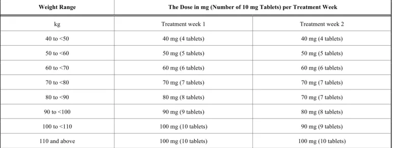
Table 3. The recommended dosing regimen for patients with different weight range receiving cladribine.
Weight Range The Dose in mg (Number of 10 mg Tablets) per Treatment Week
kg Treatment week 1 Treatment week 2
40 to <50 40 mg (4 tablets) 40 mg (4 tablets)
50 to <60 50 mg (5 tablets) 50 mg (5 tablets)
60 to <70 60 mg (6 tablets) 60 mg (6 tablets)
70 to <80 70 mg (7 tablets) 70 mg (7 tablets)
80 to <90 80 mg (8 tablets) 70 mg (7 tablets)
90 to <100 90 mg (9 tablets) 80 mg (8 tablets)
100 to <110 100 mg (10 tablets) 90 mg (9 tablets)
110 and above 100 mg (10 tablets) 100 mg (10 tablets)
as well, it induces a shift in the balance of inflammatory cy- tokines towards the anti-inflammatory side. The pack of leu- kocytes which survived the initial depleting effect of cladribine showed a sustained (for up to 58 days), altered cytokine production imprint, namely had an increased output of anti-inflammatory cytokines, such as IL-4, -5, -10. The production of other cytokines (IL-6, -8, -17A, -23, TNF- alpha and NGF-beta) remained unchanged [67].
2.1.3. Pharmacokinetics
Cladribine given orally has a bioavailability of 37-51%, ingested with meals it is approximately 40.5%, taken be- tween meals 45.6%, ingestion with food causes a net de- crease in absorption as well as a delay in the absorption of cladribine [68]. The clearance of cladribine correlates well with the patient’s creatinine clearance, the half-life of the molecule is between 5.7 and 19.4 hours depending on the renal function of the patient [68]. Mild, moderate and severe renal impairment decreases the clearance of cladribine by 19%, 30%, and 40%, respectively [69]. Protein binding of the molecule is approximately 20%, the volume of distribu- tion varies between 54-367 l/m2. The measured concentra-
tion of cladribine in the CSF is roughly 1/4th of that in the plasma [68].
2.1.4. Dosage
The currently approved and recommended cumulative dose of cladribine is 3.5 mg/kg body weight administered over 2 years, in 2 treatment courses (1 course each year).
Each treatment course includes a dose of 1.75 mg/kg cladribine. Each treatment course consists of 2 treatment weeks, one at the beginning of the first month of treatment and one at the beginning of the second month of the respec- tive treatment year. Each treatment week consists of 4 or 5 days, a patient receives 10 mg or 20 mg (one or two tablets) as a single daily dose, depending on body weight. The tablets should be taken approximately at intervals of 24 hours, if a daily dose consists of 2 tablets, then both should be taken at the same time. Following the completion of the 4 treatment courses, no further cladribine treatment is required in years 3 and 4. Re-initiation of therapy after year 4 has not been ap- proved yet [70].
Contraindications to initiating cladribine therapy are known hypersensitivity to the active substance or to any of
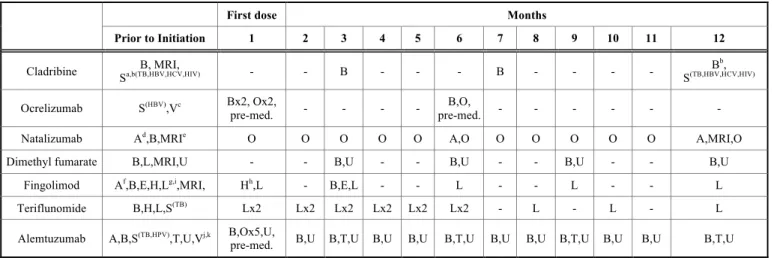
Table 4. The therapeutic burden of different DMDs.
First dose Months
Prior to Initiation 1 2 3 4 5 6 7 8 9 10 11 12
Cladribine B, MRI,
Sa,b(TB,HBV,HCV,HIV) - - B - - - B - - - - Bb,
S(TB,HBV,HCV,HIV)
Ocrelizumab S(HBV),Vc Bx2, Ox2,
pre-med. - - - - B,O,
pre-med. - - - - - -
Natalizumab Ad,B,MRIe O O O O O A,O O O O O O A,MRI,O
Dimethyl fumarate B,L,MRI,U - - B,U - - B,U - - B,U - - B,U
Fingolimod Af,B,E,H,Lg,i,MRI, Hh,L - B,E,L - - L - - L - - L
Teriflunomide B,H,L,S(TB) Lx2 Lx2 Lx2 Lx2 Lx2 Lx2 - L - L - L
Alemtuzumab A,B,S(TB,HPV),T,U,Vj,k B,Ox5,U,
pre-med. B,U B,T,U B,U B,U B,T,U B,U B,U B,T,U B,U B,U B,T,U A – antibody testing, B – blood count, E – ophthalmology consultation, H – cardiovascular monitoring, L – liver function test, MRI – brain MRI scan, O – observation, S – screen- ing, T – thyroid function test, U – urinalysis, V – vaccination. a – Blood lymphocyte counts must be normal before initiating cladribine tablets in Year 1 and at least 800/mm³ before initiating cladribine tablets in Year 2. b – Screening must be performed prior to initiation of therapy in Year 1 and Year 2. Vaccination of antibody-negative patients is recommended prior to initiation of cladribine therapy [72] c – Ocrelizumab is contraindicated in patients with active HBV confirmed by positive results for hepatitis virus B surface antigen [HBsAg]
and anti-HBV tests. For patients who are negative for HBsAg and positive for HB core antibody [HBcAb+] or are carriers of HBV [HBsAg+], consultation with liver disease expert is recommended before starting and in treatment months 2 and 6 after the start of treatment in each treatment year, and as clinically indicated thereafter [73]. d – Test for serum anti-JCV (JC virus) antibodies need to be done prior to treatment and then every 6 months. e – Before initiation of treatment, a recent (usually within 3 months) MRI should be available as a reference, and be repeated at least on a yearly basis. More frequent MRIs (e.g. on a 3–6 monthly basis) using an abbreviated protocol should be considered for patients at higher risk of PML [74]. f – It is recommended that patients without a healthcare professional confirmed the history of chickenpox or documentation of a full course of vaccination with varicella zoster virus (VZV) vaccine undergo antibody testing for VZV before initiating fingolimod therapy. A full course of vaccination for antibody-negative patients with varicella vaccine is recommended prior to commencing treatment. g – Recent (i.e. within last 6 months) transaminase and bilirubin levels should be available before initiation of treatment with fin- golimod. In the absence of clinical symptoms, liver transaminases should be monitored at months 1, 3, 6, 9 and 12 on therapy and periodically thereafter. If liver transaminases rise above 5 times the ULN, more frequent monitoring should be instituted, including serum bilirubin and alkaline phosphatase (ALP) measurement. With repeated confirmation of liver transaminases above 5 times the ULN, treatment with fingolimod should be interrupted and only re-commenced once liver transaminase values have normalized. h – Monitor ECG and BP hourly for 6 hours after the first dose and regularly throughout treatment. ECG should also be taken prior to commencing treatment. I – An ophthalmological evaluation is recommended for at-risk patients prior to initiation and for all patients at 3–4 months after treatment initiation. If patients report visual disturbances at any time while on therapy, evaluation of the fundus, including the macula, should be carried out. Patients with a history of uveitis and patients with diabetes mellitus are at increased risk of macular edema and it is recommended that these patients undergo an evaluation prior to initiating therapy and have follow-up evaluations while receiving therapy [75]. j – Varicella zoster virus vaccination of antibody-negative patients should be considered prior to treatment initiation with alemtuzumab. Patients should have complete local immunization requirements at least 6 weeks prior to treatment with alemtuzumab. Patients without a history of chickenpox or without vaccination against VZV should be tested for antibodies to VZV; k – Human papilloma virus screening should be completed annually for female patients treated with alemtuzumab [76]. Numbers indicate the number of days the monitoring tests occur within the specified month i.e. Ox5 with alemtuzumab means observation is necessary 5 times that month [77, 78].
Table 5. Dosing frequency and burden of different DMDs.
First Dose Months Total
1 2 3 4 5 6 7 8 9 10 11 12 1 Year
Cladribine 4-5 days 4-5 days - - - - - - - - - - Max.
20 doses Ocrelizumab 2 doses +
pre-med. - - - - - 1 dose +
pre-med. - - - - - 3 doses
Natalizumab 1 dose 1 dose 1 dose 1 dose 1 dose 1 dose 1 dose 1 dose 1 dose 1 dose 1 dose 1 dose 12 doses Dimethyl
fumarate 60 doses 60 doses 60 doses 60 doses 60 doses 60 doses 60 doses 60 doses 60 doses 60 doses 60 doses 60 doses 730 doses Fingolimod 30 doses 30 doses 30 doses 30 doses 30 doses 30 doses 30 doses 30 doses 30 doses 30 doses 30 doses 30 doses 365 doses Teriflunomide 30 doses 30 doses 30 doses 30 doses 30 doses 30 doses 30 doses 30 doses 30 doses 30 doses 30 doses 30 doses 365 doses Alemtuzumab 5 doses +
pre-med. - - - - - - - - - - - 5 doses
the excipients, infection with human immunodeficiency vi- rus (HIV), active chronic infection (tuberculosis or hepati- tis), immunocompromised patients of any kind, including
patients currently receiving immunosuppressive or myelo- suppressive therapy, active malignancy, moderate or severe renal impairment (creatinine clearance <60 ml/min), preg-
nancy and breastfeeding. No studies have been conducted in patients with renal or hepatic impairment, no dosage adjust- ment is necessary among patients with mild renal impair- ment (creatinine clearance between 60 and 89 ml/min) [70].
2.1.5. Therapeutic Burden
Choosing the right treatment for a patient is always a challenging task, since not only professional aspects (disease activity, concomitant morbidities, previous treatment failures, etc.) need to be taken into account, but also the patient’s per- sonal needs, the burden a given treatment places on the care- giver, the patient, and the healthcare system needs to be taken into evaluation as well, when deciding on a given DMD.
Beyond therapeutic efficacy and route of administration, there is a significant difference in the monitoring require- ments and time management burden between current thera- pies for MS. In the first four years of therapy cladribine proved to lay the smallest amount of time management bur- den on both patients and caregivers when compared to fin- golimod and alemtuzumab. Including patient workup, the drug delivery, monitoring, follow up visits, adverse event management and travel time treatment with cladribine con- sumed 25 hours, treatment with fingolimod took 103 hours and treatment with alemtuzumab required 193 hours of time in the first 4 years of treatment [71]. Monitoring require- ments differ vastly between DMDs as well. Comparing cladribine to other DMDs (excluding the injectable interfer- ons), cladribine has by far the smallest monitoring- and, ad- ministration burden, and requires the least amount of moni- toring during therapy. Another advantage is that the drug itself needs to be taken in cycles, and only for 8-10 days, therefore the chance of missing a dosing regimen (in con- trast with therapies that have to be taken more than once a day, every day), is minimal.
2.1.6. Clinical Efficacy and Phase III Studies
The CLARITY study was a pivotal, 96 weeks long, phase III, double-blind, multicenter, placebo-controlled trial. It enrolled a total of 1326 patients, they were approximately distributed in a 1:1:1 (433:456:437 patients for the 3.5 mg, 5.25 mg and placebo groups respectively) ratio to receive either placebo or a cumulative dosage of oral cladribine of 3.5 mg/kg or 5.25 mg/kg body weight [38]. The study's pri- mary endpoint was the patients' annualized relapse rate (ARR) at 96 weeks. Secondary clinical endpoints included the proportion of relapse-free patients, the time to acquire a sustained disability, the time to first relapse. Secondary MRI endpoints were the number of Gd-enhancing T1-weighted lesions, active T2-weighted lesions, and combined unique lesions at 96 weeks [38, 79]. Of the 1326 enrolled patients 1184 (89.3%) have completed the whole study (91.9% in the cladribine 3.5 mg group, 89.0% in the cladribine 5.25 mg group and 87.0% in the placebo group) and 1165 (87.9%) patients have completed treatment (91.2% in the cladribine 3.5 mg group, 86.2% in the cladribine 5.25 mg group and 86.3% in the placebo group) [38]. The mean time of partici- pation was 89.4 weeks (91.0, 89.4, 87.8 weeks respectively).
Approximately 2/3rd of patients had not received any dis- ease-modifying drugs beforehand [38].
The most commonly used drugs were intramuscular in- terferon beta-1a (Avonex, 11.2% of patients), subcutaneous interferon beta-1b (Betaferon, 10.6% of patients), subcuta- neous interferon beta-1a (Rebif, 9.4% of patients), and sub- cutaneous glatiramer acetate (Copaxone, 6.5% of patients), data are shown as mean (standard deviation (SD)), unless stated otherwise.
The primary endpoint was met in both groups receiving treatment, the ARR was found to be 0.14 in the cladribine 3.25 mg group and 0.15 in the 5.25 mg group compared to 0.33 in the placebo group meaning a relative reduction of 57.6% and 54.5% respectively (p<0.001). The cumulative number of relapses at 96 weeks was significantly lower in both cladribine groups versus placebo (109, 114 and 252 relapses respectively). The number of relapse-free patients was 345 (79.7%), 360 (78.9%) for the 3.5 and 5.25 mg groups compared to 266 (60.9%) patients in the placebo group (p<0.001). The time to first relapse was significantly longer in both groups receiving treatment (hazard ratio [HR]
in the 3.5 mg group was 0.44 with a 95% CI of 0.34 to 0.58, with the 15th percentile of time to the first relapse being 13.4 months, while the HR in the cladribine 5.25 mg group was 0.45 with a 95% CI of 0.36 to 0.60, 15th percentile to event being 13.3 months, p<0.001 for both groups) versus placebo (15th percentile being 4.6 months). [38]
Treatment with cladribine demonstrated a significant relative risk reduction in the accumulation of 3-month sus- tained progression of disability as well as compared with placebo. A reduction of 33% was seen in the 3.5 mg group (HR 0.67, 95% CI 0.48 to 0.93, p=0.02), as well as a 31%
reduction in the 5.25 mg group (HR 0.69, 95% CI 0.49 to 0.96, p=0.03). The time to 3-month sustained change in the EDSS score was significantly longer in both arms receiving treatment (13.6 months in both groups) versus 10.8 months with placebo [38, 80].
As for MRI outcomes, when assessing the whole cohort compared with placebo, cladribine treatment proved to alle- viate lesion burden in both groups. Patients in the treated groups had fewer Gd-enhancing T1 lesions (mean number 0.12 (85.7% reduction) and 0.11 (87.9% reduction) for the 3.5 mg and 5.25 mg cohorts, respectively vs. 0.91 for place- bo), active T2 lesions (0.38 [73.4% reduction] and 0.33 [76.9% reduction] vs. 1.48 respectively) as well as combined unique lesions (0.43 [74.4% reduction], 0.38 [77.9% reduc- tion] vs. 1.72 respectively), p<0.001 for all comparisons ver- sus placebo. When the patients were stratified by baseline relapse rate the reductions in combined unique lesions in the 3.5 and 5.25 mg cladribine groups were 74.8% and 75.5% (≤
1 relapse), 83.1% and 86.7% (2 relapses), and 76.7% and 90.0% (≥ 3 relapses), respectively (p < 0.001 for all compar- isons). When divided by the presence of T1 Gd-enhancing lesions at baseline a reduction of 73.5% and 75.2% was seen for patients without Gd-enhancing lesions and 80.8% and 84.9% was seen for patients with at least 1 Gd-enhancing lesion at baseline, respectively (p<0.001 for all comparisons.
The improvements in MRI outcomes seen were also con- firmed in all clinically relevant patient subgroups stratified by disease severity at baseline [38].
Table 6. Baseline demographics and disease characteristics of treatment groups in the CLARITY study.
Clarity
- - Placebo Cladribine
- - (n=437) 3.5 mg/kg (n=433) 5.25 mg/kg (n=456)
Age mean 38.7 ± 9.9 37.9±10.3 39.1±9.9
range 18-64 18-65 18-65
Females - no. (%) 288 (65.9%) 298 (68.8%) 312 (68.4%)
Mean weight - kg 70.3±15.4 68.1±14.6 69.3±14.8
Race - no. (%) White 429 (98.2%) 425 (98.2%) 446 (97.8%)
Black 1 (0.2%) 2 (0.5%) 4 (0.9%)
Other 7 (1.6%) 6 (1.4%) 6 (1.3%)
Previous DMDs - no. (%) 142 (32.5%) 113 (26.1%) 147 (32.2%)
Disease duration (years) mean 8.9±7.4 7.9±7.2 9.3±7.6
range 0.4-39.5 0.3-42.3 0.4-35.2
EDSS Score
0 - no. (%) 13 (3.0%) 12 (2.8%) 11 (2.4%)
1 - no. (%) 70 (16.0%) 75 (17.3%) 80 (17.5%)
2 - no. (%) 127 (29.1%) 133 (30.7%) 119 (26.1%)
3 - no. (%) 96 (22.0%) 108 (24.9%) 108 (23.7%)
4 - no. (%) 83 (19.0%) 71 (16.4%) 84 (18.4%)
≥5 - no. (%) 48 (11.0%) 34 (7.9%) 54 (11.8%)
Mean score 2.9 (1.3) 2.8 (1.2) 3.0 (1.4)
Gd enhancing T1-weighted lesions
Patients with lesions - no.
(%) 123 (29.3%) 138 (31.9%) 147 (32.2%)
Mean no. Of lesions 0.8 (2.1) 1.0 (2.7) 1.0 (2.3)
Mean vol. Of T2-weighted lesions - mm3 14,287.6 (13,104.8) 14,828.0 (16,266.8) 17,202.1 (17.467.7)
The annualized percentage brain volume change (PBVC/y) was evaluated [81, 82]. Treatment with cladribine had a significant reduction on the PBVC/y for both arms ([–0.56% ± 0.68; p = 0.010] for the 3.5 mg/kg group and [–0.57% ± 0.72; p = 0.019] for the 5.25mg/kg group) com- pared with placebo (–0.70% ± 0.79). When the patients were stratified by the presence of Gd enhancing lesions at baseline no significant difference could be demonstrated in the PBVC/y between the three arms (−0.92% ± 1.02%, n = 110;
−1.00% ± 1.08%, n = 115; −0.97% ± 0.97%, n = 106< 3.5 mg/kg, 5.25 mg/kg cladribine and placebo, respectively) [82]. Subdividing the patients by PBVC/y the patients with the lowest brain volume loss (BVL) (PBVC/y>-0.4%) showed the highest chance of being free from disability pro- gression, meanwhile patients with the highest BVL (PBVC/y<-1.08%) had the lowest probability of being disa- bility free at the end of the second year (HR 0.67, 95% CI 0.571 to 0.787; p<0.001) [82].
Various posthoc analyses were conducted after the com- pletion of the CLARITY study. One assessed the NEDA (no evidence of disease activity) status of the participants [83].
Over 24, 48 and 96 weeks 67.3%, 69.7% versus 38.9%;
54.2% and 56.1% versus 23.9% and 44.2% and 46.0% ver- sus 15.8% of the patients were found to be free of disease activity (3.5 mg/kg, 5.25 mg/kg and placebo, respectively, p<0.001 for all comparisons). In regard to previous treatment status (already treated with other DMDs vs. treatment naïve patients), disease duration significant difference was seen between the proportion of patients fulfilling the NEDA crite- ria. 37.2% and 42.9% vs. 16.2% of patients who already have received another DMD(s) were confirmed to have no evidence of activity (3.5 mg/kg, 5.25 mg/kg, placebo, re- spectively), this ratio in the case of treatment naïve patient was to be found 45.0%, 45.0% vs. 15.9%). When stratifying base on baseline disease duration 38.1%, 46.5% vs. 8.2% of patients fulfilled the NEDA criteria with less than 3 years of disease history, with 3 to 10 years the ratio was 47.0%, 40.3% vs. 15.5%, meanwhile with a longer history than 10 years it was 41.8%, 47.8% vs. 22.1% (3.5 mg/kg, 5.25 mg/kg, placebo, respectively, p<0.001 for all comparisons).
The ORs for remaining disease activity free while on treat- ment versus no treatment 3.99 [95% CI 2.89 to 5.49] and 4.24 (95% CI 3.09 to 5.82) for 3.5 mg/kg and 5.25 mg/kg doses respectively, p < 0.001 for each comparison [84].
Another posthoc analysis of the 3.5 mg/kg treatment arm sought to assess the treatment effect of the approved dosage of cladribine based on different baseline patient characteris- tics compared with the overall study population [85]. In this posthoc analysis patients with high disease activity (HDA), (the European Medicines Agency [EMA] has previously defined patients with HDA as those with ‘rapidly evolving severe relapsing-remitting multiple sclerosis defined by 2 or more disabling relapses in one year, and with 1 or more Gadolinium-enhancing (Gd+) lesions on brain MRI or a sig- nificant increase in T2 lesion load as compared to a previous recent MRI) were further subgrouped [85-91]. New defini- tions were introduced, one being “high relapse activity”
(HRA - patients with ⩾2 relapses during the year prior to study entry, whether on DMD treatment or not), the other being HRA+“disease activity on treatment” (HRA+ DAT - patients with ⩾2 relapses during the year prior to study en- try, whether on DMD treatment or not, PLUS patients with
⩾1 relapse during the year prior to study entry while on ther- apy with other DMDs and ⩾1 T1 Gd+ or ⩾9 T2 lesions).
The overall 3.5 mg/kg base population consisted of 437 par- ticipants, the HRA group had 131 patients, the non-HRA consisted of 306 patients, the HRA+DAT subgroup involved 149 patients, while the non-HRA+DAT subgroup comprised of 288 subjects. The ARR in the overall (treated with 3.5 mg/kg cladribine, HRA+non-HRA) population was 0.35 (95% CI 0.31 to 0.39), in the HRA group it was 0.50 (95%
CI 0.41 to 0.60), in the non-HRA group 0.29 (95% CI 0.24 to 0.34), in the HRA+DAT group 0.47 (95% CI 0.40 to 0.57) in the non-HRA+DAT group 0.29 (95% CI 0.24 to 0.34). In the original study treatment with 3.5 mg/kg cladribine in comparison with placebo reduced the risk of 6-month- confirmed EDSS worsening by 47% (HR = 0.53, 95% CI = 0.36 to 0.79, p = 0.0016). In the posthoc analysis then HRA and HRA+DAT patients were compared to non-HRA pa- tients the risk reduction was even greater, 82% (interaction p-value 0.0036 for HRA vs. non-HRA, and p = 0.0037 for HRA + DAT vs. non-HRA + DAT). The time for first quali- fying relapse was also shorter in HRA and HRA+DAT pa- tients than in non-HRA and non-HRA+DAT patients. Odds ratio assessment was also conducted to evaluate the probabil- ity of achieving NEDA state of the patients in the HRA and HRA+DAT groups. Odds ratios were 8.02, (95% CI = 3.93 to 16.35; p<0.0001) and 7.82 (95% CI = 4.03 to 15.19;
p<0.0001) respectively. MRI findings indicated that the rela- tive risk for T2 active lesions, Gd-enhancing lesions, and combined unique lesions were low in all assessed subgroups.
Overall safety data for the HRA and HRA+DAT subgroups did not differ from the data obtained from the overall study population. The analysis concluded that subjects with high disease activity responded better to treatment both clinically and radiologically than the overall study population [85].
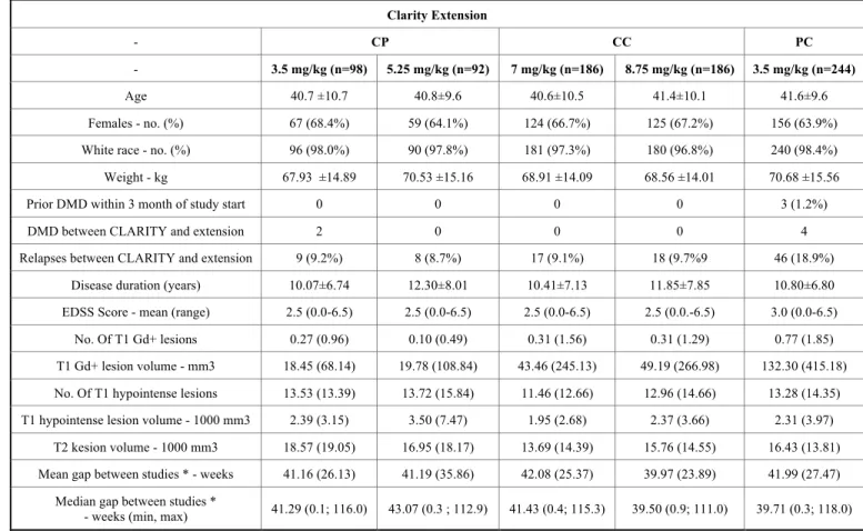
Of the 1184 patients that have completed the CLARITY trial, 867 were enrolled in the CLARITY Extension study [92]. 61 patients who were not eligible were solely followed, the rest 806 patients were re-randomized or assigned to treatment if previously received placebo. Of the 806 treated patients, 738 (91.6%) completed the 2-year trial. The socio- demographic and disease characteristics of patients enrolled were similar across all arms to the base trial's, with the ex- ception that patients who had received placebo in the CLAR-
ITY study had higher disease activity than the rest of the population. The patients participating in the study were re- randomized. All patients taking placebo in CLARITY re- ceived a 3.5 mg/kg cumulative dosage of cladribine (PC 3.5 mg/kg, n=244), patients taking 3.5 mg/kg in the original study either received placebo (CP 3.5 mg/kg, n=98) or treatment (CC, cumulative dosage 7.0 mg/kg over the 2+2 years, n=186), patients treated with 5.25 mg/kg cladribine were either given placebo (CP 5.25 mg/kg, n=92) or cladribine in 3.5 mg/kg dosage (CC, 8.75 mg/kg cumulative dosage, n=186). To assess the potential effect of temporal delay and incremental effect of treatment with cladribine for an extended duration and larger doses additional compari- sons were conducted than in the original trial. The assessed and compared groups in the extensions were CP 3.5 mg/kg vs. PC 3.5 mg/kg (to investigate the effect of early versus late treatment with the same dosage), CC 7 mg/kg vs. CP 3.5 mg/kg, CC 8.75 mg/kg vs. CP 5.25 mg/kg (192 weeks of treatment vs. 96 weeks of treatment), and CC 7 mg/kg + CC 8.75 mg/kg (combined data for patients who received treat- ment with cladribine tablets in CLARITY and CLARITY Extension) vs. CP 3.5 mg/ kg + CP 5.25 mg/kg (combined data for placebo in Years 3 and 4) [92].
The primary and secondary clinical endpoints in the ex- tension study were the same as in the pivotal trial. No MRI endpoints were specified in the extension study. Between- group analysis of the clinical efficacy endpoints (ARR, time to 3 months confirmed EDSS progression, the proportion of relapse-free patients) showed no significant differences be- tween the evaluated treatment groups (p>0.05 for all com- parisons) [92].
The ARR reduction seen in the extension study was simi- lar in all groups to that seen in the pivotal study, between- group comparison has proven no significant differences be- tween treatment arms. Furthermore, there was no significant difference between treatment groups in time to first relapse [92]. The proportion of relapse-free patients approximately matched the magnitude seen in the treated arms of the CLARITY study and was roughly equivalent in all treated groups in the extension study. The only significant differ- ence between the study groups was seen in the PC 3.5 mg/kg group (n=244), in which the proportion of relapse-free pa- tients was 58.0% while on placebo, meanwhile it was 79.6%
at the end of the extension study (p<0.0001), which was ap- proximately equivalent to the values seen in other treatment arms [92]. Also, the proportion of patients who remained free of 3-month confirmed EDSS progression was similar across all treatment arms (~75%), however, it was slightly lower (6.3%-13.3%) than the results seen in the original study (~85%). Roughly 4/5th of patients were free of relapses in the pivotal trial, who received 3.5 mg/kg cladribine, 75%
of the patients were still free of relapses at the end of week 120 who were afterward randomized to placebo in the exten- sion study (i.e. the CP 3.5 mg/kg treatment group) [92]. This indicates that treatment produced a clinical response, that was maintained for at least 2 years after treatment discontin- uation [92]. At the end of the extension study, no evidence of disease rebound effect was seen in the groups which were re- randomized to placebo (from either treatment groups), after treatment with cladribine in the original study, a side effect
Table 7. Baseline demographics and disease characteristics of treatment groups in the CLARITY Extension study. Data are shown as mean (standard deviation (SD)) unless stated otherwise.
Clarity Extension
- CP CC PC
- 3.5 mg/kg (n=98) 5.25 mg/kg (n=92) 7 mg/kg (n=186) 8.75 mg/kg (n=186) 3.5 mg/kg (n=244)
Age 40.7 ±10.7 40.8±9.6 40.6±10.5 41.4±10.1 41.6±9.6
Females - no. (%) 67 (68.4%) 59 (64.1%) 124 (66.7%) 125 (67.2%) 156 (63.9%)
White race - no. (%) 96 (98.0%) 90 (97.8%) 181 (97.3%) 180 (96.8%) 240 (98.4%)
Weight - kg 67.93 ±14.89 70.53 ±15.16 68.91 ±14.09 68.56 ±14.01 70.68 ±15.56
Prior DMD within 3 month of study start 0 0 0 0 3 (1.2%)
DMD between CLARITY and extension 2 0 0 0 4
Relapses between CLARITY and extension 9 (9.2%) 8 (8.7%) 17 (9.1%) 18 (9.7%9 46 (18.9%)
Disease duration (years) 10.07±6.74 12.30±8.01 10.41±7.13 11.85±7.85 10.80±6.80
EDSS Score - mean (range) 2.5 (0.0-6.5) 2.5 (0.0-6.5) 2.5 (0.0-6.5) 2.5 (0.0.-6.5) 3.0 (0.0-6.5)
No. Of T1 Gd+ lesions 0.27 (0.96) 0.10 (0.49) 0.31 (1.56) 0.31 (1.29) 0.77 (1.85)
T1 Gd+ lesion volume - mm3 18.45 (68.14) 19.78 (108.84) 43.46 (245.13) 49.19 (266.98) 132.30 (415.18) No. Of T1 hypointense lesions 13.53 (13.39) 13.72 (15.84) 11.46 (12.66) 12.96 (14.66) 13.28 (14.35) T1 hypointense lesion volume - 1000 mm3 2.39 (3.15) 3.50 (7.47) 1.95 (2.68) 2.37 (3.66) 2.31 (3.97)
T2 kesion volume - 1000 mm3 18.57 (19.05) 16.95 (18.17) 13.69 (14.39) 15.76 (14.55) 16.43 (13.81) Mean gap between studies * - weeks 41.16 (26.13) 41.19 (35.86) 42.08 (25.37) 39.97 (23.89) 41.99 (27.47)
Median gap between studies *
- weeks (min, max) 41.29 (0.1; 116.0) 43.07 (0.3 ; 112.9) 41.43 (0.4; 115.3) 39.50 (0.9; 111.0) 39.71 (0.3; 118.0)
*The duration of time in weeks between the last visit date in the CLARITY study treatment period and the randomization date in the CLARITY Extension trial.
seen in other therapies with a direct effect on lymphocytes.
The length of the treatment gap between the original and extension study (which varied between patients, but was roughly equivalent [median of 40.3 weeks] across all treat- ment groups) had no effect on the ARR, the proportion of relapse-free patients, time to first qualifying relapse, nor the time to 3-month confirmed EDSS progression. Nonetheless, when both studies were considered together treatment delay had a negative effect on the time to first qualifying relapse;
the shortest time was seen in the PC 3.5 mg/kg treatment group. Similarly, when the same patient groups were re- evaluated at the end of the extension study from the original trial there was no difference between the groups in the matter of ARR, except in the PC 3.5 mg/kg group. In this group, a significant reduction was seen in the ARR after treatment (0.26 in the original trial, receiving placebo, 0.10 in the ex- tension study, 60.7% relative reduction, p < 0.0001). This difference in ARR reduction is on par with the reduction seen in the original study between the placebo and 3.5 mg/kg treatment groups (57.6% relative reduction, p<0.001) [92].
2.1.7. Safety and Safety Management
The safety and tolerability profile of cladribine has been established over several well-constructed studies and was shown to be consistent throughout these evaluations [93].
Through the CLARITY and CLARITY EXTENSION stud-
ies, there was a relatively low proportion of discontinuation due to adverse or serious adverse events (AE, SAE) [38, 92].
Most of the AEs were categorized as mild or moderate and were distributed relatively evenly through the treatment groups [92, 94]. The exception from this rule was the ap- pearance of lymphopenia, however, it was expected consid- ering cladribine’s mechanism of action [92, 94]. The most rapid and underlined reduction was observed in the CD19+
B-cell subtypes [95, 96]. Also, it was shown, that the reduc- tion of CD8+ T-cells was less severe and had a faster rate of recovery than CD4+ T-cells [95, 96].
The rate of infections was not higher in the cladribine treatment groups as compared to placebo in the CLARITY and CLARITY EXTENSION studies, with the exception of herpes zoster infections [38, 92-94].
The rate of malignancies was slightly higher in the treat- ment groups than in the placebo arm in the CLARITY study;
throughout CLARITY EXTENSION there was no difference between the groups [38, 92, 93]. This observed increase in the rate of malignancy in the CLARITY study and the fact that only one pivotal phase III study was initially done were the primary reason the EMA refused to approve oral cladribine in 2011 [93]. However, since then data from the second phase III study (ORACLE) - a continuous data stream from the safety register – and the change in the ad- ministration schedule of cladribine when encountering seri-
ous lymphopenia and also solid epidemiological data indicat- ing that there is no increased risk for secondary malignancies has led to the approval of cladribine for RRMS on second resubmission [93].
There were 4 fatal cases during the study and 2 cases after withdrawal [93, 94]. The 4 fatal cases during the study had no connection whatsoever to the treatment, just like 1 case of metastatic pancreatic carcinoma after withdrawal [93, 94]. Yet, 1 case, an acute cardiopulmonary arrest was linked to cladribine. A 21-years old woman experienced pancyto- penia one week after a single course of cladribine 5.25 mg/kg. The patient was immediately withdrawn from the study. The second phase of pancytopenia was observed ap- proximately 2 months after stopping the treatment; an X-ray performed that time, revealed bilateral alveolar-interstitial infiltrates. Bone marrow biopsy was performed, which con- firmed "slight myelodysplasia". Five months after treatment the patient went through another period of pancytopenia, the chest X-ray performed at that time revealed the recurrence of the bilateral infiltration and bone-marrow biopsy confirmed myelodysplastic syndrome. The patient died 6 months after receiving one single course of cladribine 5.25 mg/kg. The post-mortem diagnosis of tuberculosis was made, and it was proved that the disease was long-standing and has been al- ready present at the time of the initiation of cladribine treat- ment [93, 94]. It was concluded that treatment with cladribine sped up the progression of an already present in- fection [93, 94].
Hence, the two most pivotal safety management issues are the constant monitoring of the patients' lymphocyte counts and screening for VZV infections and tuberculosis.
Varicella negative patients must be inoculated prior to the initiation of therapy with cladribine, which must be post- poned 4-6 weeks to allow the vaccination to take full effect [70]. If a patient’s lymphocyte count drops below 500 cells/mm3 active monitoring for infections, particularly for herpes zoster should begin [70]. If a patient develops grade IV lymphopenia herpes prophylaxis according to local prac- tice should be considered during the time of lymphopenia [70]. Every patient must be screened for latent tuberculosis before initiation of treatment (both in the first and second year) [70]. The commencement of treatment with cladribine should be delayed until the infection has been adequately treated [70].
Also, in light of cladribine’s mechanism of action and its potential for aggravating infections, attenuated live vaccines are contraindicated during treatment and after until the pa- tients' white blood cell counts reach normal levels [93].
For further, detailed information of the adverse and seri- ous adverse events and side effects that have occurred during the pivotal and the extension study - which is beyond the scope of this review - can be found in separate articles de- voted to the matter [38, 92, 94].
CONCLUSION
As a conclusion, we can state that cladribine proved to be a highly effective treatment choice for RRMS. Furthermore, despite the pivotal study being terminated early, 3.5 mg/kg
cladribine tablets proved to be effective in halting the con- version of CIS patients into definite MS and in the future cladribine could become a potent choice for first-line thera- py. It is the first DMD for MS for which the mechanism of action targets both the T- and B-cells, thus taking into ac- count the complex pathomechanism of the disease. In spite of this dual action, the safety profile of the drug is clearly good; with some easily managed precautions, serious ad- verse events can be avoided. It has some other clear ad- vantages as well. It is an orally administered DMD, which can enhance the adherence of the patients. Also, it is admin- istered through 2x2 short courses which can be an attractive choice over constant drug-taking or infusions; and patients do not need to be admitted to the hospital for the administra- tion and requires no long observation periods in contrast to other therapies.
Yet there are two important limitations right now, which need to be taken seriously. Firstly, we do not have any data yet on the long-term effectiveness of cladribine, as these data are yet to be revealed. Secondly, a limitation stemming from the mentioned lack of data above, we do not have any clear information on how to proceed, if the 2x2 courses of the drug fail to be effective enough. More data is needed for the satisfactory answer on how and when to administer another course of cladribine in such an event, or how and when to potentially change to another DMD (and which) if cladribine fails to halt the progression of the disease.
LIST OF ABBREVIATIONS
ADA = Adenosine Deaminase
AE = Adverse Event
AIF = Apoptosis-Inducing Factor ARR = Annualised Relapse Rate BBB = Blood-Brain Barrier BCR = B-Cell Receptor BVL = Brain Volume Loss
CC = Cladribine Tablets in Both Studies CDK-2 = Cyclin-Dependent Kinase 2 CI = Confidence Interval
CIS = Clinically Isolated Syndrome CNS = Central Nervous System CP = Cladribine then Placebo CSF = Cerebrospinal Fluid
2-CdADP = 2-Chloro-2’-deoxy-ß-D-adenosine diphos- phate
2-CdAMP = 2-Chloro-2’-deoxy-ß-D-adenosine mono- phosphate
2-CdATP = 2-Chloro-2’-deoxy-ß-D-adenosine tri- hosphate
DA = Deoxyadenosine