Visceral Adiposity Elevates the Risk of Critical Condition in COVID-19: A Systematic Review and Meta-Analysis
Mária Földi 1,2,3, Nelli Farkas1,4, Szabolcs Kiss 1,2,3, Fanni Dembrovszky 1,2, Zsolt Szakács1,2, Márta Balaskó1,2, Bálint Erőss1,2, Péter Hegyi1,2,3, and Andrea Szentesi 1,2,3
Objective: A higher BMI has become acknowledged as one of the impor-
tant risk factors for developing critical condition in coronavirus disease 2019 (COVID-19). In addition to BMI, body composition, and particularly visceral adiposity, might be an even more accurate measure to stratify patients. Therefore, the aim of this study was to evaluate the association between the distributions of computed-tomography-quantified fat mass and critical condition of patients with COVID-19.
Methods:
A systematic search was conducted in five databases for studies published until November 17, 2020. In the meta-analysis, pooled mean difference (standardized mean difference [SMD]) of visceral fat area (VFA; in square centimeters) was calculated between patients in the in- tensive care unit and those in general ward and between patients with the requirement for invasive mechanical ventilation (IMV) and those without the IMV requirement.
Results:
The quantitative synthesis revealed that patients requiring in- tensive care had higher VFA values (SMD = 0.46, 95% CI: 0.20-0.71,
P < 0.001) compared with patients on the general ward. Similarly, patientsrequiring IMV had higher VFA values (SMD = 0.38, 95% CI: 0.05-0.71,
P = 0.026) compared with patients without the IMV requirement.Conclusions: VFA values were found to be significantly higher in patients
with critical condition. Therefore, abdominal adiposity seems to be a risk factor in COVID-19, and patients with central obesity might need special attention.
Obesity (2021) 29, 521-528.
Introduction
With the escalation of the pandemic caused by severe acute respiratory syndrome coro- navirus 2 (SARS-CoV-2), recognizing risk factors is of utmost importance. Among other risk factors such as age and comorbidities (1,2), a higher BMI has been acknowledged as a risk factor for developing critical condition in coronavirus disease 2019 (COVID-19) in our former analysis and in other articles since then (3-6). Because the obesity epidemic is rapidly spreading worldwide, it is vital to accurately identify patients with a higher risk for developing critical condition in COVID-19.
© 2021 The Authors. Obesity published by Wiley Periodicals LLC on behalf of The Obesity Society.
This is an open access article under the terms of the Creative Commons Attribution-NonCommercial-NoDerivs License, which permits use and distribution in any medium, provided the original work is properly cited, the use is non-commercial and no modifications or adaptations are made.
Received: 16 October 2020; Accepted: 24 November 2020; Published online 2 February 2021. doi:10.1002/oby.23096
1 Institute for Translational Medicine, Medical School, University of Pécs, Pécs, Hungary. Correspondence: Andrea Szentesi (szentesiai@gmail.com) 2 Szentágothai Research Centre, University of Pécs, Pécs, Hungary 3 Centre for Translational Medicine, Department of Medicine, University of Szeged, Szeged, Hungary
4 Institute of Bioanalysis, Medical School, University of Pécs, Pécs, Hungary.
Study Importance What is already known?
► A higher BMI was found to be a risk fac- tor for developing critical condition in COVID-19.
► Because the prevalence of obesity is in- creasing worldwide, it is vital to identify patients at a higher risk.
► Several studies have proposed that vis- ceral adiposity might be a risk factor.
What does this study add?
► We performed a comprehensive search, selection, and quantitative and qualita- tive analysis concerning the association between computed-tomography-quan- tified fat mass distribution and critical condition among COVID-19 patients.
► Pooled analysis of three studies revealed that patients requiring intensive care or invasive mechanical ventilation had higher visceral fat area values compared with patients without the need for them.
How might these results change the direction of research or the focus of clinical practice?
► Although BMI is widely used to define obesity, further phenotyping of patients, for example by assessing body com- position and central obesity, might be recommended.
► Considering the potential role of visceral adipose tissue, it might also be worth studying adipose-tissue-related sub- stances as potential pharmacological targets.
Although BMI is widely used to diagnose obesity (7), body com- position, and especially visceral adiposity, might be an even more accurate measure to stratify patients (8-12). Therefore, we aimed to evaluate the association between the distributions of computed tomography (CT)-quantified fat mass and critical condition of patients with COVID-19.
Methods
We report this meta-analysis and systematic review according to the Preferred Reporting Items for Systematic Reviews and Meta-Analyses Statement (13).
Search strategy
A systematic search was conducted in MEDLINE (via PubMed), Embase, Cochrane Library (CENTRAL), Scopus, and Web of Science for studies published until November 17, 2020. The following search terms were used in all databases: ((“covid 19”) OR (“Wuhan virus”) OR (“coronavirus”) OR (“2019 nCoV”) OR (“SARS-cov-2”)) AND ((fat OR obes* OR adipos*) AND (visceral OR intraabdominal OR abdominal OR central)). There was no restriction applied to the search.
Selection and eligibility criteria
We selected clinical studies reporting on patients hospitalized with confirmed SARS-CoV-2 infection (based on the World Health Organization case definition) and on the distribution of body fat mass assessed by CT. Studies were included in the meta-analysis if data on the following variables were reported: the number of patients with and without critical condition (defined as the need for invasive me- chanical ventilation [IMV] or admission to intensive care unit [ICU]) and distribution of body fat mass (total adipose tissue [TAT], vis- ceral adipose tissue [VAT], subcutaneous adipose tissue [SAT]). The latter parameters could be reported as thickness (millimeters), area (square centimeters), or volume (cubic centimeters). Abstracts and grey literature (preprints and other non-peer-reviewed material) were excluded from the analysis.
The yield of the search was combined in a reference manager software (EndNote X9; Clarivate Analytics, Philadelphia, Pennsylvania). After automatic and manual removal of duplicate records, full texts of all studies were evaluated by two independent review authors. Two review authors decided to include a study in the meta-analysis if they agreed. A third author resolved the disagreements. Reference lists of the included studies were screened for additional eligible articles.
Data extraction
Two independent review authors extracted data into a standardized data collection form (Microsoft Excel 365, Microsoft Corporation, Redmond, Washington). The following data were extracted from each eligible article: first and second authors; publication year; study site;
sex; age; the number of patients with and without critical condition;
and the means, standard deviations, medians, ranges, and interquartile ranges related to body fat mass (the article provided thickness in mil- limeters, area in square centimeters, and volume in cubic centimeters of TAT, VAT, and SAT). Odds ratios (ORs) and risk ratios (with the corresponding confidence intervals) relating to the association between
body fat mass and critical condition were also extracted. A third party resolved discrepancies. The authors of the eligible articles were not contacted for further information.
Quality assessment
Quality of the eligible studies was evaluated by using the Quality in Prognosis Studies (QUIPS) tool by two independent review authors (14). Any disagreement was resolved by third-party arbitration.
Statistical analysis
Cohen’s kappa coefficient (κ) was calculated to measure interrater reli- ability during the selection process. κ ≤ 0 is interpreted as no agreement, 0.01-0.20 as none to slight agreement, 0.21-0.40 as fair agreement, 0.41- 0.60 as moderate agreement, 0.61-0.80 as substantial agreement, 0.81- 1.00 as almost perfect agreement, and 1.00 as perfect agreement (15).
We calculated pooled mean difference (standardized mean difference [SMD]) for continuous variables because the CT examinations were performed at different vertebral levels among studies. We used random effect model with the DerSimonian-Laird estimation (16). Statistical heterogeneity was calculated performing the I2 test, and we also carried out χ2 tests to acquire probability values: P < 0.1 indicated significant heterogeneity. The interpretation of I2 was as follows: 0% to 40%: not important; 30% to 60%: moderate heterogeneity; 50% to 90%: substan- tial heterogeneity; and 75% to 100%: considerable heterogeneity (17).
If the mean with standard deviation could not be extracted, we esti- mated them from median, interquartiles, and range using the method by Wan et al. (18).
Results
Systematic search and selection
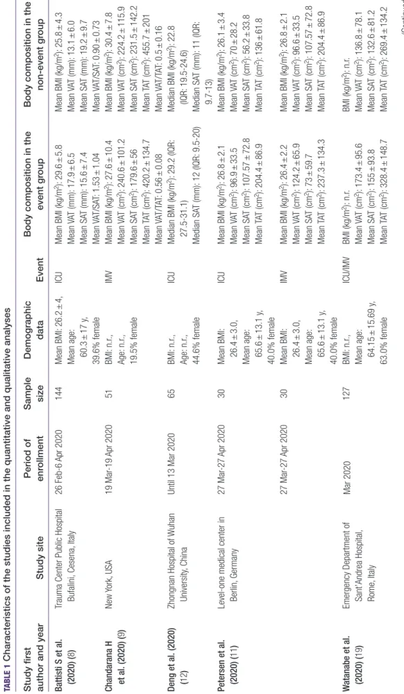
The results of the systematic search and selection are shown in the Preferred Reporting Items for Systematic Reviews and Meta-Analyses Flowchart (Figure 1). The calculated κ value based on the full-text se- lection was 1.00, which was interpreted as perfect agreement. On com- pletion of the selection, six studies, including data from 560 patients, were eligible. The characteristics of the studies included are summa- rized in Table 1. Five out of six studies, including 509 patients, reported on ICU admission rate, which ranged between 18.5% and 43.3%. Three studies with 208 patients reported on IMV requirement in the associ- ation with body composition metrics. The visceral fat area (VFA; in cubic centimeres) values ranged between 70.9 cm2 and 240 cm2. The results of the quantitative and qualitative synthesis are shown in Table 1, Table 2, and Figure 2.
VAT mass and COVID-19
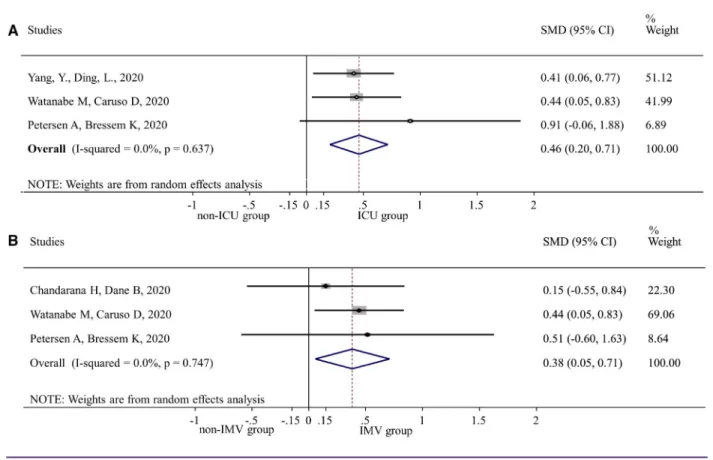
Our quantitative synthesis revealed that patients requiring inten- sive care had higher VFA values (SMD = 0.46, 95% CI: 0.20-0.71, P < 0.001) compared with patients on the general ward. Similarly, pa- tients requiring IMV had higher VFA values (SMD = 0.38, 95% CI:
0.05-0.71, P = 0.022) compared with patients without IMV require- ment. Statistical heterogeneity might not be important in any analysis (I2 = 0.0%, P = 0.637 and I2 = 0.0%, P = 0.747 for ICU admission and IMV, respectively). These results are depicted in Figure 2. Two stud- ies found, in age- and gender-adjusted analyses, that an increased VFA carries a higher risk for ICU admission (11,19). Yang et al. did
not identify VFA > 100 mm2 as a significant risk factor for ICU admis- sion (OR = 1.94, 95% CI: 0.95-4.05). (32). Battisti et al. also found a higher visceral fat thickness (in millimeters) among patients admitted to the ICU compared with patients on the general ward (13.1 ± 6 mm vs.
17.9 ± 6.5 mm, P < 0.001) (8).
SAT mass and COVID-19
Watanabe et al. found that an increased subcutaneous fat area (SFA) is not associated with a higher risk for ICU admission (19). Yang et al. did not identify SFA > 100 mm2 as a risk factor for ICU admission
(OR = 1.06, 95% CI: 0.52-2.17) (32). However, a high VFA/SFA ratio was found to be associated with an increased risk for ICU admission (OR = 2.47, 95% CI = 1.05-5.98) (9).
TAT mass and COVID-19
Two studies evaluated total fat area (TFA) in COVID-19 patients.
Petersen et al. revealed that every additional 10 cm2 of TFA carries 1.13 times (95% CI: 1.03-1.29) and 1.28 times (95% CI: 1.06-1.80) additional risk for ICU admission and IMV requirement, respectively (11). Another study identified an increased TFA as a risk factor for ICU
Figure 1 PRISMA flowchart for the study selection process. PRISMA, Preferred Reporting Items for Systematic Reviews and Meta- Analyses. [Color figure can be viewed at wileyonlinelibrary.com]
TABLE 1 Characteristics of the studies included in the quantitative and qualitative analyses Study first author and yearStudy sitePeriod of enrollmentSample sizeDemographic dataEventBody composition in the event groupBody composition in the non-event group Battisti S et al. (2020) (8)Trauma Center Public Hospital Bufalini, Cesena, Italy26 Feb-6 Apr 2020144Mean BMI: 26.2 ± 4, Mean age: 60.3 ± 17 y, 39.6% female ICUMean BMI (kg/m2): 29.6 ± 5.8 Mean VAT (mm): 17.9 ± 6.5 Mean SAT (mm): 15.6 ± 7.4 Mean VAT/SAT: 1.53 ± 1.04
Mean BMI (kg/m2): 25.8 ± 4.3 Mean VAT (mm): 13.1 ± 6.0 Mean SAT (mm): 19.2 ± 9.7 Mean VAT/SAT: 0.90 ± 0.73 Chandarana H et al. (2020) (9)New York, USA19 Mar-19 Apr 202051BMI: n.r., Age: n.r., 19.5% female
IMVMean BMI (kg/m2): 27.6 ± 10.4 Mean VAT (cm2): 240.6 ± 101.2 Mean SAT (cm2): 179.6 ± 56 Mean TAT (cm2): 420.2 ± 134.7 Mean VAT/TAT: 0.56 ± 0.08
Mean BMI (kg/m2): 30.4 ± 7.8 Mean VAT (cm2): 224.2 ± 115.9 Mean SAT (cm2): 231.5 ± 142.2 Mean TAT (cm2): 455.7 ± 201 Mean VAT/TAT: 0.5 ± 0.16 Deng et al. (2020) (12)Zhongnan Hospital of Wuhan University, ChinaUntil 13 Mar 202065BMI: n.r., Age: n.r., 44.6% female
ICUMedian BMI (kg/m2): 29.2 (IQR: 27.5-31.1) Median SAT (mm): 12 (IQR: 9.5-20)
Median BMI (kg/m2): 22.8 (IQR: 19.5- 24.6) Median SAT (mm): 11 (IQR: 9.7-13) Petersen et al. (2020) (11)Level-one medical center in Berlin, Germany27 Mar-27 Apr 202030Mean BMI: 26.4 ± 3.0, Mean age: 65.6 ± 13.1 y, 40.0% female
ICUMean BMI (kg/m2): 26.8 ± 2.1Mean BMI (kg/m2): 26.1 ± 3.4 Mean VAT (cm2): 96.9 ± 33.5Mean VAT (cm2): 70 ± 28.2 Mean SAT (cm2): 107.57 ± 72.8Mean SAT (cm2): 56.2 ± 33.8 Mean TAT (cm2): 204.4 ± 86.9Mean TAT (cm2): 136 ± 61.8 27 Mar-27 Apr 202030Mean BMI: 26.4 ± 3.0, Mean age: 65.6 ± 13.1 y, 40.0% female
IMVMean BMI (kg/m2): 26.4 ± 2.2Mean BMI (kg/m2): 26.8 ± 2.1 Mean VAT (cm2): 124.2 ± 65.9Mean VAT (cm2): 96.6 ± 33.5 Mean SAT (cm2): 73 ± 59.7Mean SAT (cm2): 107.57 ± 72.8 Mean TAT (cm2): 237.3 ± 134.3Mean TAT (cm2): 204.4 ± 86.9 Watanabe et al. (2020) (19)Emergency Department of Sant’Andrea Hospital, Rome, Italy
Mar 2020127BMI: n.r., Mean age: 64.15 ± 15.69 y, 63.0% female ICU/IMVBMI (kg/m2): n.r. Mean VAT (cm2): 173.4 ± 95.6 Mean SAT (cm2): 155 ± 93.8 Mean TAT (cm2): 328.4 ± 148.7 BMI (kg/m2): n.r. Mean VAT (cm2): 136.8 ± 78.1 Mean SAT (cm2): 132.6 ± 81.2 Mean TAT (cm2): 269.4 ± 134.2 (Continued)
admission in univariate and age- and sex-adjusted multivariate logistic regression analysis (19).
Risk of bias assessment
The overall risk of bias was low to moderate in the studies included.
Detailed results of the quality assessment are found in Supporting Information.
Discussion
Our most important finding is that VFA was higher in patients admitted to the ICU and requiring IMV, which draws attention to the importance of abdominal adiposity in COVID-19.
A recent meta-analysis by Huang et al. has come to the same conclu- sion (20); they have found higher VAT values in patients with critical condition, as well. However, their search interval was shorter, and we included two additional studies in the meta-analyses (9,11). They did not change the direction of the results but rather confirmed the previ- ous findings. A further strength of our study is that, unlike Huang et al., we pooled only those studies that reported on VFA (in square cen- timeters), whereas we excluded from the meta-analysis those stud- ies that reported visceral fat thickness (in millimeters), preventing biases resulting from combining values reported in different units of measures. Thereby, the possible distortion owing to indirectness was minimized. In addition, we performed a qualitative synthesis con- cerning the effects of fat distribution (VAT, SAT, and TAT values) on the outcomes.
There are several theories proposed to explain how abdominal obesity leads to adverse outcomes in COVID-19.
The malfunction of VAT can impair the immune system by producing different inflammatory substances and adipokines (21). The unhealthy expansion of adipose tissue is associated with endoplasmic reticulum stress, adipose tissue fibrosis, and localized hypoxia (22). In turn, it is associated with adipocyte cell death and inflammatory response initi- ation (23). An increase in monocyte chemoattractant protein-1 in VAT contributes to macrophages’ infiltration, predominantly M1 macro- phages, which promote inflammation, generate reactive oxygen spe- cies, and release pro-inflammatory cytokines, such as tumor necrosis factor-α and interleukin-6. In contrast, lean adipose tissue contains M2 macrophages predominantly, showing anti-inflammatory activity.
Besides, in obesity, inflammation-inducing leptin release increases, whereas protective adiponectin production declines.
The emerging low-grade chronic inflammation may contribute to the
“cytokine storm” in severe COVID-19 cases (24) and increases the vulnerability to infections as well as to metabolic and cardiovascular complications, such as insulin resistance, type 2 diabetes mellitus, microvascular disorders, or progressive atherosclerosis (25). These dis- eases are also recognized as risk factors in COVID-19 (1). Angiotensin- converting enzyme 2 receptors are also abundant in adipose tissue, contributing to more severe infection and disease course (26).
Moreover, visceral obesity is associated with a complex pro-coagulant and a suppressed fibrinolytic profile (because of, among other reason, extensive endothelial damage, enhanced estrogen, and plasmin activa- tor inhibitor-1 production), which can lead to thrombotic complications in COVID-19 (27).
Study first author and yearStudy sitePeriod of enrollmentSample sizeDemographic dataEventBody composition in the event groupBody composition in the non-event group Yang Y et al. (2020) (32)Tongji Hospital in Wuhan, China1 Jan-30 Mar 2020143Median BMI: 23.4 (IQR: 21.9- 25.3),ICUMedian BMI (kg/m2): 24.8 (IQR: 22.5-26.1)Median BMI (kg/m2): 23.0 (IQR: 21.4-24.8) Median age: 66 y (IQR: 56- 73.5), 51.0% female
Median VAT (cm2): 131.9 (IQR: 79.2-185.7) Median SAT (cm2): 108.2 (IQR: 66-138.5) Median VAT/SAT: 1.31 (IQR: 0.79-1.76) Median VAT (cm2): 90.5 (IQR: 51.3-156.1) Median SAT (cm2): 108.8 (IQR: 83.2- 175.2) Median VAT/SAT: 1.31 (IQR: 0.79-1.76) ICU, intensive care unit admission; IMV, invasive machanical ventilation; IQR, interquartile range; n.r., not reported; SAT, subcutaneous adipose tissue; TAT, total adipose tissue; VAT, visceral adipose tissue.
TABLE 1 (continued).
Severe abdominal obesity also leads to restrictive ventilator disorders with decreased chest compliance and low respiratory reserve (reduced vital capacity and forced expiratory volume in 1 second), which may aggravate lung complications in COVID-19 (28).
In addition to CT-quantified VAT, other indicators of abdominal obesity, such as waist circumference and waist-hip ratio, would be worth further investigation since they are easily applicable in routine clinical practice (10,11,29-31).
Our study has some limitations. First, this difference mentioned ear- lier is difficult to interpret without adjusting for BMI and other risk factors such as age, gender, or comorbidities. Nonetheless, most of
the included studies performed multivariate analysis, and they mostly agreed on that visceral adiposity might be an independent risk factor for adverse outcomes. Second, we could not perform meta-analyses related to TAT, SAT, VAT/TAT ratio, or VAT/SAT ratio in the absence of a sufficient number of studies. However, our qualitative synthesis suggested that SAT does not carry a risk of critical condition. Third, we could only combine those studies in the meta-analysis that evalu- ated VAT by area, which resulted in the exclusion of a study reporting on VAT thickness (8). Finally, the small number of included studies remained a limitation.
In summary, we found that VFA values were significantly higher in patients with critical condition. In light of the high prevalence of TABLE 2 Summary of the qualitative synthesis and results of the individual studies
Study first author and
year Risk factor Outcome
Sample
size Results of the study
Battisti et al. (2020) (8) VAT thickness (per mm increase) ICU 144 OR = 1.16 (95% CI: 1.07-1.26)*
*Multivariate analysis: adjusted for gender, age, and BMI
20% VAT/SAT increase ICU 144 OR = 1.25 (95% CI: 1.1-1.42)*
*Multivariate analysis: adjusted for gender, age, and BMI
Deng et al. (2020) (12) SAT higher than 10 mm ICU 65 P = 1.0
VAT CT density higher than 107 HU ICU 65 P = 0.706
Petersen et al. (2020) (11) VAT (per 10 cm2) ICU 30 OR = 1.36 (95% CI: 1.08-1.86)
OR = 1.37 (95% CI: 1.07-1.89)*
*Multivariate analysis: adjusted for gender and age
TAT (per 10 cm2) ICU 30 OR = 1.11 (95% CI: 1.02-1.28)
OR = 1.13 (95% CI: 1.03-1.29)*
*Multivariate analysis: adjusted for gender and age
VAT (per 10 cm2) IMV 30 OR = 1.30 (95% CI: 1.05-1.81)
OR = 1.32 (95% CI: 1.04-1.91)*
*Multivariate analysis: adjusted for gender and age
TAT (per 10 cm2) IMV 30 OR = 1.08 (95% CI: 0.99-1.19)
OR = 1.28 (95% CI: 1.06-1.80)*
*Multivariate analysis: adjusted for gender and age Watanabe et al. (2020)
(19)
VAT (mm2) ICU/IMV 127 OR = 3.13 (95% CI: 1.36-7.19)
OR = 1.57 (95% CI: 1.0.5-2.37)*
OR = 2.47 (95% CI: 1.02-6.02)**
*Multivariate analysis: adjusted for gender and age
**Multivariate analysis: adjusted for age, gender, diabetes, hypertension, ACEi/ARB use
SAT (mm2) ICU/IMV 127 OR = 1.6 (95% CI: 0.73-3.5)
TAT (mm2) ICU/IMV 127 OR = 2.22 (95% CI: 0.99-4.93)
OR = 1.59 (95% CI: 1.06-2.39)*
*Multivariate analysis: adjusted for gender and age
Yang Y et al. (2020) (32) >100 cm2 VAT ICU 143 OR = 1.94 (95% CI: 0.95-4.05)
>100 cm2 SAT ICU 143 OR = 1.06 (95% CI: 0.52-2.17)
High VAT/SAT ratio ICU 143 OR = 2.32 (95% CI: 1.13-4.89)
OR = 2.47 (95% CI: 1.05-5.98)*
*Multivariate analysis: adjusted for age and gender
ACEi/ARB, angiotensin- converting enzyme inhibitors/angiotensin II receptor blockers; CI, confidence interval; CT, computed tomography; HU, Hounsfield unit; ICU, intensive care unit admission; IMV, invasive mechanical ventilation; OR, odds ratio; SAT, subcutaneous adipose tissue; TAT, total adipose tissue; VAT, visceral adipose tissue.
obesity, this area of research should be further investigated. Besides the distribution of body fat, adipose-tissue-related substances as potential pharmacological targets might be worth studying as well.O
Acknowledgments
The analysis was conducted on behalf of the Translational Action and Research Group against Coronavirus (KETLAK) Study Group.
Funding agencies: This work was funded by the Economic Development and Innovation Operational Programme Grant (GINOP-2.3.2-15-2016-00048–STAY ALIVE and GINOP-2.3.4-15-2020-00010 Competence Center for Health Data Analysis, Data Utilisation and Smart Device and Technology Development at the University of Pécs), the Human Resources Development Operational Programme Grant (EFOP 3.6.2-16- 2017-00006–LIVE LONGER), the Medical School of University of Pécs, and the ÚNKP- 20-3, a New National Excellence Program of the Ministry for Innovation and Technology from the source of the National Research, Development, and Innovation Fund.
Disclosure: The authors declared no conflict of interest.
Supporting information: Additional Supporting Information may be found in the on- line version of this article.
References
1. Zádori N, Váncsa S, Farkas N, Hegyi P, Erőss B. The negative impact of comorbidities on the disease course of COVID-19. Intensive Care Med 2020;46:1784-1786.
2. Yang J, Zheng Y, Gou X, et al. Prevalence of comorbidities and its effects in patients infected with SARS-CoV-2: a systematic review and meta-analysis. Int J Infect Dis 2020;94:91-95.
3. Chang TH, Chou CC, Chang LY. Effect of obesity and body mass index on coronavirus disease 2019 severity: a systematic review and meta- analysis. Obes Rev 2020;21:e13089.
doi:10.1111/obr.13089
4. Hussain A, Mahawar K, Xia Z, Yang W, El- Hasani S. Obesity and mortality of COVID- 19. Meta- analysis. Obes Res Clin Pract 2020;14:295- 300.
5. Copin MC, Parmentier E, Duburcq T, et al. Time to consider histologic pattern of lung injury to treat critically ill patients with COVID- 19 infection. Intensive Care Med 2020;46:1124- 1126.
6. Foldi M, Farkas N, Kiss S, et al. Obesity is a risk factor for developing critical con- dition in COVID- 19 patients: a systematic review and meta- analysis. Obes Rev 2020;21:e13095. doi:10.1111/obr.13095
7. Borga M, West J, Bell JD, et al. Advanced body composition assessment: from body mass index to body composition profiling. J Investig Med 2018;66:1- 9. doi:10.1136/
jim- 2018- 000722
8. Battisti S, Pedone C, Napoli N, et al. Computed tomography highlights increased visceral adiposity associated with critical illness in COVID- 19. Diabetes Care 2020;43:e129- e130. doi:10.2337/dc20- 1333
9. Chandarana H, Dane B, Mikheev A, Taffel MT, Feng Y, Rusinek H. Visceral adipose tis- sue in patients with COVID-19: risk stratification for severity [published online August 3, 2020]. Abdom Radiol (NY) 2020. doi:10.1007/s0026 1-020-02693 -2
10. Gualtieri P, Falcone C, Romano L, et al. Body composition findings by computed to- mography in SARS-CoV-2 patients: increased risk of muscle wasting in obesity. Int J Mol Sci 2020;21:4670. doi:10.3390/ijms2 1134670
11. Petersen A, Bressem K, Albrecht J, et al. The role of visceral adiposity in the sever- ity of COVID-19: highlights from a unicenter cross-sectional pilot study in Germany.
Metabolism 2020;110:154317. doi:10.1016/j.metab ol.2020.154317
12. Deng M, Qi Y, Deng L, et al. Obesity as a potential predictor of disease severity in young COVID-19 patients: a retrospective study. Obesity (Silver Spring) 2020;28:1815-1825.
13. LA Moher D, Tetzlaff J, Altman DG, PRISMA Group. Preferred reporting items for systematic reviews and meta-analyses: the PRISMA Statement. PLoS Med 2009;6:e1000097. doi:10.1371/journ al.pmed.1000097
14. Hayden JA, Côté P, Bombardier C. Evaluation of the quality of prognosis studies in systematic reviews. Ann Intern Med 2006;144:427-437.
15. McHugh ML. Interrater reliability: the kappa statistic. Biochemia Medica 2012;22:276-282.
16. DerSimonian R, Laird N. Meta-analysis in clinical trials. Control Clin Trials 1986;7:177-188.
17. Deeks JJ, Higgins JPT, Altman DG, eds. Analysing data and undertaking meta- analyses.
In: Higgins JPT, Thomas J, Chandler J, et al, eds. Cochrane Handbook for Systematic Reviews of Interventions. Version 6.1. Updated September 2020. www.train ing.cochr ane.org/handbook
Figure 2 SMD of visceral adipose tissue in patients needing ICU or IMV compared with visceral adipose tissue in patients not needing these. (A) Non-ICU versus ICU. (B) Non-IMV versus IMV. ICU, intensive care unit; IMV, invasive mechanical ventilation; SMD, standardized mean difference. [Color figure can be viewed at wileyonlinelibrary.com]
18. Wan XWW, Liu J, Tong T. Estimating the sample mean and standard deviation from the sample size, median, range and/or interquartile range. BMC Med Res Methodol 2014;14:135. doi:10.1186/1471- 2288- 14- 135
19. Watanabe M, Caruso D, Tuccinardi D, et al. Visceral fat shows the strongest as- sociation with the need of intensive care in patients with COVID- 19. Metabolism 2020;111:154319. doi:10.1016/j.metab ol.2020.154319
20. Huang Y, Lu Y, Huang YM, et al. Obesity in patients with COVID- 19: a system- atic review and meta- analysis. Metabolism 2020;113:154378. doi:10.1016/j.metab ol.2020.154378
21. Dhurandhar NV, Bailey D, Thomas D. Interaction of obesity and infections. Obes Rev 2015;16:1017- 1029.
22. Rutkowski JM, Stern JH, Scherer PE. The cell biology of fat expansion. J Cell Biol 2015;208:501- 512.
23. Lee YS, Kim JW, Osborne O, et al. Increased adipocyte O2 consumption triggers HIF- 1α, causing inflammation and insulin resistance in obesity. Cell 2014;157:1339- 1352.
24. Dicker D, Bettini S, Farpour- Lambert N, et al. Obesity and COVID- 19: the two sides of the coin. Obes Facts 2020;13:430- 438.
25. Wiebe N, Stenvinkel P, Tonelli M. Associations of chronic inflammation, insulin re- sistance, and severe obesity with mortality, myocardial infarction, cancer, and chronic pulmonary disease. JAMA Netw Open 2019;2:e1910456. doi:10.1001/jaman etwor kopen.2019.10456
26. Goossens GH, Dicker D, Farpour- Lambert NJ, et al. Obesity and COVID- 19: a per- spective from the European Association for the Study of Obesity on Immunological Perturbations, therapeutic challenges, and opportunities in obesity. Obes Facts 2020;13:439- 452.
27. Vilahur G, Ben- Aicha S, Badimon L. New insights into the role of adipose tissue in thrombosis. Cardiovasc Res 2017;113:1046- 1054.
28. Rastogi D, Bhalani K, Hall CB, Isasi CR. Association of pulmonary function with adi- posity and metabolic abnormalities in urban minority adolescents. Ann Am Thorac Soc 2014;11:744- 752.
29. De Lorenzo A, Tarsitano MG, Falcone C, et al. Fat mass affects nutritional status of ICU COVID- 19 patients. J Transl Med 2020;18:299. doi:10.1186/s1296 7- 020- 02464 - z 30. Iacobellis G, Secchi F, Capitanio G, et al. Epicardial fat inflammation in severe
COVID- 19. Obesity (Silver Spring) 2020;28:2260- 2262.
31. Zhu Z, Hasegawa K, Ma B, Fujiogi M, Camargo CA, Liang L. Association of obe- sity and its genetic predisposition with the risk of severe COVID- 19: analysis of population- based cohort data. Metabolism 2020;112:154345. doi:10.1016/j.metab ol.2020.154345
32. Yang Y, Ding L, Zou X, et al. Visceral adiposity and high intramuscular fat deposi- tion independently predict critical illness in patients with SARS- CoV- 2. Obesity (Silver Spring) 2020;28:2040– 2048. doi:10.1002/oby.22971