Pediatric multiple sclerosis and fulminant disease course: features and approaches to treatment – a case report and review of the literature
Dániel Sandi1, Edit Bereg2, Tamás Biernacki1, Erika Vörös3, Péter Klivényi1, Csaba Bereczki2, László Vécsei1,4, Krisztina Bencsik1
1Department of Neurology, University of Szeged, Szeged, Hungary
2Department of Pediatrics, University of Szeged, Szeged, Hungary
3Department of Radiology, University of Szeged, Szeged, Hungary
4MTA-SZTE Neuroscience Research Group, University of Szeged, Szeged, Hungary
Corresponding author:
Dr. Krisztina Bencsik M.D., Ph.D, habil.
E-mail: bencsik.krisztina@med.u-szeged.hu 6720 Szeged, Semmelweis str. 6, Hungary Tel.: +36-62-545-356; Fax: +36-62-545-597
Abstract
Multiple Sclerosis (MS) is the autoimmune, neurodegenerative disease of the central nervous system (CNS). Typically, it affects the young adult population, however, up to 10% of the cases, it can
develop in childhood. Atypical manifestations, such as the tumefactive variant (tMS) or acute disseminated encephalomyelitis (ADEM), especially coupled with fulminant disease course, are even more rare and pose a considerable differential diagnostic and therapeutic challenge. Recently, the therapeutic strategy on the use of disease modifying therapies (DMTs) in MS has shifted to the direction of a more individualized approach, that takes the personal differences heavily into account, in particular regard to the activity and prognosis of the disease. Despite this change has only been applied to adults yet, it is plausible to predict, that it will soon be applied to pediatric patients as well, particularly, as several randomized studies are under way concerning DMTs in pediatric populations.
To our best knowledge, we are the first to report a successful natalizumab treatment of pediatric fulminant tMS, in case of a 13.5 years old girl. We feel that this report demonstrates the need of early and adequate treatment in such an aggressive case, because it can reverse the course of a possibly fatal disease.
Keywords: pediatric multiple sclerosis, fulminant multiple sclerosis, tumefactive lesions, acute disseminated encephalomyelitis, disease modifying treatment, Hungary
Introduction
Multiple Sclerosis (MS) is an autoimmune, demyelinating, neurodegenerative disease of the central nervous system (CNS). Usually it starts in young adulthood, as the majority of the patients are aged between 20-45 years at disease onset (1, 2). However, in a smaller proportion of cases, the disease manifests itself before the age of 18 years. Atypical manifestations of demyelination are even more rare in children: there are very few data regarding these phenomena. The options of treatment in pediatric onset MS (POMS), atypical manifestations variants in particular, are by far not as established as in adults, mainly due to the relatively low number of patients and the difficulty of pediatric studies (both non-interventional and interventional).
In this short review, we aim to give a comprehensive summary of the latest findings in POMS, atypical variants and therapeutic approaches. Also, we aim to share our own experience on the field via a case-report.
Pediatric-onset multiple sclerosis versus adult-onset multiple sclerosis
EpidemiologyThe reported prevalence of POMS varies between 0.4-10.4% of all MS cases, with the later
examinations yielding higher rates, possibly due to the development of better diagnostic tools and criteria (3). MS onset before puberty (before 10-12 years of age) is extremely rare, accounting for less than 1% of all cases (4-6). The mean age at disease onset tends to be around 14 years (4). The annual incidence rate of POMS varies between 0.07-2.90/100.000 (7, 8). The female-male ratio tends to be even higher than in adults, approximately 4-5:1 in POMS regarding onset around puberty, but interestingly in younger onset patients (before the age of 10-12 years) the distribution is near equal (4, 9). This highlights the possible role of sex hormones in the development of the disease (10).
Clinical manifestations
Overwhelming majority of pediatric MS patients develop the relapsing-remitting (RRMS) course of the disease, up to 98% of the patients; the primary progressive course (PPMS) is extremely rare (4, 11). Relapses occur more frequently in POMS than in adults, the annualized relapse rate was
consequently shown to be higher in many assessments, even during longer follow-up periods (up to 6 years) (12, 13). There are also evidences, that the first attack interval is shorter. The relapses tend to be severe more often in children than in adults (8, 14). Yet, the recovery from these relapses are usually far better: nearly all children reach complete or near-complete recovery from the attacks (8, 14-16). Moreover, several examinations consequently reported, that the median time to conversion to SPMS is approximately 10 years longer in POMS as well as the time to acquire EDSS score 4 (4, 5, 17-19). However, once EDSS score 4 is reached, there is no difference in the disability progression between children and adults (4, 5, 17-19). It was also evident from these data, that POMS patients reach the secondary progressive (SPMS) stage 10 years younger than adult-onset MS patients (4, 5, 17-19). There are also some differences regarding the clinical symptoms between children and adults. The initial relapses tend to be monofocal, and attacks involving the brain-stem and the cerebellum are more often seen in POMS (8, 17, 20). However, in children under the age of 10 years, attacks can be multifocal often, leading to difficulties in differential diagnosis from ADEM and other neurological conditions (4, 7, 9, 21). Other signs, as fever, headache, vomiting, altered mental status and seizures are present far more often in POMS than in adults (4, 22). Cognitive impairment is a frequent and substantial symptom of MS both in children and adults. Yet, the psychological profile is different than in adults: linguistic skills are much more often affected in children, as are several executive functions, yet dysfunction in conceptual reasoning, which is considered to be a rather MS-
specific phenomenon, is less prevalent in children (23-28). In addition, POMS patients seem to be vulnerable to deficits in mathematic skills, which are not seen in adults (25).
MRI presentation
The MRI characteristics of pediatric MS are not as clear as in adult-onset MS. Initially, it was established that children with MS suffer from a lower lesion burden (11). Some reports state even, that pediatric patients less often meet the criteria for dissemination in space (29). However, recent assessments proved that the T2 lesion burden is similar in POMS and in adults (21, 30). Furthermore, Waubant et al concluded that lesion burden on MRI scans at presentation and disease activity on follow-up scans in POMS patients is significantly higher, than adults at the same disease stage in the same population (31). They also concluded, that infratentorial lesions (including the brainstem) are significantly more prevalent in children (31). In accordance to this, another assessment found that despite the overall T1 lesion burden is lower in POMS, when evaluated regionally, POMS patients showed a significantly higher T1 lesion burden in the infratentorial area (21). These findings correlate very well with the fact, that brainstem signs are more frequently seen in children, than in adults (9).
Several studies proved, that the negative effect on brain volume and on brain maturation is severe in POMS: global brain volume (as well as in adult onset MS) is significantly lower compared to the healthy population (30, 32). It seems that on the regional level, the thalamus is the most vulnerable in pediatric patients (32). Also, it seems, that this relative thalamic brain volume loss (and of the corpus callosum) is also indicative of cognitive dysfunction in POMS (33).
Table 1.: The summary of differences of pediatric-onset multiple sclerosis in epidemiology, clinical manifestations and MRI features as compared to adult-onset multiple sclerosis
Pediatric-onset MS Adult-onset MS
Epidemiology Sex ratio Before the age of 12 years, female-male ratio 1:1, after the age of 12 years, female-male ratio 4-5:1
The female-male ratio is approximately 3:1
Clinical course
98% of the patients have RRMS, PPMS is extremely rare
The rate of PPMS patients is approximately 10%
Clinical presentation
Somatic symptoms
Relapses tend to be monofocal, polyfocal relapses occur more oftenly before the age 12 years.
Brainstem symptoms are seen oftenly.
Brainstem symptoms are not as often present as in POMS
Cognitive symptoms
Linguistic skills, executive
functions are more often present, problems with mathematic skills are unique to pediatric-onset patients. Conceptual reasoning is not affected.
The most commonly affected cognitive domains are information processing speed, visual and verbal memory and attention.
Conceptual reasoning may be affected
Relapse rate The annualized relapse rate is higher.
The annualized relapse rate is lower.
Severity of, and recovery from relapses
Relapses tend to be more severe, yet complete or near-complete recovery from relapses is more often in pediatric patients.
Severe relapses occur less often than in POMS, yet the recovery from the relapses tend to be worse.
Disability The time to reach the threshold associated with irreversible neurologic damage is ~10 years longer, patients reach SPMS state
~10 years younger
Patients reach EDSS: 4 points
~10 years earlier than in POMS.
MRI
parameters
Localisation Infratentorial (mainly brainstem) lesion load is higher in POMS patients.
Infratentorial lesions occur less often in adults.
Atrophy Regional atrophy, most
prominently the atrophy of the thalami is more pronounced.
Global atrophy is the most prominent in adults.
Management DMTs There are no data from
randomized-control studies; most of the DMTs considered off-label in most countries.
There are several DMTs available with data from randomized-control studies.
MS, multiple sclerosis; RRMS, relapsing-remitting multiple sclerosis; SPMS, secondary progressive multiple sclerosis; POMS, pediatric-onset multiple sclerosis; DMT, disease modifying therapy.
Management
When discussing the therapy of MS, the goal is to achieve NEDA: no evidence of disease activity. This concept has been continuously under further development in the last couple of years: currently, NEDA-3 is the most widely accepted aim. NEDA-3, as the acronym suggests, defines three needs to be met in order to call the disease inactive: no clinical relapses, no confirmed disability progression and no MRI progression defined by the lack of new or enlarging T2 and T1 lesions (34). Until very recently, the DMTs used in MS were categorized into first-line (interferon-ß agents, glatiramer- acetate, teriflunomide, dimethyl-fumarate) and second-line therapies (fingolimod, natalizumab, alemtuzumab; and the recently approved, or underway to being approved cladribine, ocrelizumab and daclizumab) based on the proved capability in effectiveness in comparison with the possible side-effects (35). Generally, first-line therapies are of moderate effectiveness with few and highly tolerable side-effects. In contrast, second (and third)-line therapies were characterized by strong effectiveness with possible side-effects being far more severe (35). Usually, first-line therapies were administered, and in case they were ineffective, the treatment was escalated to second-line DMTs (35). However, this way of thinking is changing: the new guideline, established by the European Committee for Treatment and Research in Multiple Sclerosis (ECTRIMS) and the European Academy of Neurology (EAN) suggest a novel approach to the use of DMTs. In the consensus statement of the members of the panel, it is stated, that “For active relapsing-remitting MS, choosing between the
wide range of available drugs (interferon beta-1b, interferon beta-1a subcutaneously,
intramuscularly, peginterferon beta-1a, glatiramer acetate, teriflunomide, dimethyl fumarate, cladribine, fingolimod, daclizumab, natalizumab and alemtuzumab) from the modestly effective to the highly efficacious will depend on the following factors, in discussion with the patient: 1. patient characteristics and comorbidities; 2. disease severity/activity; 3. drug safety profile; 4. accessibility of the drug” (36, 37). This statement means that the choice of the DMT is based on the individual characteristic of the patient at the start of the treatment and that drug safety profile is just one of the factors that should be considered, rather than being the most important.
The therapeutic approach in POMS patients pose even more obstacles than in adults. As there has been a lack of randomized-clinical trials, there are far less data and thus, strong evidence supporting the use of DMTs (1, 38). The most reliable data considering pediatric patients are about the classical injectable therapies: interferon-ß agents and glatiramer-acetate (39, 40). There were sparse, but promising results considering the use of natalizumab (41, 42). Yet, none of these data came from randomized control studies. Until now, no valuable results were published regarding the efficacy and (even more importantly) safety of the orally administered DMTs or the newer monoclonal antibody therapies. In the last year, at the joint ECTRIMS-ACTRIMS meeting in Paris, some evidences emerged from retrospective studies concerning fingolimod, and it was revealed that several randomized studies are being conducted on several DMTs concerning pediatric patients, although we will gain those highly needed results years in the future (43, 44).
However, despite the aforementioned issues, guidelines nonetheless state, that similarly to adults, children with MS should be started on DMTs as soon as possible, to lower the risk of early disability (39). Yet, in many countries, most of the DMTs are considered off-label, and thus require special permission to use in children. This fact complicates the management of pediatric patients, even more so, when the disease activity is high, culminating in the fulminant course of the disease.
Fulminant course of demyelination is characterized by very rapid disability progression measured in days or a couple of weeks, with symptoms that may involve mental state alterations, increasing intracranial pressure, seizures and even cardiac and respiratory compromise, which can lead to a need for Intensive Care Unit (ICU) admission (45). Without treatment, it may be fatal in a short period of time. Many times, the attacks are resistant to steroid therapy (45). In these cases, plasma- exchange therapy is needed to halt the rapid progression (45). First-line disease modifying therapies (DMTs) are usually not sufficient to lower the disease activity, and usually second-line DMTs should be used, in some cases even as an acute relapse-treatment (45, 46).
Atypical demyelination
Despite CNS demyelinating diseases are usually well defined, there are atypical manifestations, which are rare and can cause serious differential diagnostic challenge. These manifestations include acute disseminating encephalomyelitis (ADEM) and its hemorrhagic variant, acute hemorrhagic
leukoencephalitis (AHLE or Weston-Hurst Syndrome); rare MS manifestations such as tumefactive MS (tMS), the Marburg variant, Balo’s concentric sclerosis and Schilder’s disease; and neuromyelitis optica spectrum disease (NMO-SD). In this review, we focus on ADEM and tMS, which are the most common and pose the most serious differential diagnostic problems.
Acute disseminated encephalomyelitis
ADEM is the most common atypical CNS demyelinating disorder in children. The incidence is approximately 0.4-0.8/100000, commonly it appears in children under the age of 10 (median age at disease onset is 5-8 years) (7, 47-51). It is more common in male patients (50, 51). It is observed, that
the disease usually manifests after an episode of an infectious disease (usually banal) or after
immunization (47, 52, 53). There are rare cases of adult-onset ADEM, yet there are no incidence-data available (45).
MRI imaging usually shows bilateral, asymmetric, often confluating, poorly demarcated hyperintense lesions on the T2-weighted sequences (54). However, on T1-weighted sequences, usually no
hypointensity can be detected (55). Typically, the central and subcortical white matter, as well as the cortical white-gray matter junction, the thalami, the basal ganglia, the cerebellum and the brainstem are involved – infratentorial lesions can be seen in approximately 50% of the patients (54, 56, 57). In 30-85% of the cases, lesions can be detected in the spinal cord (58). Characteristically, the
periventricular and juxtacortical white matter is spared, in contrast to MS (59, 60). However, in some cases tumefactive lesions has been described as well (49). Oligoclonal gammopathy (OGP) is rarely seen in the cerebrospinal fluid (CSF) (56).
The diagnostic criteria have last been defined in 2013 (61):
1. ADEM should be the first CNS event in the history of the patient with a presumed immunological, demyelinating cause.
2. Symptoms are polyfocal.
3. Encephalopathy is present, and it cannot be explained by fever, any systemic illness, or postictal symptoms.
4. The presence of brain MRI abnormalities is required, that consistent with demyelination during the acute phase.
5. The acute phase lasts no longer than 3 months.
6. No new clinical or MRI findings appear 3 months or more after the clinical onset.
Characteristically, the disease is monophasic with no resurgences of the symptoms and no new symptoms appearing after the initial event (61). In 2013, multiphasic disseminated encephalomyelitis (MDEM) was defined as two episodes of CNS attacks consistent with ADEM, separated by at least 3 months (61). If a third relapse occurs, the suspected diagnosis is one of the chronic, relapsing CNS disorders, such as MS or NMO-SD (61).
Clinically, the disease is characterized by polyfocal neurological symptoms (pyramid signs, ataxia, seizures, cranial nerve involvement, optic neuritis, speech disturbances or spinal cord signs) and the presence of encephalopathy (51). Sometimes prodromal symptoms (headache, nausea, vomiting, irritability, somnolence etc.) precedes the onset (51). Usually the course of the disease is fast, the patient reaches maximum disability within 2-5 days (54).
In up to 25% of the cases, patients are admitted to the Intensive Care Unit (ICU) because of severe symptoms (62). Historically, the mortality rate of such patients was between 10-30%, due to
respiratory failure, pneumonia, sepsis or refractory intracranial hypertension (63). Despite that, most of the patients recover with minimal or no neurological disability, even after comatose state or long period of high intracranial pressure (45). Yet, in about 30% of pediatric cases, persistent cognitive impairment was detected (64).
Tumefactive multiple sclerosis
Tumefactive MS is characterized by cerebral mass lesions with a diameter larger than 2 cm, often bilateral/multifocal (65). Usually they are accompanied by perilesional oedema (77%) and ring-like gadolinium enhancement on MRI scans (95-100%) (65, 66). In almost half of the cases, the lesion presents with mass effect (65). CSF examination reveals oligoclonal gammopathy (OGP) in only 11- 33% of the cases (65). Visually evoked potential (VEP) testing is positive in approximately one-third of
the cases, while somato-sensory evoked potential (SSEP) can be positive in up to 60% of the cases (65).
The etiology of tMS is largely unknown, but a B-cell mediated mechanism behind the lesions has been suggested in some evaluations (67). Also, both Fingolimod-therapy and the cessation of it has been reported as a causative agent in the development of tMS (68). The reason behind it may be some paradoxical CD8+ memory cell response, causing a shift from the inhibitory subset of cells to a subset initializing a more active immune response in the first, and an unusually strong rebound effect, causing a high influx of T-cells into the CNS in the second scenario (68-75).
It is the most common type of rare MS variants, with an incidence rate of 0.3/100000 (76). In overwhelming majority of the cases, tumefactive lesions appear as the first attack of the disease, only in a low number of cases was MS diagnosed beforehand (65). The female-male ratio is similar to
“conventional” MS, about 3:1 (68). It was proposed in a publication that tumefactive lesions may be overrepresented in pediatric population, yet other evaluations did not find any evidence supporting this claim (77, 78). Earlier studies found tMS to be extremely rare in the pediatric population (65, 78).
Clinically, tMS is usually polysymptomatic or even presents with diffuse CNS signs (65, 79). In a course of just 1-2 weeks or even a couple of days the patients develop severe symptoms: usually motor, sensory, cerebellar and cognitive signs appear, but often aphasia, mental disturbances and convulsions are also present, in some cases even the intracranial pressure increases (45, 65, 79). In some very scarce cases, beside the CNS presentation, Guillan-Barré syndrome-like peripheral polyneuropathy can accompany tumefactive demyelination (53, 80). Differential diagnoses mostly include CNS neoplasms, malignancies, abscesses (65). The gold standard diagnostic tool is the MRI scan, but in some cases, even biopsy may be needed, if the diagnosis is unclear (56).
Approximately 50% of tMS patients are proved to be resistant to steroid therapy and develop fulminant attacks (81). In these cases, plasma-exchange therapy is administered in order to halt the rapid progression (82). There were some sporadic reports of successful treatment with
immunosuppresive agents (cyclophosphamide, rituximab), but the data are sparse and scattered (45). In 2013, natalizumab and mitoxantrone were also offered as potential therapies for fulminant MS variants (45). All in all, the management of fulminant attacks is not well established in tMS.
Case presentation
As it is becoming clear from the data described above, the management of POMS, atypical, fulminant variants in particular, is by far not as established as in “classical” MS. The evidences are sparse and sometimes even controversial. Because of this, we feel that any new information regarding these issues are highly valuable. Thus, we would like to share a case of a young girl, born in 2001 (13.5 years old at the time of disease onset) diagnosed with fulminant tMS who received natalizumab therapy after the initial therapeutic approaches failed. We believe that this case is demonstrative for both the aforementioned issues of pediatric MS treatment and the potential of success even in aggressive cases.
The patient was admitted to the ICU of the Department of Pediatrics on 3rd March 2015, after waking up with a left-side central facial palsy and hemiparesis (muscle strength at the time of admission was 4/5). She had no history of any serious infectious disease and had no perinatal complications
whatsoever in her history. Cranial MRI -performed immediately after admission- revealed multiple demyelinating lesions from which one, located in the right hemisphere, had a diameter of 3 cm and
showed ring-like gadolinium enhancement. Visually Evoked Potential (VEP), lumbar puncture (LP) and blood tests on autoimmune vasculitis were performed. The VEP was normal, the LP showed no signs of acute bacterial or viral infections. Initially the girl was diagnosed with acute disseminated
encephalomyelitis (ADEM) and mega-dose parenteral steroid therapy (3 grams over 3 days, then commencing a step-wise building down) was administered. She was moved from the ICU to the Unit of Pediatric Neurology.
On the 8th day after admission, during the building down of corticosteroid therapy, her condition abruptly worsened: her left-side muscle weakness progressed from 4/5 to 1-2/5 (Expanded Disability Status Scale [EDSS] score: 6.5 points). An acute brain MRI was performed which showed the
enlargement of the right hemispherical lesion and the appearance of another, vast tumefactive lesion in the left frontal lobe. MRI spectroscopy ruled out mitochondrial cytopathies. Meanwhile, the immunoblot assessment of the cerebrospinal fluid (CSF) revealed oligoclonal gammopathy (OGP).
The blood tests on autoimmune vasculitis came back negative.
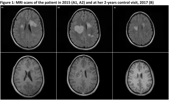
Immediately after this worsening period, adult neurologists were consulted. Considering the anamnestic data, the test results, the MRI picture and the resistance to steroid therapy, the patient was diagnosed with the fulminant variant of tMS and plasma-exchange therapy was started. After 5 plasma-exchanges, her neurological state stagnated. On the 16th day of admission, her second control MRI showed that the right hemispherical tumefactive lesion again increased in size and the tumefactive lesion in the left frontal lobe developed gadolinium-enhancement (Figure 1, A1).
Because of the rapid progression and the therapy resistance of the disease, adult neurologists
recommended natalizumab therapy for the treatment of the patient. As natalizumab is considered an off-label treatment in children, after thoroughly informing the patient’s family on the possible side- effects and acquiring their written consent, the approval procedure for natalizumab was
commenced. Meanwhile the patient received four more plasma-exchange therapies to stop the progression.
The National Institute of Quality and Organizational Development in Healthcare and Medicines of Hungary permitted the use of natalizumab in this case. The patient received the first natalizumab infusion on the 27th, March 2015, the 24th day of admission. She tolerated the treatment well, no side-effects or allergic reactions were observed. Her condition steadily improved: the muscle strength increased to 4/5 again. A third control MRI was performed, which still showed the enlargement of the right hemispherical lesion, but also the decrease of the gadolinium
enhancement. In improving condition, the girl was released from the Department of Pediatrics.
In April, 2015, after the second natalizumab infusion therapy, the patient showed no sign of paresis whatsoever; and apart from slight hyperreflexia, Babinski-tendency and slight cerebellar ataxia on the left side, her neurological state was normal, the EDSS score was 1.5 points. She scored 5.5 seconds on the Timed-25 Foot Walk (T25-FW) and 6 minutes 43 seconds on the Timed-500 Meter Walk Test (T500-MW). She was assessed with the 9-Hole Peg Test (9-HPT): she scored 18.4 and 20.6 seconds with the dominant (right) hand and 39.2 and 43.4 seconds with the non-dominant (left) hand respectively. Her cognitive state was evaluated with the Symbol Digit Modalities Test (SDMT) and the Paced Auditory Serial Addition Test (PASAT): she scored 45 points on SDMT (z-score: -0,75) and 35 points on PASAT. She scored 33 points on the Fatigue Impact Scale (FIS) implying a mild fatigue, and 8 points on the Beck’s Depression Inventory (BDI-II).
In 2016, on her 12-months control visit, a control brain MRI was performed, which showed the decrease in size of the right hemispherical lesion, revealed no gadolinium-enhancement in any lesions and confirmed no signs of PML or other anomalies (Figure 1, A2). Her EDSS score was 1.0
point. Her performance vastly increased as compared to the previous assessments: she finished the T25-FW in 4.6 seconds and the T500-MW in exactly 5 minutes. The 9-HPT scores also showed great improvement: her score was 22.3 and 19.6 seconds with the dominant hand and 14.5 and 23.6 seconds with the non-dominant hand respectively. Her SDMT score increased to 63 points (z-score:
0.87) and her PASAT performance to 54 points. Her fatigue de facto disappeared: her FIS score was 6, and she received only 4 points on the BDI-II assessment.
Figure 1: MRI scans of the patient in 2015 (A1, A2) and at her 2-years control visit, 2017 (B)
A1: Brain MRI taken on 3rd, March, 2015. The FLAIR sequence shows multiple demyelinating lesions and the left hemispherical tumefactive lesion. The T1 sequence reveals no gadolinium
enhancement.
A2: Brain MRI taken on 19th March, 2015. The FLAIR sequence shows multiple tumefactive lesions developed as compared to the previous MRI scan, the largest one being in the right hemisphere, largest diameter 9 cm. The T1 sequence reveals diffuse gadolinium-enhancement of the lesion in the right hemisphere.
B: Brain MRI taken on 3rd, February, 2017. The FLAIR shows that the lesions vastly decreased in size as compared to 2 years earlier and the T1 sequence reveals no sign of gadolinium-
enhancement.
As of February, 2018, the patient has received 37 natalizumab infusions. Her EDSS score remains steadily on 1.0 point. She is regularly tested for JCV, so far, all her tests came back negative. On her 24-months control MRI, the size of the lesions stagnated, there was no evidence of gadolinium- enhancement and no sign of PML or any other anomalies, whatsoever (Figure 1, B). She did not worsen in any other assessed areas: she finished the T25-FW in 5 seconds, the T500-MW in 4
minutes 22 seconds. She further improved on the 9-HPT: her scores were 18.3 and 19.9 seconds with the dominant hand and 21.8 and 19.3 seconds with the non-dominant hand. Her SDMT score
increased to 73 points (z-score: 1.98), but her PASAT performance slightly lowered to 49 points.
There was no sign of return of fatigue or depression (both FIS and BDI-II scores were 1).
Conclusions
Though MS is primarily the disease of adulthood, diagnosis with pediatric onset is becoming more and more often. The affected patients of POMS may number in the hundred-thousands worldwide, according to new epidemiological data (3). Despite being rare, atypical manifestations and fulminant disease course pose a serious differential diagnostic and therapeutic challenge. However, the global view on MS therapy is changing: we are moving closer to individual therapy day by day. Guidelines recommend to commence DMTs as early as possible, and the choice of the drugs are now based on several factors, the activity and severity of the disease course being one of the key points (36, 37).
This shift in perspective was brought on by hard evidence demonstrating the benefit of early
treatment, early change to more effective therapies and in case of aggressive disease course, the use of more effective DMTs as initial choice (36, 37).
This perspective may very well be applied to POMS patients in the near future. Despite the lack of data from randomized control studies, existing guidelines already recommend the initiation of DMTs as early as possible, to halt the disease progression and reduce or stop the accumulation of disability in children, as existing data shows the benefit of such action (39). Luckily however, several studies are being conducted on pediatric patients right now, to improve our knowledge on the benefits and potential risks of DMTs (43).
The presented case highlights the difficulties in diagnosis and treatment of POMS, especially in case of a highly active, atypical manifestation. Such an aggressive, fulminant course of MS may very well be fatal in a short period of time if left untreated or treated with inadequately effective methods (45). As many DMTs are still considered off-label in the pediatric population, sometimes the choice of treatment becomes hard and require additional paper-work and special permissions. However, the case also demonstrates the great success of such choices in these situations. To our best knowledge, we are the first to report successful treatment of fulminant pediatric tMS with natalizumab.
The reviewed data and the presented case leads us to conclude, that the choice of MS therapy should very well be based on the individual requirements of the patient (in accordance with the new ECTRIMS/EAN guidelines), regardless of the patients age, because early diagnosis and effective treatment may reverse the disease course even in highly aggressive, potentially fatal cases.
Conflict of Interest
The authors declare they do not have any conflicts of interest regarding the research, authorship and/or the publication of the article.
Financial Disclosure
This report received funding from the Economic Development and Innovation Operative Program of the European Structural and Investment Funds (Gazdaságfejlesztési és Innovációs Operatív Program;
GINOP-2.3.2-15-2016-00034).
References
1. Jancic J, Nikolic B, Ivancevic N, Hencic B, Samardzic J. Multiple Sclerosis Therapies in Pediatric Patients: Challenges and Opportunities. In: Zagon IS, McLaughlin PJ, editors. Multiple Sclerosis:
Perspectives in Treatment and Pathogenesis. Brisbane (AU)2017.
2. Confavreux C, Aimard G, Devic M. Course and prognosis of multiple sclerosis assessed by the computerized data processing of 349 patients. Brain. 1980;103(2):281-300.
3. Ness JM, Chabas D, Sadovnick AD, Pohl D, Banwell B, Weinstock-Guttman B, et al. Clinical features of children and adolescents with multiple sclerosis. Neurology. 2007;68(16 Suppl 2):S37-45.
4. Renoux C, Vukusic S, Mikaeloff Y, Edan G, Clanet M, Dubois B, et al. Natural history of multiple sclerosis with childhood onset. N Engl J Med. 2007;356(25):2603-13.
5. Boiko A, Vorobeychik G, Paty D, Devonshire V, Sadovnick D, University of British Columbia MSCN. Early onset multiple sclerosis: a longitudinal study. Neurology. 2002;59(7):1006-10.
6. Boster AL, Endress CF, Hreha SA, Caon C, Perumal JS, Khan OA. Pediatric-onset multiple sclerosis in African-American black and European-origin white patients. Pediatr Neurol.
2009;40(1):31-3.
7. Banwell B, Kennedy J, Sadovnick D, Arnold DL, Magalhaes S, Wambera K, et al. Incidence of acquired demyelination of the CNS in Canadian children. Neurology. 2009;72(3):232-9.
8. Waldman A, Ness J, Pohl D, Simone IL, Anlar B, Amato MP, et al. Pediatric multiple sclerosis:
Clinical features and outcome. Neurology. 2016;87(9 Suppl 2):S74-81.
9. Huppke B, Ellenberger D, Rosewich H, Friede T, Gartner J, Huppke P. Clinical presentation of pediatric multiple sclerosis before puberty. Eur J Neurol. 2014;21(3):441-6.
10. Bove R, Chitnis T. The role of gender and sex hormones in determining the onset and outcome of multiple sclerosis. Mult Scler. 2014;20(5):520-6.
11. Waldman A, Ghezzi A, Bar-Or A, Mikaeloff Y, Tardieu M, Banwell B. Multiple sclerosis in children: an update on clinical diagnosis, therapeutic strategies, and research. Lancet Neurol.
2014;13(9):936-48.
12. Gorman MP, Healy BC, Polgar-Turcsanyi M, Chitnis T. Increased relapse rate in pediatric- onset compared with adult-onset multiple sclerosis. Arch Neurol. 2009;66(1):54-9.
13. Benson LA, Healy BC, Gorman MP, Baruch NF, Gholipour T, Musallam A, et al. Elevated relapse rates in pediatric compared to adult MS persist for at least 6 years. Mult Scler Relat Disord.
2014;3(2):186-93.
14. Fay AJ, Mowry EM, Strober J, Waubant E. Relapse severity and recovery in early pediatric multiple sclerosis. Mult Scler. 2012;18(7):1008-12.
15. Cossburn M, Ingram G, Hirst C, Ben-Shlomo Y, Pickersgill TP, Robertson NP. Age at onset as a determinant of presenting phenotype and initial relapse recovery in multiple sclerosis. Mult Scler.
2012;18(1):45-54.
16. Al-Hamadani HA, Abdalla AS, Al-Saffar AJ. The course of early-onset multiple sclerosis in Iraqi children. World J Pediatr. 2012;8(1):47-51.
17. Ghezzi A, Deplano V, Faroni J, Grasso MG, Liguori M, Marrosu G, et al. Multiple sclerosis in childhood: clinical features of 149 cases. Mult Scler. 1997;3(1):43-6.
18. Simone IL, Carrara D, Tortorella C, Liguori M, Lepore V, Pellegrini F, et al. Course and prognosis in early-onset MS: comparison with adult-onset forms. Neurology. 2002;59(12):1922-8.
19. Harding KE, Liang K, Cossburn MD, Ingram G, Hirst CL, Pickersgill TP, et al. Long-term outcome of paediatric-onset multiple sclerosis: a population-based study. J Neurol Neurosurg Psychiatry. 2013;84(2):141-7.
20. Ruggieri M, Iannetti P, Polizzi A, Pavone L, Grimaldi LM, Italian Society of Paediatric
Neurology Study Group on Childhood Multiple S. Multiple sclerosis in children under 10 years of age.
Neurol Sci. 2004;25 Suppl 4:S326-35.
21. Ghassemi R, Antel SB, Narayanan S, Francis SJ, Bar-Or A, Sadovnick AD, et al. Lesion distribution in children with clinically isolated syndromes. Ann Neurol. 2008;63(3):401-5.
22. Etemadifar M, Abtahi SH, Tabrizi N. Epileptic seizures in early-onset multiple sclerosis. Arch Iran Med. 2012;15(6):381-3.
23. Suppiej A, Cainelli E. Cognitive dysfunction in pediatric multiple sclerosis. Neuropsychiatr Dis Treat. 2014;10:1385-92.
24. Amato MP, Goretti B, Ghezzi A, Lori S, Zipoli V, Moiola L, et al. Cognitive and psychosocial features in childhood and juvenile MS: two-year follow-up. Neurology. 2010;75(13):1134-40.
25. Till C, Deotto A, Tipu V, Sled JG, Bethune A, Narayanan S, et al. White matter integrity and math performance in pediatric multiple sclerosis: a diffusion tensor imaging study. Neuroreport.
2011;22(18):1005-9.
26. MacAllister WS, Belman AL, Milazzo M, Weisbrot DM, Christodoulou C, Scherl WF, et al.
Cognitive functioning in children and adolescents with multiple sclerosis. Neurology.
2005;64(8):1422-5.
27. Banwell BL, Anderson PE. The cognitive burden of multiple sclerosis in children. Neurology.
2005;64(5):891-4.
28. Rao SM. Neuropsychology of multiple sclerosis: a critical review. J Clin Exp Neuropsychol.
1986;8(5):503-42.
29. Hahn CD, Shroff MM, Blaser SI, Banwell BL. MRI criteria for multiple sclerosis: Evaluation in a pediatric cohort. Neurology. 2004;62(5):806-8.
30. Yeh EA, Weinstock-Guttman B, Ramanathan M, Ramasamy DP, Willis L, Cox JL, et al.
Magnetic resonance imaging characteristics of children and adults with paediatric-onset multiple sclerosis. Brain. 2009;132(Pt 12):3392-400.
31. Waubant E, Chabas D, Okuda DT, Glenn O, Mowry E, Henry RG, et al. Difference in disease burden and activity in pediatric patients on brain magnetic resonance imaging at time of multiple sclerosis onset vs adults. Arch Neurol. 2009;66(8):967-71.
32. Aubert-Broche B, Fonov V, Ghassemi R, Narayanan S, Arnold DL, Banwell B, et al. Regional brain atrophy in children with multiple sclerosis. Neuroimage. 2011;58(2):409-15.
33. Till C, Ghassemi R, Aubert-Broche B, Kerbrat A, Collins DL, Narayanan S, et al. MRI correlates of cognitive impairment in childhood-onset multiple sclerosis. Neuropsychology. 2011;25(3):319-32.
34. Giovannoni G, Turner B, Gnanapavan S, Offiah C, Schmierer K, Marta M. Is it time to target no evident disease activity (NEDA) in multiple sclerosis? Mult Scler Relat Disord. 2015;4(4):329-33.
35. Comi G, Radaelli M, Soelberg Sorensen P. Evolving concepts in the treatment of relapsing multiple sclerosis. Lancet. 2017;389(10076):1347-56.
36. Montalban X, Gold R, Thompson AJ, Otero-Romero S, Amato MP, Chandraratna D, et al.
ECTRIMS/EAN Guideline on the pharmacological treatment of people with multiple sclerosis. Mult Scler. 2018:1352458517751049.
37. Montalban X, Gold R, Thompson AJ, Otero-Romero S, Amato MP, Chandraratna D, et al.
ECTRIMS/EAN guideline on the pharmacological treatment of people with multiple sclerosis. Eur J Neurol. 2018;25(2):215-37.
38. Rose K, Muller T. Children with multiple sclerosis should not become therapeutic hostages.
Ther Adv Neurol Disord. 2016;9(5):389-95.
39. Chitnis T, Tenembaum S, Banwell B, Krupp L, Pohl D, Rostasy K, et al. Consensus statement:
evaluation of new and existing therapeutics for pediatric multiple sclerosis. Mult Scler.
2012;18(1):116-27.
40. Waldman AT, Gorman MP, Rensel MR, Austin TE, Hertz DP, Kuntz NL, et al. Management of pediatric central nervous system demyelinating disorders: consensus of United States neurologists. J Child Neurol. 2011;26(6):675-82.
41. Jancic J, Nikolic B, Ivancevic N, Djuric V, Zaletel I, Stevanovic D, et al. Multiple Sclerosis in Pediatrics: Current Concepts and Treatment Options. Neurol Ther. 2016;5(2):131-43.
42. Kornek B, Aboul-Enein F, Rostasy K, Milos RI, Steiner I, Penzien J, et al. Natalizumab therapy for highly active pediatric multiple sclerosis. JAMA Neurol. 2013;70(4):469-75.
43. Waubant E. Treatment of Peadiatric MS (Abstract). Mult Scler. 2017;23(S3):8-84.
44. Huppke PH, B.; Ellenberger, D.; Rostasy, K.; Hummel, H.; Stark, W.; Brück, W.; Gärtner, J.
Treatment of highly active multiple sclerosis in pediatric patients (poster abstract). Mult Scler.
2017;27(S3):427-679.
45. Rahmlow MR, Kantarci O. Fulminant demyelinating diseases. Neurohospitalist. 2013;3(2):81- 91.
46. Gobbin F, Marangi A, Orlandi R, Richelli S, Turatti M, Calabrese M, et al. A case of acute fulminant multiple sclerosis treated with alemtuzumab. Mult Scler Relat Disord. 2017;17:9-11.
47. Menge T, Hemmer B, Nessler S, Wiendl H, Neuhaus O, Hartung HP, et al. Acute disseminated encephalomyelitis: an update. Arch Neurol. 2005;62(11):1673-80.
48. Leake JA, Albani S, Kao AS, Senac MO, Billman GF, Nespeca MP, et al. Acute disseminated encephalomyelitis in childhood: epidemiologic, clinical and laboratory features. Pediatr Infect Dis J.
2004;23(8):756-64.
49. Alper G, Heyman R, Wang L. Multiple sclerosis and acute disseminated encephalomyelitis diagnosed in children after long-term follow-up: comparison of presenting features. Dev Med Child Neurol. 2009;51(6):480-6.
50. Pohl D, Hennemuth I, von Kries R, Hanefeld F. Paediatric multiple sclerosis and acute disseminated encephalomyelitis in Germany: results of a nationwide survey. Eur J Pediatr.
2007;166(5):405-12.
51. Pohl D, Alper G, Van Haren K, Kornberg AJ, Lucchinetti CF, Tenembaum S, et al. Acute disseminated encephalomyelitis: Updates on an inflammatory CNS syndrome. Neurology. 2016;87(9 Suppl 2):S38-45.
52. Koelman DL, Mateen FJ. Acute disseminated encephalomyelitis: current controversies in diagnosis and outcome. J Neurol. 2015;262(9):2013-24.
53. Marchioni E, Ravaglia S, Piccolo G, Furione M, Zardini E, Franciotta D, et al. Postinfectious inflammatory disorders: subgroups based on prospective follow-up. Neurology. 2005;65(7):1057-65.
54. Tenembaum S, Chamoles N, Fejerman N. Acute disseminated encephalomyelitis: a long-term follow-up study of 84 pediatric patients. Neurology. 2002;59(8):1224-31.
55. Marin SE, Callen DJ. The magnetic resonance imaging appearance of monophasic acute disseminated encephalomyelitis: an update post application of the 2007 consensus criteria.
Neuroimaging Clin N Am. 2013;23(2):245-66.
56. Hardy TA, Reddel SW, Barnett MH, Palace J, Lucchinetti CF, Weinshenker BG. Atypical inflammatory demyelinating syndromes of the CNS. Lancet Neurol. 2016;15(9):967-81.
57. Atzori M, Battistella PA, Perini P, Calabrese M, Fontanin M, Laverda AM, et al. Clinical and diagnostic aspects of multiple sclerosis and acute monophasic encephalomyelitis in pediatric patients: a single centre prospective study. Mult Scler. 2009;15(3):363-70.
58. Rossi A. Imaging of acute disseminated encephalomyelitis. Neuroimaging Clin N Am.
2008;18(1):149-61; ix.
59. Callen DJ, Shroff MM, Branson HM, Li DK, Lotze T, Stephens D, et al. Role of MRI in the differentiation of ADEM from MS in children. Neurology. 2009;72(11):968-73.
60. Verhey LH, Branson HM, Shroff MM, Callen DJ, Sled JG, Narayanan S, et al. MRI parameters for prediction of multiple sclerosis diagnosis in children with acute CNS demyelination: a prospective national cohort study. Lancet Neurol. 2011;10(12):1065-73.
61. Krupp LB, Tardieu M, Amato MP, Banwell B, Chitnis T, Dale RC, et al. International Pediatric Multiple Sclerosis Study Group criteria for pediatric multiple sclerosis and immune-mediated central nervous system demyelinating disorders: revisions to the 2007 definitions. Mult Scler.
2013;19(10):1261-7.
62. Absoud M, Parslow RC, Wassmer E, Hemingway C, Duncan HP, Cummins C, et al. Severe acute disseminated encephalomyelitis: a paediatric intensive care population-based study. Mult Scler. 2011;17(10):1258-61.
63. Sonneville R, Demeret S, Klein I, Bouadma L, Mourvillier B, Audibert J, et al. Acute
disseminated encephalomyelitisin the intensive care unit:clinical features and outcome of 20 adults.
Intensive Care Med. 2008;34(3):528-32.
64. Ketelslegers IA, Visser IE, Neuteboom RF, Boon M, Catsman-Berrevoets CE, Hintzen RQ.
Disease course and outcome of acute disseminated encephalomyelitis is more severe in adults than in children. Mult Scler. 2011;17(4):441-8.
65. Lucchinetti CF, Gavrilova RH, Metz I, Parisi JE, Scheithauer BW, Weigand S, et al. Clinical and radiographic spectrum of pathologically confirmed tumefactive multiple sclerosis. Brain. 2008;131(Pt 7):1759-75.
66. Comi G. Multiple sclerosis: pseudotumoral forms. Neurol Sci. 2004;25 Suppl 4:S374-9.
67. Hardy TA, Chataway J. Tumefactive demyelination: an approach to diagnosis and management. J Neurol Neurosurg Psychiatry. 2013;84(9):1047-53.
68. Algahtani H, Shirah B, Alassiri A. Tumefactive demyelinating lesions: A comprehensive review.
Mult Scler Relat Disord. 2017;14:72-9.
69. Pilz G, Harrer A, Wipfler P, Oppermann K, Sellner J, Fazekas F, et al. Tumefactive MS lesions under fingolimod: a case report and literature review. Neurology. 2013;81(19):1654-8.
70. Visser F, Wattjes MP, Pouwels PJ, Linssen WH, van Oosten BW. Tumefactive multiple sclerosis lesions under fingolimod treatment. Neurology. 2012;79(19):2000-3.
71. Jander S, Turowski B, Kieseier BC, Hartung HP. Emerging tumefactive multiple sclerosis after switching therapy from natalizumab to fingolimod. Mult Scler. 2012;18(11):1650-2.
72. Castrop F, Kowarik MC, Albrecht H, Krause M, Haslinger B, Zimmer C, et al. Severe multiple sclerosis relapse under fingolimod therapy: incident or coincidence? Neurology. 2012;78(12):928-30.
73. Faissner S, Hoepner R, Lukas C, Chan A, Gold R, Ellrichmann G. Tumefactive multiple sclerosis lesions in two patients after cessation of fingolimod treatment. Ther Adv Neurol Disord.
2015;8(5):233-8.
74. Gross CM, Baumgartner A, Rauer S, Stich O. Multiple sclerosis rebound following herpes zoster infection and suspension of fingolimod. Neurology. 2012;79(19):2006-7.
75. Salam S, Mihalova T, Siripurapu R. Severe tumefactive rebound of multiple sclerosis following fingolimod cessation. BMJ Case Rep. 2016;2016.
76. Frederick MC, Cameron MH. Tumefactive Demyelinating Lesions in Multiple Sclerosis and Associated Disorders. Curr Neurol Neurosci Rep. 2016;16(3):26.
77. Chabas D, Castillo-Trivino T, Mowry EM, Strober JB, Glenn OA, Waubant E. Vanishing MS T2- bright lesions before puberty: a distinct MRI phenotype? Neurology. 2008;71(14):1090-3.
78. Yiu EM, Laughlin S, Verhey LH, Banwell BL, Canadian Pediatric Demyelinating Disease N.
Clinical and magnetic resonance imaging (MRI) distinctions between tumefactive demyelination and brain tumors in children. J Child Neurol. 2014;29(5):654-65.
79. Wattamwar PR, Baheti NN, Kesavadas C, Nair M, Radhakrishnan A. Evolution and long term outcome in patients presenting with large demyelinating lesions as their first clinical event. J Neurol Sci. 2010;297(1-2):29-35.
80. Krivickas LS, Hochberg FH, Freeman S. Chronic inflammatory demyelinating
polyradiculoneuropathy with tumefactive central demyelination. Muscle Nerve. 2006;33(2):283-8.
81. Nagappa M, Taly AB, Sinha S, Bharath RD, Mahadevan A, Bindu PS, et al. Tumefactive demyelination: clinical, imaging and follow-up observations in thirty-nine patients. Acta Neurol Scand. 2013;128(1):39-47.
82. Weinshenker BG, O'Brien PC, Petterson TM, Noseworthy JH, Lucchinetti CF, Dodick DW, et al.
A randomized trial of plasma exchange in acute central nervous system inflammatory demyelinating disease. Ann Neurol. 1999;46(6):878-86.