III./5.3.: Multiple sclerosis
Epidemiology
Multiple sclerosis (MS) is the most common neuroimmunological disorder of the central nervous system. This chronic illness begins in early adulthood, mostly at age of 20 to 40 years, significantly affecting the quality of life. The prevalence of MS in continental climate is 30-70/100,000. There are approximately 2.5 million MS patients worldwide; in Hungary, the estimated number of MS patients is 8-10,000.
Etiology
MS is a multifactorial disorder, both genetic and environmental factors play a role in the development of the disease. The prevalence of MS increases from the equator to the poles. Migration studies have shown that the prevalence of MS among people who emigrated at a young age - around puberty – is similar to that of their new country, whereas the prevalence of MS among people who emigrated at an older age is similar to that of their country of origin. A connection between viral infections and the frequency of relapses is also well known. All these facts emphasize the importance of environmental factors in the development of MS.
Family- and twin-studies have shown the role of genetic factors in the development of MS. Twenty percent of the MS patients have a relative with MS; concordance of MS in monozygotic twins is 25%, while in dizygotic twins it is 5%.
Pathology
MS is basically an autoimmune inflammation in the central nervous system; first T-cells are activated in the periphery, which then cross-react with certain
components of the myelin in the central nervous system. This results in myelin degradation and secondary axon degeneration. The demyelinated regions in the brain or spinal cord are called plaques.
Active plaques are characterized by blood-brain barrier dysfunction, perivascular infiltration of lymphocytes and plasma cells, and segmental demyelination.
In chronic plaques, scar tissue is formed (sclerosis) due to the proliferation of astrocytes. Axonal damage occurs already in the acute inflammatory plaques (early stage of the disease), but clinical symptoms of chronic axonal degeneration become manifest in the second stage of the disease (secondary progressive). Although MS is predominantly a white matter disorder, cortical involvement may also be detected (Fig. 5).
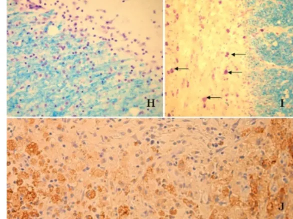
Fig. 5: Multiple sclerosis – pathology
H: Chronic active plaque. Hypocellular central core can be seen in the right upper corner, and myelinated fibers stained blue in the left lower corner. Elevated number of macrophages and lymphocytes at the margins of these regions indicates ongoing demyelination.
I: The structure of neurons is intact inside the plaques (shown by arrows).
J: Active plaque from a biopsy with extensive CD68-positive macrophage
infiltration (brown). H, I: Luxol fast blue-Nissl staining (Klüver-Barrea): cells are purple, myelin is blue. J: CD68-immunohistochemical staining.
The pathomechanism of MS is not homogeneous; based on the results of recent studies, four pathological subtypes are differentiated. In a given patient, all plaques are of the same subtype, indicating either a predominant immunological or
degenerative process. Further characterization of each subtype may be crucial for the development of subtype specific treatments (Fig. 6).
Fig. 6: Pathological subtypes of multiple sclerosis
Clinical subtypes
Sixty-five to eighty percent of MS patients show a relapsing-remitting (RR-MS) course, which is characterized by relapses followed by periods of disease inactivity (remission). At the beginning of the disease, complete remission is achieved after the first few relapses. A relapse is defined as a progressive deterioration of
neurological function when the neurological deficit is sustained for longer than 24 hours, and which is followed by a relapse period lasting for at least a month. In most patients, after the initial relapsing-remitting period lasting for years or decades, disease course becomes secondary progressive (SP-MS). In this subtype, there are no more relapses and slow progression is seen. Ten to fifteen percent of patients show the primary progressive (PP-MS) subtype, which is characterized by a continuous slow deterioration of function. The relapsing-progressive (RP-MS) form is a rare subtype, affecting 5-6% of patients, where the continuous decline of function is punctuated by relapses.
Diagnosis
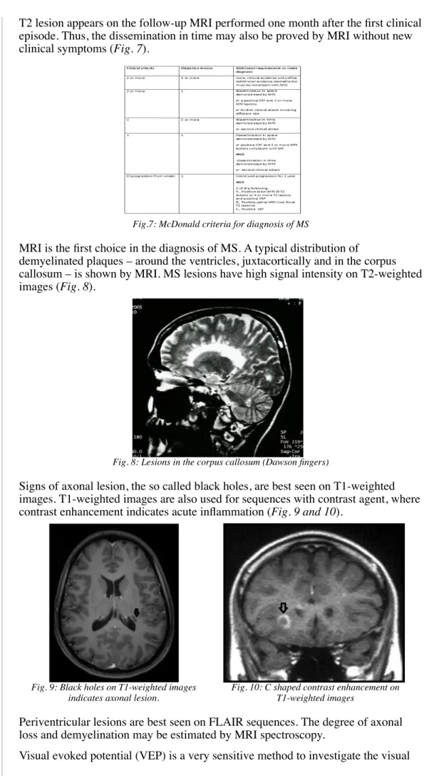
Currently, the McDonald criteria are used for the diagnosis of MS. This diagnostic algorithm is based on the demonstration of dissemination of MS lesions in time and space using clinical, laboratory and radiological data.
Dissemination in time is defined as the occurrence of two relapses, separated by at least one month. The two relapses should be associated with neurological
symptoms explained by two separate lesions. Furthermore, MRI lesions must meet the criteria defined in the McDonald criteria. According to the recently revised version of McDonald criteria (2005), the diagnosis of MS is confirmed if a new
T2 lesion appears on the follow-up MRI performed one month after the first clinical episode. Thus, the dissemination in time may also be proved by MRI without new clinical symptoms (Fig. 7).
Fig.7: McDonald criteria for diagnosis of MS
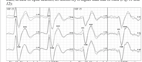
MRI is the first choice in the diagnosis of MS. A typical distribution of
demyelinated plaques – around the ventricles, juxtacortically and in the corpus callosum – is shown by MRI. MS lesions have high signal intensity on T2-weighted images (Fig. 8).
Fig. 8: Lesions in the corpus callosum (Dawson fingers)
Signs of axonal lesion, the so called black holes, are best seen on T1-weighted images. T1-weighted images are also used for sequences with contrast agent, where contrast enhancement indicates acute inflammation (Fig. 9 and 10).
Fig. 9: Black holes on T1-weighted images indicates axonal lesion.
Fig. 10: C shaped contrast enhancement on T1-weighted images
Periventricular lesions are best seen on FLAIR sequences. The degree of axonal loss and demyelination may be estimated by MRI spectroscopy.
Visual evoked potential (VEP) is a very sensitive method to investigate the visual
tract; in case of optic neuritis, its sensitivity is higher than that of MRI (Fig. 11 and 12):
Fig. 11: Visual evoked potential (VEP) – normal findings; (no P100 latency difference between the
eyes)
Fig. 12: Visual evoked potential (VEP) –optic neuritis on the right side; (delayed P100 latency on
the right side)
Somatosensory and motor evoked potentials play an important role in the detection of subclinical lesions and in predicting the course of the disease. MS may be diagnosed based on clinical signs and MRI findings alone, however examination of the CSF may further support the diagnosis. CSF protein content and cell count is normal in MS, but mild pleiocytosis may be seen. An elevated IgG index or the presence of oligoclonal gammopathy (OGP) are characteristic in MS.
Clinical signs and symptoms
MS may involve all tracts of the central nervous system, but longer tracts are preferentially affected. Optic nerve lesion (optic neuritis) is often the first sign of the disease. Optic neuritis is characterized by loss of vision developing within days and pain on movement of the affected eye. Concerning the sensory tracts, both the proprioceptive and spinothalamic tracts may be affected. Tingling, pricking, numbness, pain or disturbed proprioception are the most common sensory signs.
The most common motor symptom is paraparesis, but hemi-, mono- or tetraparesis may also occur. Brainstem involvement is often manifested in gaze palsy,
internuclear ophthalmoplegia or trigeminal pain. Cerebellar signs may include ataxia, coordination and balance problems, or tremor. Detrusor hyperactivity and detrusor-sphincter dyssynergia are the dominant autonomic symptoms. Chronic fatigue, cognitive decline, subcortical dementia, and depression may also appear during the course of the disease. Paroxysmal signs (e.g. Lhermitte’s sign, trigeminal neuralgia, dystonia, hemiataxia) may occur several hundred times a day, with a sudden onset and short duration, lasting for less than a minute.
Fig. 13: Clinical signs in multiple sclerosis
Treatment
Relapses are treated with high-dose (500-1000 mg) intravenous corticosteroids (methyl-prednisolone) given for three to five days. If corticosteroids are
contraindicated, plasma exchange is recommended. As a symptomatic treatment, spasticity can be reduced by baclofen or tizanidine. Detrusor hyperactivity is treated with anticholinergic drugs, and intermittent self-catheterization is recommended in detrusor-sphincter dyssynergia. Fatigue may be treated by amantadine. Carbamazepine or gabapentin may be effective in paroxysmal symptoms.
Self-injection of interferon beta-1a (Avonex, Rebif), interferon beta-1b (Betaferon, Extavia), or glatiramer acetate (Copaxone) is the first choice of long-term
immunosuppressive treatment. Clinical studies have shown that interferons and glatiramer acetate have similar clinical efficacy. Both treatments reduce relapse rate in the relapsing-remitting form of MS. Interferons may cause initial flu-like side effects (fever, shivering, muscle pain), but these tend to diminish during further treatment. Interferon treatment is contraindicated in severe depression; neither of the immunosuppressive treatments are recommended during pregnancy or breast- feeding.
Natalizumab (Tysabri) is a monoclonal antibody against the cellular adhesion molecule called integrin, which is predominantly expressed on T cells. Natalizumab is used in rapidly progressive relapsing-remitting MS, or in cases where interferons are ineffective. It significantly diminishes relapse rate and the number of MRI lesions. Long-term natalizumab treatment caused progressive multifocal leukoencephalopathy (PML) in 0.001% of patients, which is related to the activation of latent form of JC virus present in the body. Therefore, close monitoring and regular clinical and MRI examinations are recommended during natalizumab treatment, especially in JC-antibody positive patients.
It is important to consider when to start a long-term immunomodulant treatment.
The main argument for early treatment is that degenerative processes leading to disability start parallel with the inflammation, already at the beginning of the illness. Clinical studies have shown that early treatment is associated with a better outcome.
Mitoxantrone infusion – administered every three months - is a treatment option in relapsing-progressive MS (RP-MS). It has a cardiotoxic side effect, therefore regular cardiologic control with echocardiography is necessary during treatment.
The drugs mentioned above are ineffective in the primary progressive form of MS.
On-going research and clinical trials focus on monoclonal antibody treatments, on oral treatments, and on neuroprotective drugs. Combined treatments are also under investigation.
Special recommendations
Relapse rate decreases during pregnancy, but it increases during the first six months after childbirth. Re-introduction of previous immunotherapy is recommended after childbirth. It is generally advised to avoid dehydration, extreme physical exhaustion or elevation of body temperature due to fever, sauna or hot shower. A diet rich with unsaturated fatty acids is recommended.
Differential diagnosis
Differential diagnosis includes conditions, which have a similar clinical course or MRI pathology as MS, such as autoimmune disorders (Sjögren’s syndrome, SLE, CIDP, etc.), vascular diseases, vasculitis, tumors or subacute combined
degeneration due to vitamin B12 deficiency (Fig. 14).
Fig. 14: Differential diagnosis of multiple sclerosis
Acute disseminated encephalomyelitis (ADEM) is a monophasic disorder of unknown origin, however it can develop after a vaccination or infection. Contrary to MS, ADEM is more common in childhood. Headache, fever, seizures, altered consciousness, personality changes and myelopathy are among the leading symptoms. Pleiocytosis, elevated protein content, normal IgG index and lack of OGP are seen in the CFS. MRI shows well-defined single or multiple merging demyelinating lesions with contrast enhancement. ADEM is monophasic and follow-up MRI examinations show the disappearance of MRI lesions. Potential treatments include high-dose corticosteroids, plasma exchange and IVIG.
Neuromyelitis optica (Devic’s disease) is also discussed here. It is an autoimmune, inflammatory disorder, causing inflammation in one or both of optic nerves (optic neuritis) and/or in the spinal cord (myelitis). It’s onset may be anywhere from childhood to late adulthood, but typical age of onset is around the early thirties, ten years later than in MS. Eighty percent of cases show a relapsing course. MRI typically shows large lesions with increased signal intensity on T2-weighted images in the spinal cord, extending over more than three vertebras. CSF examination shows pleiocytosis, elevated protein content and lack of OGP, but these findings are non-specific. The presence of antibodies against aquaporin-4 (AQP4), a water channel, confirms the diagnosis.
The acute phase of Devic’s disease is treated with high-dose corticosteroids or plasma exchange, and oral corticosteroids or azathioprine are used for chronic treatment.