Ritta Jubran1,*, Judit Kocsis2,*,**, Nóra Garam2, Éva Maláti2,***, Tímea Gombos2, Lóránd Barabás3, László Gráf2, Zoltán Prohászka2, * and Zvi Fishelson1,4, *
1 Department of Cell and Developmental Biology, Sackler Faculty of Medicine, Tel Aviv University, Tel Aviv 69978, Israel
23rd Department of Internal Medicine, 3and 2nd Department of Surgery, Semmelweis University, Kútvölgyi út 4, Budapest 1125, Hungary
*,# R.T and J.K. contributed equally to this study. Z.P. and Z. F. are joint senior authors.
** Current address: Debrecen University, Institute of Oncology, Debrecen, Hungary
*** Current address: János Balassa Hospital, County Hospital Tolna, Szekszárd, Hungary
4 Corresponding author at: Department of Cell and Developmental Biology, Sackler School of Medicine, Tel Aviv University, Tel Aviv 69978, Israel (e-mail: lifish@post.tau.ac.il).
Short title: Circulating mortalin and Hsp70 in colorectal cancer patients
Key Words: colorectal cancer, mortalin, GRP75, Hsp70, overall survival, risk factor Category: Tumor markers and signatures
Description of the work: Circulating levels of mitochondrial mortalin and cytosolic Hsp70 are elevated in sera of colorectal cancer patients. Elevated mortalin and Hsp70 levels indicate a lower patients’ survival rate, irrespective of cancer stage. High mortalin and Hsp70 significantly enhances death risk score even after considering age, number of affected lymph nodes, CEA and CA19-9 levels, disease stage and perioperative therapy. It is suggested that mortalin/Hsp70 levels in blood be examined as prognostic markers in other cancer types.
Grant sponsor: The Israel Science Foundation and the Israel Cancer Association.
Abstract
Mitochondrial mortalin and cytosolic Hsp70 are essential chaperones overexpressed in cancer cells. Our goals were to reproduce our earlier findings of elevated circulating levels of mortalin and Hsp70 in colorectal cancer (CRC) patients with a larger patient cohort, to compare death risk assessment of mortalin, Hsp70, CEA and C19-9 and to assess their prognostic value in various CRC stages. Mortalin, Hsp70, CEA and CA19-9 levels were determined in sera of 235 CRC patients enrolled in the study and followed-up 5 years after surgery. Association between their concentrations and patients’ survival was analyzed by Kaplan-Meier estimator and subjected to Cox Proportional hazards analysis.
Serum level of mortalin was independent of that of Hsp70, CEA and CA19-9, whereas Hsp70 level weakly correlated with CEA and CA19-9 levels. Improved short-term survival was found in early or advanced disease stages associated with lower mortalin and Hsp70 levels. Cox regression analysis showed a high mortality hazard (HR=3.7, P<0.001) in patients with both high mortalin and Hsp70 circulating levels. Multivariate analysis showed that high mortalin and Hsp70 significantly enhances risk score over a baseline model of age, number of affected lymph nodes, CEA, CA19-9, disease stage and perioperative therapy. Analysis of mortalin and Hsp70 in CRC patients’ sera adds a high prognostic value to TNM stage and to CEA and CA19-9 and identifies patients with a lower or higher survival probability in all CRC stages. Determination of mortalin and Hsp70 in blood could be a useful additive prognostic tool in guiding clinical management of patients.
Introduction
The heat shock proteins family (HSP) is composed of evolutionary conserved stress proteins and chaperones that play numerous roles in cell growth, homeostasis and stress response 1. Family members are involved in cancer proliferation, invasion and metastasis2-
5. Two of the HSPs are presented here: the cytoplasmic heat shock protein 70 (Hsp70)6 and the mitochondrial stress 70 protein (mortalin, GRP75) 7-9. Cytoplasmic Hsp70 supports proper folding of misfolded proteins, assembly of protein complexes, protein trafficking and inhibition of death promoting proteins10,11. It confers protection from various stressogenic factors and conditions such as reactive oxygen species, toxins, hypoxia, hyperthermia and others5. Mortalin plays a major role in import and refolding of mitochondrial proteins 7-9. It protects cells from glucose deprivation and ROS accumulation 12 and from serum deprivation 13 and blocks apoptosis induction via p53 14. It also promotes tumorigenesis 15. In contrast, knock down of mortalin caused senescence- like growth arrest in immortalized cells 16. The main cellular location of mortalin is in mitochondria, yet it was described also in other cellular compartments17. Hsp7018 and mortalin19,20 play a role in protection of cancer cells from complement-dependent cytotoxicity. Following complement attack, both Hsp7018 and mortalin 19 appear on the surface of the targeted cells. Under non-lytic conditions, mortalin relocates rapidly from mitochondria to the cell surface 21 and is also shed out from the cells 20,19,22. Release of cytosolic Hsp70 from cells in a well-known phenomenon 23.
Cytoplasmic Hsp70 is overexpressed in cancer and this is correlated with accelerated proliferation, aggressiveness and chemoresistance2,24,25. Thus, elevated expression of Hsp70 is a marker for advanced disease and poor prognosis in colon cancer, breast cancer, melanoma and bladder cancer4,5. In a prospective study, baseline serum concentration of soluble Hsp70 was associated with an increased risk of lung cancer 26.
Plasma level of soluble Hsp70 was found to be a potential biomarker for small cell lung cancer and prostate cancer, although its clinical utility is still undefined27,28. Extracellular soluble Hsp70 was also identified in blood plasma of patients with colorectal cancer 29, and was found to be a stage-independent prognostic marker in colorectal cancer, especially in patients without distant metastasis. These findings were reproduced recently with a larger group of CRC patients (Gráf et al., manuscript in preparation).
Mortalin is also overexpressed in cancer15,30. Thus, in human colorectal adenocarcinoma, higher mortalin expression in the cancer cells correlates with poor patient survival 31. Elevated expression of mortalin in gastric cancer is associated with deeper invasion and more metastases 32. In our earlier study performed on 175 colorectal carcinoma (CRC) patients, a higher concentration of soluble mortalin in blood serum was found to correlate with shorter patients' survival33. Serum levels of mortalin and cytosolic Hsp70 were found to be independent variables in these patients. Concurrence of high mortalin and high Hsp70in the CRC patients’ sera was associated with a rapid mortality 33. Here, this study was repeated with a larger cohort of independent CRC patients and extended to include measurements of CEA, and CA19-9 levels. As shown here, serum levels of mortalin, CEA and CA19-9 vary in patients independently. CRC patients with high mortalin and high Hsp70 in their serum have a poor prognosis, irrespective of their cancer stage. Importantly, multivariate analysis indicated that determination of mortalin and Hsp70 levels in CRC patients’ sera is adding a high prognostic value to the TNM stage and the levels of CEA and CA19-9.
Material and Methods
Patients, pathology and serum samples
235 patients diagnosed with colorectal cancer were involved in the study between January 2011 and June 2013 in the oncology ward of the 3rd Department of Internal Medicine, Semmelweis University. The study was carried out at the 3rd Department of Internal Medicine, Semmelweis University, Budapest, based on a study protocol approved by the Hungarian Medical Research Council Scientific and Research Committee and by the ethical committee of Tel Aviv University, Tel Aviv. Patients were consented consecutively after confirmation of invasive colorectal cancer with any stage, then clinical data and blood samples were collected before commencing therapy. Clinical data and blood samples were taken after 6 hours of fasting between 8 and 10 am by antecubital venipuncture into native, EDTA, or sodium citrate anticoagulated tubes before starting any anticancer therapy, were aliquoted and stored at -700C for further analysis. Baseline demographic and clinical characteristics of patients are summarized in Table 1. After diagnosis and adequate surgery, patients were treated and followed in the oncology ward according to the stage of their disease and to the current guidelines. Patients with locally advanced rectal cancer (cT3 or N+ disease) received radiochemotherapy before definitive surgery. Patients who were offered postoperative chemotherapy by a multidisciplinary tumor board decision received 6 month adjuvant chemotherapy starting 4-6 weeks after surgery. Twenty-two patients who had unresectable and/or metastatic disease received upfront primary systemic treatment without definitive surgery. Lymphovascular and/or venous invasion (LVI) and perineural invasion (PNI) were assessed by a qualified pathologist and included in the routine pathological report. Vascular invasion was regarded positive when tumor cells
were detected in the lumen of any peritumoral vein or lymphatic vessel. These indicative findings are referred together as LVI. PNI was indicated as positive when tumor cells were seen in circular fashion around any peritumoral nerve. During follow-up of at least 5 years, overall survival data was collected.
Mortalin and Hsp70 ELISA
The capture ELISA used for measuring serum levels of mortalin was described earlier33. Briefly, microtiter plate wells (Thermo Fisher Scientific, Rochester, NY) were coated overnight at 40C with 1 mg/ml mouse monoclonal antibody directed to human mortalin (Abcam) in TB (50 mM Tris Base pH 7.0, 100 mM NaCl). Following blocking of the wells with bovine serum albumin in TBS for 1 h at 37°C, diluted CRC patients’ serum samples (diluted 1:3 in Tris buffer saline) were added to the wells. Next, the wells were washed with TBT (TB with 0.05% Tween-20) and treated with a goat anti-mortalin polyclonal antibody (Santa Cruz Biotechnology, Santa Cruz, CA) diluted in TB for 1 h at 37°C. Finally, after a wash with TBT, peroxidase-conjugated donkey anti-goat IgG (Jackson ImmunoResearch Laboratories, West Grove, PA) diluted in TB was added for 1 h at 25°C. Antibody binding was quantified by using TMB One Component Microwell Substrate (SouthernBiotech, Birmingham, AL) and absorbance was read at 450 nm in a Microplate Reader (Spectrafluor plus, Tecan, Austria). In each ELISA test plate, a linear calibration of increasing mortalin concentrations was performed with normal human serum supplemented with 0-2.5 ng recombinant mortalin 34. Normal human serum without added mortalin gave no signal above background controls. Furthermore, the signal observed with normal human serum was not affected by pre-adsorption of the serum over an affinity column bearing anti-mortalin antibodies. Soluble Hsp70 levels in the serum were measured using the ELISA kit of R&D Systems (Minneapolis, MN,USA), according to the
manufacturer’s instructions as described earlier 29. Serum Hsp70 levels in healthy individuals (median, 25-75 percentile) were determined in our two earlier studies. Young (non-pregnant) women had 0.29 (0.20-0.35) ng/ml serum Hsp7035 and healthy adults had 0.59 (0.43-0.89) ng/ml serum Hsp7036.
Statistical analysis
Statistical analysis was performed using the SPSS 15.0 software (SPSS Inc., Chicago, IL). Patients’ survival was tested by the Kaplan-Meier survival analysis, using the Log Rank test. The impact of various variables on patients’ survival was tested by the Cox Proportional hazards analysis (univariate and multivariate analyses). The results of the Cox regression models are presented as hazard ratios, the corresponding 95% confidence intervals (CI) and the Wald chi-square and p values of likelihood ratio tests.
Results
The levels of mortalin were determined in the sera of the patients. Mortalin concentration varied in the sera of this cohort of CRC patients between 0-7,753 ng/ml with a median value of 30 ng/ml. Hsp70, CEA and CA19-9 levels were determined in our recent study (Gráf et al., manuscript in preparation). Spearman’s rank correlation coefficient analysis showed that serum levels of mortalin in the patients did not correlate with the Hsp70, CEA and CA19-9 levels (Table S1). Serum levels of Hsp70 (median value 1.65 ng/ml) showed modest, but significant correlation with CEA (r=0.19; P=0.005) and CA19- 9 (r=0.21; P=0.002) levels. CEA and CA19-9 showed a good correlation (r=0.48; P<0.001) in these patients. The Chi-squared distribution of serum Hsp70 and mortalin levels among CRC stages I-IV was also examined. A weak correlation was observed between Hsp70
levels and CRC stages (P=0.013) and no correlation between mortalin levels and CRC stages (P=0.236) (Table S2).
Records of lymphatic vascular invasion (LVI) and perineural invasion (PNI) were also analyzed. By using the Mann-Whitney U Test, the distribution of circulating Hsp70 and mortalin levels were compared between patients with negative and positive LVI or PNI. No correlation was found between Hsp70 levels (P=0.065; P=0.353) or mortalin levels (P=0.844; P=0.660) and LVI or PNI (respectively).
Kaplan Meier survival analysis of the patients in stage I, stage II, Stage III and stage IV (Figure 1A) showed the clear expected correlation between stage and the death of the patients during follow-up after therapy (Log Rank (Mantel-Cox): stage II, P=0.063;
stage III, P=0.008; stage IV, P<0.001, when compared to stage I). Analysis of patients with low (≤30 ng/ml) vs. high (>30 ng/ml) mortalin level in serum during the follow-up, gave a Log Rank overall comparison of P<0.05 between the two groups (Figure 1B). In the Kaplan Meier survival analysis shown in Figure 1C, the patients were divided into 4 groups according to mortalin and Hsp70 levels: patients with low mortalin and Hsp70 (Low/Low), patients with low mortalin and high Hsp70 (Low/High), patients with high mortalin and low Hsp70 (High/Low) and patients with high mortalin and Hsp70 (High/High). Survival of the Low/Low patients was much better than the Low/High (P=0.002), High/Low (P=0.01) and High/High (P<0.001) patients. Cox regression analysis of the 4 sub-groups of patients according to mortalin and Hsp70 also supported the observation that having high Hsp70 and/or mortalin (>median values) is associated with faster disease progression (Table 2). Thus, High/High patients had a hazard of 3.7 (95% CI 1.85-7.35, P<0.001) relative to Low/Low patients.
Sub-dividing the patients according to their stages (Figure 2), showed that having a High/High mortalin/Hsp70 phenotype was a worse prognosis for all. Regarding early
stages, 3 stage I/II CRC patients with high mortalin and high Hsp70 (out of 15 patients with high/high phenotype) survived less than a year despite surgery and conventional therapy whereas all patients with low mortalin and low Hsp70 (26 patients with low/low phenotype) survived at least 2 years (Fig. 2 and Table S3). In Stage III patients, after 6 years, 100% of Low/Low patients survived but only 52% of the patients who had either high mortalin or Hsp70 or both survived. In Stage IV patients, after 6 years, 40% of Low/Low patients survived but only 8% of the patients who had either high mortalin or Hsp70 or both survived.
Univariate analysis by Cox regression of the variables of the patients: sex, age, grade and stage of the disease as well as serum CEA, CA19_9, mortalin and Hsp70 concentrations are presented in Table 3. Only patients without missing data were included in this analysis (n=207-231). Age was found to be a highly significant factor with a hazard of 1.04 (P<0.001). High Hsp70 serum concentration (>1.65 ng/ml) was associated with mortality with a HR of 1.92 (P<0.001). High mortalin level (>30 ng/ml) was also found to be a significant factor (P=0.04) with a hazard ratio of 1.53.
Multivariate analysis of the risk of mortality was performed on 185 patients for whom all information was available. The added value of high Hsp70 and high mortalin to a baseline model of age, CEA, CA19-9, TNM staging and systemic perioperative therapy was analyzed. It significantly (P<0.001) increased the hazard ratio to 4.8 (95% CI 2.10- 11.18) (Table 4). We conclude that analysis of mortalin and Hsp70 in CRC patients’ sera adds a high prognostic value to the TNM staging and to the levels of CEA and CA19-9 and will identify colorectal cancer patients at low risk as well as high risk of short survival.
Discussion
Colorectal cancer patients were shown to have circulating soluble mortalin and Hsp70 in their blood29,33. In earlier studies, we demonstrated that patients with high levels of Hsp70 and/or mortalin are at risk of shorter survival33. The results shown here reinforce this claim in a second larger cohort of colorectal patients and compares their prognostic strength with that of tumor stage, grade and traditional tumor markers such as CEA and CA19_9. Importantly, the concentrations of Hsp70, CEA and CA19-9 distributed differently from mortalin in patients’ sera, thus allowing testing of prognosis based on their combined expression. Univariate Cox regression analysis confirmed that the four variables were significant prognostic markers of mortality.
To detect Hsp70, this study utilized an assay that was optimized and validated for the detection of free, circulating Hsp7029. More recently, a novel assay was developed to detect both free Hsp70 and Hsp70 bound to lipid vesicles (lipHsp70 ELISA) 37. It will be interesting to examine if the findings reported here may be reproduced with a novel lipHsp70 ELISA. The latter assay was recently used to show in non-small cell lung cancer patients a positive correlation between tumor volume and serum Hsp70. 38. Tumor volume was not measured in the CRC patients described here, but lymphatic vascular invasion and perineural invasion were recorded. No correlation was found between Hsp70 or mortalin levels and the extent of LVI or PNI.
Survival curves clearly demonstrated that above-median concentrations of either mortalin or Hsp70 categorizes colorectal cancer patients as patients at risk of shorter survival. This is even more pronounced when both mortalin and Hsp70 are elevated.
Strikingly, percent surviving patients after 4 years was ~50% in the High/High patients group and ~80% in the Low/Low patients group. Analysis of the added value of measurements of mortalin and Hsp70 concentrations in blood serum on top of the clinical value of the conventional tests extensively performed in the clinics (Table 4) is
demonstrating unequivocally that identification of patients with high mortalin and high mortalin is essential since their risk of mortality is much increased (HR= 4.84, 2.10-11.18, P<0.001).
An important conclusion from our findings (Figure 2) is that determinations of mortalin and Hsp70 concentrations in blood may be an extremely valuable non-invasive test for assessment and management of both early stage and advanced stage colorectal cancer patients. Kaplan-Meier curves (Figure 2) and Cox regression analyses (not shown) of survival of early stage patients clearly demonstrate that stages I and II patients with the High/High phenotype should be regarded as high risk and may require adjuvant treatment, similar to Stage III and stage II traditionally high risk patients. In our last cohort of CRC patients, 3 out of 15 patients with High/High phenotype died within 3 years. In contrast, for stages III and IV CRC patients, having a Low/Low mortalin/Hsp70 phenotype is clearly indicative of longer survival times. Thus, 5 years after their operation, all 17 ‘Low/Low’
stage III CRC patients were alive, whereas 25 of the other 48 stage III patients have died.
Collectively, the findings presented in this and our earlier report 33, strongly indicate that measurements of mortalin and Hsp70 levels in serum of CRC patients have an important added value for clinical management of these patients.
It still remains to be tested whether or not high mortalin and/or high Hsp70 in serum are predictive markers to one or more of the prevailing therapeutic strategies. Since CRC is a prevalent cause of death from cancer, the impact of any novel and effective predictive marker will likely have a major impact on cancer death. Clearly39 , this is a priority area for future research on CRC and on cancer at large. Several circulating proteins have been proposed as diagnostic, predictive and prognostic biomarkers for CRC, including CEA, CA19-9, p53 and VEGF39-42. We propose that for the benefit of successful precision cancer therapy, mortalin and Hsp70 should be considered and examined as predictive biomarkers
to assess success of conventional, targeted- and immuno-therapies in early and advanced metastatic CRC. This will enable identification beforehand of patients who are going to benefit from each therapy. The source for circulating mortalin and Hsp70 is still not clear.
They are known to be released in vitro from cells undergoing stress. In the cancer patients, they may originate from the cancer cells and/or from affected organs and tissues. Future research will have to determine whether, besides being biomarkers, circulating mortalin and Hsp70 have any impact on tumor progression and through which mechanism.
Nowadays, cellular chaperones are considered major targets for cancer therapy and numerous targeted therapies are under development4,11. Treatment modalities targeting the chaperones at the protein level will have to take into account the circulating levels of these chaperones that will likely affect their therapeutic efficacy.
Acknowledgements
We thank Ilana Gelernter for her valuable assistance with the statistical analysis of the results and Ilona Szigeti and Judit Varga for their continuous support to this study.
Tables
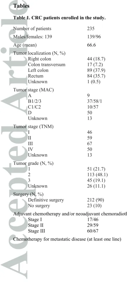
Table 1. CRC patients enrolled in the study.
Number of patients 235
Males/females: 139 139/96
Age (mean) 66.6
Tumor localization (N, %)
Right colon 44 (18.7)
Colon transversum 17 (7.2)
Left colon 89 (37.9)
Rectum 84 (35.7)
Unknown 1 (0.5)
Tumor stage (MAC)
A 9
B1/2/3 37/58/1
C1/C2 10/57
D 50
Unknown 13
Tumor stage (TNM)
I 46
II 59
III 67
IV 50
Unknown 13
Tumor grade (N, %)
1 51 (21.7)
2 113 (48.1)
3 45 (19.1)
Unknown 26 (11.1)
Surgery (N, %)
Definitive surgery 212 (90)
No surgery 23 (10)
Adjuvant chemotherapy and/or neoadjuvant chemoradiotherapy
Stage I 17/46
Stage II 29/59
Stage III 60/67
Chemotherapy for metastatic disease (at least one line) 44/50
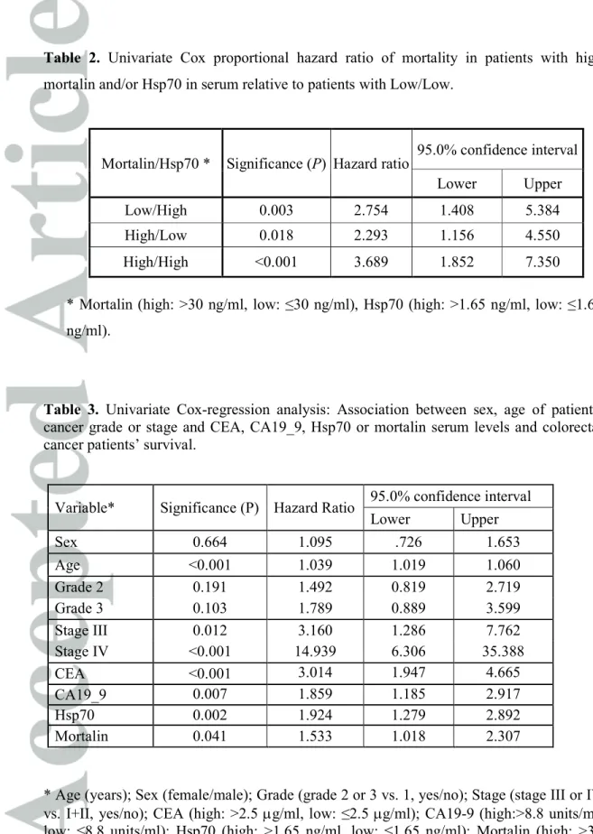
Table 2. Univariate Cox proportional hazard ratio of mortality in patients with high mortalin and/or Hsp70 in serum relative to patients with Low/Low.
Mortalin/Hsp70 * Significance (P) Hazard ratio 95.0% confidence interval
Lower Upper
Low/High 0.003 2.754 1.408 5.384
High/Low 0.018 2.293 1.156 4.550
High/High <0.001 3.689 1.852 7.350
* Mortalin (high: >30 ng/ml, low: ≤30 ng/ml), Hsp70 (high: >1.65 ng/ml, low: ≤1.65 ng/ml).
Table 3. Univariate Cox-regression analysis: Association between sex, age of patients, cancer grade or stage and CEA, CA19_9, Hsp70 or mortalin serum levels and colorectal cancer patients’ survival.
Variable* Significance (P) Hazard Ratio 95.0% confidence interval
Lower Upper
Sex 0.664 1.095 .726 1.653
Age <0.001 1.039 1.019 1.060
Grade 2 0.191 1.492 0.819 2.719
Grade 3 0.103 1.789 0.889 3.599
Stage III 0.012 3.160 1.286 7.762
Stage IV <0.001 14.939 6.306 35.388
CEA <0.001 3.014 1.947 4.665
CA19_9 0.007 1.859 1.185 2.917
Hsp70 0.002 1.924 1.279 2.892
Mortalin 0.041 1.533 1.018 2.307
* Age (years); Sex (female/male); Grade (grade 2 or 3 vs. 1, yes/no); Stage (stage III or IV vs. I+II, yes/no); CEA (high: >2.5 µg/ml, low: ≤2.5 µg/ml); CA19-9 (high:>8.8 units/ml, low: ≤8.8 units/ml); Hsp70 (high: >1.65 ng/ml, low: ≤1.65 ng/ml); Mortalin (high: >30 ng/ml, low: ≤30 ng/ml).
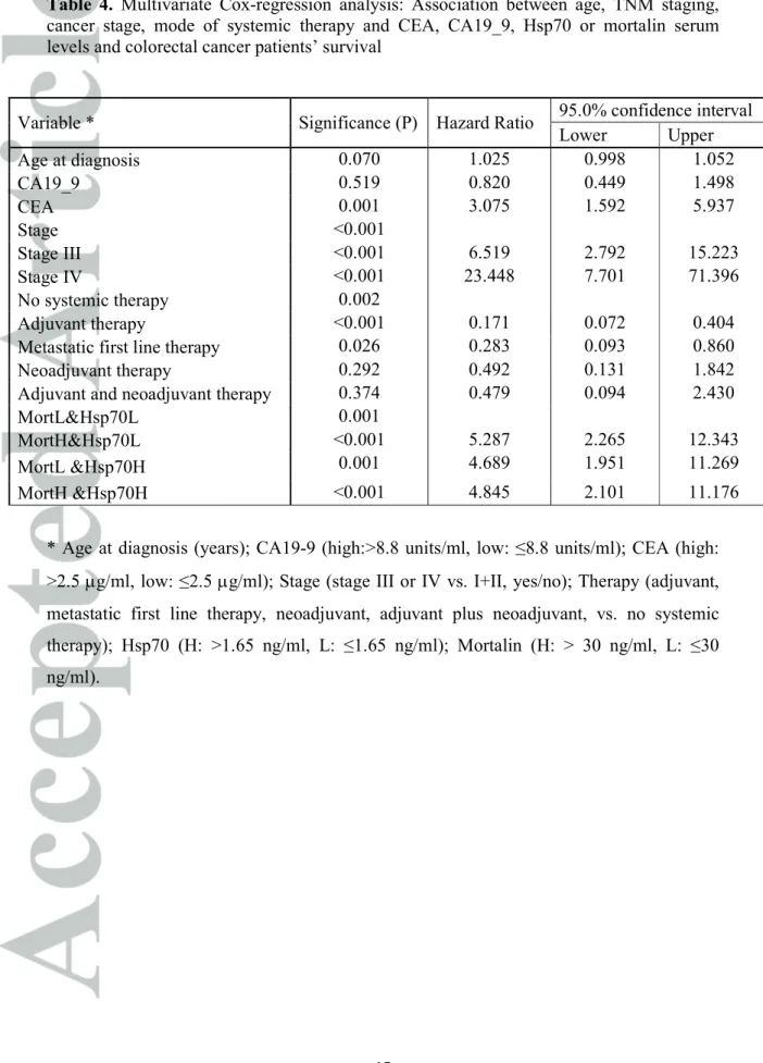
Table 4. Multivariate Cox-regression analysis: Association between age, TNM staging, cancer stage, mode of systemic therapy and CEA, CA19_9, Hsp70 or mortalin serum levels and colorectal cancer patients’ survival
* Age at diagnosis (years); CA19-9 (high:>8.8 units/ml, low: ≤8.8 units/ml); CEA (high:
>2.5 µg/ml, low: ≤2.5 µg/ml); Stage (stage III or IV vs. I+II, yes/no); Therapy (adjuvant, metastatic first line therapy, neoadjuvant, adjuvant plus neoadjuvant, vs. no systemic therapy); Hsp70 (H: >1.65 ng/ml, L: ≤1.65 ng/ml); Mortalin (H: > 30 ng/ml, L: ≤30 ng/ml).
Variable * Significance (P) Hazard Ratio 95.0% confidence interval
Lower Upper
Age at diagnosis 0.070 1.025 0.998 1.052
CA19_9 0.519 0.820 0.449 1.498
CEA 0.001 3.075 1.592 5.937
Stage <0.001
Stage III <0.001 6.519 2.792 15.223
Stage IV <0.001 23.448 7.701 71.396
No systemic therapy 0.002
Adjuvant therapy <0.001 0.171 0.072 0.404
Metastatic first line therapy 0.026 0.283 0.093 0.860
Neoadjuvant therapy 0.292 0.492 0.131 1.842
Adjuvant and neoadjuvant therapy 0.374 0.479 0.094 2.430
MortL&Hsp70L 0.001
MortH&Hsp70L <0.001 5.287 2.265 12.343
MortL &Hsp70H 0.001 4.689 1.951 11.269
MortH &Hsp70H <0.001 4.845 2.101 11.176
Legends to Figures
Figure 1. Survival (Kaplan-Meier) of colorectal cancer patients. (A) Patients divided according to stages I-IV of disease. (B) Patients divided according to mortalin level in their serum; high (≥30 ng/ml) or low (<30 ng/ml). Log Rank overall comparison showed significant equality of survival distribution (P=0.042). (C) Patients divided according to their mortalin (low: ≤30 ng/ml, high: >30 ng/ml) and Hsp70 (low: <≤1.65 ng/ml, high:
>1.65 ng/ml) levels. Patients sub-divided into Low/Low (blue), High/ Low (green), Low/High (beige) and High/High (purple) levels of mortalin/Hsp70 in their sera. Number of patients (survivors/all) and significance of difference between survival curves of High and Low (B, C) or High/High and Low/Low (D) patients are shown.
Figure 2. Survival (Kaplan-Meier) of stage I (A), stage II (B), stage III (C) and stage IV (D) colorectal cancer patients. Patients divided according to their mortalin (low: ≤30 ng/ml, high: >30 ng/ml) and Hsp70 (low: ≤1.65 ng/ml, high: >1.65 ng/ml) levels. Patients sub-divided into Low/Low (blue), High/ Low (green), Low/High (beige) and High/High (purple) levels of mortalin/Hsp70 levels in their sera. Number of patients (survivors/all) and significance of difference between survival curves of High/High and Low/Low patients are shown.
References
1. Richter K, Haslbeck M, Buchner J. The heat shock response: life on the verge of death. Mol Cell 2010; 40: 253-66.
2. Calderwood SK, Gong J. Heat Shock Proteins Promote Cancer: It's a Protection Racket. Trends Biochem Sci 2016; 41: 311-23.
3. Daugaard M, Rohde M, Jaattela M. The heat shock protein 70 family: Highly homologous proteins with overlapping and distinct functions. FEBS Lett 2007; 581: 3702- 10.
4. Lianos GD, Alexiou GA, Mangano A, Mangano A, Rausei S, Boni L, Dionigi G, Roukos DH. The role of heat shock proteins in cancer. Cancer Lett 2015; 360: 114-8.
5. Murphy ME. The HSP70 family and cancer. Carcinogenesis 2013; 34: 1181-8.
6. Radons J. The human HSP70 family of chaperones: where do we stand? Cell Stress Chaperones 2016; 21: 379-404.
7. Kang PJ, Ostermann J, Shilling J, Neupert W, Craig EA, Pfanner N. Requirement for hsp70 in the mitochondrial matrix for translocation and folding of precursor proteins.
Nature 1990; 348: 137-43.
8. Bhattacharyya T, Karnezis AN, Murphy SP, Hoang T, Freeman BC, Phillips B, Morimoto RI. Cloning and subcellular localization of human mitochondrial hsp70. J Biol Chem 1995; 270: 1705-10.
9. Wiedemann N, Frazier AE, Pfanner N. The protein import machinery of mitochondria. The Journal of biological chemistry 2004; 279: 14473-6.
10. Mayer MP, Bukau B. Hsp70 chaperones: cellular functions and molecular mechanism. Cell Mol Life Sci 2005; 62: 670-84.
11. Jego G, Hazoume A, Seigneuric R, Garrido C. Targeting heat shock proteins in cancer. Cancer Lett 2013; 332: 275-85.
12. Liu Y, Liu W, Song XD, Zuo J. Effect of GRP75/mthsp70/PBP74/mortalin overexpression on intracellular ATP level, mitochondrial membrane potential and ROS accumulation following glucose deprivation in PC12 cells. Mol Cell Biochem 2005; 268:
45-51.
13. Taurin S, Seyrantepe V, Orlov SN, Tremblay TL, Thibault P, Bennett MR, Hamet P, Pshezhetsky AV. Proteome analysis and functional expression identify mortalin as an antiapoptotic gene induced by elevation of [Na+]i/[K+]i ratio in cultured vascular smooth muscle cells. Circ Res 2002; 91: 915-22.
14. Lu WJ, Lee NP, Kaul SC, Lan F, Poon RT, Wadhwa R, Luk JM. Mortalin-p53 interaction in cancer cells is stress dependent and constitutes a selective target for cancer therapy. Cell Death Differ 2011; 18: 1046-56.
15. Wadhwa R, Takano S, Kaur K, Deocaris CC, Pereira-Smith OM, Reddel RR, Kaul SC. Upregulation of mortalin/mthsp70/Grp75 contributes to human carcinogenesis. Int J Cancer 2006; 118: 2973-80.
16. Wadhwa R, Takano S, Taira K, Kaul SC. Reduction in mortalin level by its antisense expression causes senescence-like growth arrest in human immortalized cells. J Gene Med 2004; 6: 439-44.
17. Ran Q, Wadhwa R, Kawai R, Kaul SC, Sifers RN, Bick RJ, Smith JR, Pereira- Smith OM. Extramitochondrial localization of mortalin/mthsp70/PBP74/GRP75. Biochem Biophys Res Commun 2000; 275: 174-9.
18. Fishelson Z, Hochman I, Greene LE, Eisenberg E. Contribution of heat shock proteins to cell protection from complement-mediated lysis. Int Immunol 2001; 13: 983-91.
19. Pilzer D, Fishelson Z. Mortalin/GRP75 promotes release of membrane vesicles from immune attacked cells and protection from complement-mediated lysis. Int Immunol 2005; 17: 1239-48.
20. Pilzer D, Saar M, Koya K, Fishelson Z. Mortalin inhibitors sensitize K562 leukemia cells to complement-dependent cytotoxicity. Int J Cancer 2010; 126: 1428-35.
21. Mazkereth N, Rocca F, Schubert JR, Geisler C, Hillman Y, Egner A, Fishelson Z.
Complement triggers relocation of Mortalin/GRP75 from mitochondria to the plasma membrane. Immunobiology 2016; 221: 1395-406.
22. Pilzer D, Gasser O, Moskovich O, Schifferli JA, Fishelson Z. Emission of membrane vesicles: roles in complement resistance, immunity and cancer. Springer Semin Immunopathol 2005; 27: 375-87.
23. De Maio A. Extracellular heat shock proteins, cellular export vesicles, and the
damage. It is never known how far a controversial finding will go! Dedicated to Ferruccio Ritossa. Cell Stress Chaperones 2011; 16: 235-49.
24. Yoshidomi K, Murakami A, Yakabe K, Sueoka K, Nawata S, Sugino N. Heat shock protein 70 is involved in malignant behaviors and chemosensitivities to cisplatin in cervical squamous cell carcinoma cells. J Obstet Gynaecol Res 2014; 40: 1188-96.
25. Alexiou GA, Vartholomatos G, Stefanaki K, Patereli A, Dova L, Karamoutsios A, Lallas G, Sfakianos G, Moschovi M, Prodromou N. Expression of heat shock proteins in medulloblastoma. J Neurosurg Pediatr 2013; 12: 452-7.
26. Suzuki K, Ito Y, Wakai K, Kawado M, Hashimoto S, Seki N, Ando M, Nishino Y, Kondo T, Watanabe Y, Ozasa K, Inoue T, Tamakoshi A. Serum heat shock protein 70 levels and lung cancer risk: a case-control study nested in a large cohort study. Cancer Epidemiol Biomarkers Prev 2006; 15: 1733-7.
27. Abe M, Manola JB, Oh WK, Parslow DL, George DJ, Austin CL, Kantoff PW.
Plasma levels of heat shock protein 70 in patients with prostate cancer: a potential biomarker for prostate cancer. Clin Prostate Cancer 2004; 3: 49-53.
28. Balazs M, Zsolt H, Laszlo G, Gabriella G, Lilla T, Gyula O, Balazs D, Eva M, Zoltan B, Zoltan P, Judit K. Serum Heat Shock Protein 70, as a Potential Biomarker for Small Cell Lung Cancer. Pathol Oncol Res 2017; 23: 377-83.
29. Kocsis J, Madaras B, Toth EK, Fust G, Prohaszka Z. Serum level of soluble 70-kD heat shock protein is associated with high mortality in patients with colorectal cancer without distant metastasis. Cell Stress Chaperones 2010; 15: 143-51.
30. Takano S, Wadhwa R, Yoshii Y, Nose T, Kaul SC, Mitsui Y. Elevated levels of mortalin expression in human brain tumors. Exp Cell Res 1997; 237: 38-45.
31. Dundas SR, Lawrie LC, Rooney PH, Murray GI. Mortalin is over-expressed by colorectal adenocarcinomas and correlates with poor survival. J Pathol 2005; 205: 74-81.
32. Ando K, Oki E, Zhao Y, Ikawa-Yoshida A, Kitao H, Saeki H, Kimura Y, Ida S, Morita M, Kusumoto T, Maehara Y. Mortalin is a prognostic factor of gastric cancer with normal p53 function. Gastric Cancer 2014; 17: 255-62.
33. Rozenberg P, Kocsis J, Saar M, Prohaszka Z, Fust G, Fishelson Z. Elevated levels of mitochondrial mortalin and cytosolic HSP70 in blood as risk factors in patients with
34. Saar Ray M, Moskovich O, Iosefson O, Fishelson Z. Mortalin/GRP75 binds to complement C9 and plays a role in resistance to complement-dependent cytotoxicity. J Biol Chem 2014; 289: 15014-22.
35. Molvarec A, Rigo J, Jr., Nagy B, Walentin S, Szalay J, Fust G, Karadi I, Prohaszka Z. Serum heat shock protein 70 levels are decreased in normal human pregnancy. J Reprod Immunol 2007; 74: 163-9.
36. Jenei ZM, Szeplaki G, Merkely B, Karadi I, Zima E, Prohaszka Z. Persistently elevated extracellular HSP70 (HSPA1A) level as an independent prognostic marker in post-cardiac-arrest patients. Cell Stress Chaperones 2013; 18: 447-54.
37. Breuninger S, Erl J, Knape C, Gunther S, Regel I, Rödel F, Gaipl US, Thorsteinsdottir J, Giannitrapani L, Dickinso AM, Multhoff G. Quantitative Analysis of Liposomal Heat Shock Protein 70 (Hsp70) in the Blood of Tumor Patients Using a Novel Liphsp70 ELISA. J Clin Cell Immunol 2014; 5: 264.
38. Gunther S, Ostheimer C, Stangl S, Specht HM, Mozes P, Jesinghaus M, Vordermark D, Combs SE, Peltz F, Jung MP, Multhoff G. Correlation of Hsp70 Serum Levels with Gross Tumor Volume and Composition of Lymphocyte Subpopulations in Patients with Squamous Cell and Adeno Non-Small Cell Lung Cancer. Front Immunol 2015; 6: 556.
39. Duffy MJ. Personalized treatment for patients with colorectal cancer: role of biomarkers. Biomark Med 2015; 9: 337-47.
40. Hundt S, Haug U, Brenner H. Blood markers for early detection of colorectal cancer: a systematic review. Cancer Epidemiol Biomarkers Prev 2007; 16: 1935-53.
41. Aghagolzadeh P, Radpour R. New trends in molecular and cellular biomarker discovery for colorectal cancer. World J Gastroenterol 2016; 22: 5678-93.
42. Das V, Kalita J, Pal M. Predictive and prognostic biomarkers in colorectal cancer:
A systematic review of recent advances and challenges. Biomed Pharmacother 2017; 87:
8-19.
Figure 1
178x135mm (300 x 300 DPI)
Figure 2
172x113mm (300 x 300 DPI)