I
Central European Journal of Gastroenterology and Hepatology 172
Volume 7, Issue 4 / December 2021
Eredeti közlemény / Original paper
171 Central European Journal of Gastroenterology and Hepatology Volume 7, Issue 4 / December 2021
Eredeti közlemény / Original paper
IBD-related Malignancies Observed in 2015–2019
4 Years’ Results from the Prospective, Nationwide Hungarian Registry IBD-related Malignancies – Hungarian Registry
Ágnes Milassin MD1, Mariann Rutka MD1, Mónika Szűcs MD2, Klaudia Farkas MD1, Mark Marcus MD1, Renáta Bor MD1, Kata Judit Szántó MD1, Ferenc Nagy MD1, Zoltán Szepes MD1, László Lakatos MD3, Zsuzsanna Erdélyi MD3, László Sze
gedi MD4, Eszter Schäfer MD5, Attila Zaránd MD6, Anna Fábián MD1, Anita Bálint MD1, Tamás Molnár MD1
1Department of Medicine, University of Szeged, Albert Szent-Györgyi Faculty of Medicine and Health Center, Hungary;
2Department of Medical Physics and Informatics, University of Szeged, Albert Szent-Györgyi Medical School, Faculty of Science and Informatics, Hungary; 3Department of Internal Medicine, Csolnoky Ferenc Regional Hospital, Veszprém, Hungary; 4First Department of Internal Medicine, András Jósa Teaching Hospital, Nyíregyháza, Hungary; 5Military Hospital Medical Center, Hungarian Defense Forces, Budapest, Hungary; 6First Department of Surgery, Semmelweis University, Budapest, Hungary
Correspondence: milagn422@hotmail.com
Introduction: Patients with inflammatory bowel disease (IBD) have an increased risk to develop malignant neoplasms. Neither the exact mechanism, nor the frequency of different malignancies is completely clear.
Our aim was to assess the IBD associated malignancies, to collect clinical and mortality data and to ana- lyse possible risk factors.
Methods: Data on malignancies developed between January 2015 and May 2019 in Hungarian IBD pa- tients was recorded. Every member of the Hungarian Society of Gastroenterology was prospectively in- terviewed. The following data were collected: demographic data, disease characteristics, previous thera- py, patient adherence, type and localisation of malignancies.
Results: 140 IBD patients with newly diagnosed malignancies were reported. 61.4%, 35.7%, and 2.9% of the patients had ulcerative colitis (UC), Crohn’s disease (CD) and indeterminate colitis, respectively. The mean la- tency was 15.2±10.5 years. Colorectal cancer (CRC) was the most common cancer (49.6%, 70). 72.9% (51/70) of them was associated with UC, more than 80% had extensive (50.9%, 26) and left-sided (31.2%, 16) colitis.
The most frequent CRC localisation was the rectosigmoid colon in UC (54.9%), and the rectum in CD (38.9%).
The most common non-CRC malignancies were non-melanotic skin-cancer, haematological and pulmonary cancer. Disease duration at the time of the diagnosis of malignancy was lower (17.9±10.7 versus 12.6±9.7 years); mean age at the time of the death was higher (49.3±9.4 versus 64.3±16.4 years); and survival was longer after the diagnosis of extraintestinal malignancy than CRC (0.73±1.01 versus 1.2±0.8).
Summary: CRC presented typically in the distal part of the colon by male UC patients with pancolitis or left-sid- ed colitis with a long-standing disease course of IBD. The most common non-CRC malignancies were non-mel- anotic skin cancer, haematological cancer and lung cancer. Non-CRC malignancies developed typically in fe- male patients, older than CRC patients with shorter disease-course of IBD and longer survival times.
KEYWORDS: inflammatory bowel disease, colorectal cancer, malignancy, prospective
IBD-vel összefüggésben megfigyelt daganatos megbetegedések 2015–2019 között – Az IBD-vel kapcsolatos rosszindulatú
daganatos megbetegedések prospektív, országos regiszterének 4 éves eredményei – Magyar Regiszter
Bevezetés: A gyulladásos bélbetegségben (IBD) szenvedő betegeknél fokozott a rosszindulatú daganatok kialakulásának kockázata. Sem a pontos mechanizmus, sem a különböző malignitások gyakorisága nem teljesen tisztázott. Célunk az IBD-vel összefüggő rosszindulatú daganatok felmérése, klinikai és halálozá- si adatok gyűjtése, valamint a lehetséges kockázati tényezők elemzése volt.
Módszerek: A 2015 januárja és 2019 májusa között, magyar IBD-s betegeknél kialakult rosszindulatú da- ganatok adatait rögzítettük. A Magyar Gasztroenterológiai Társaság minden tagját prospektív módon kérdeztük. A következő adatokat gyűjtöttük: demográfiai adatok, betegségjellemzők, korábbi terápia, betegadherencia, a malignitások típusa és lokalizációja.
Eredmények: 140 IBD-s betegről számoltak be, akiknél újonnan diagnosztizált rosszindulatú daganatot állapítottak meg. A betegek 61,4%-a colitis ulcerosában (UC), 35,7%-a Crohn-betegségben (CD), 2,9%-a pedig indeterminált colitisben szenvedett. Az átlagos latenciaidő 15,2±10,5 év volt. A vastagbélrák (CRC) volt a leggyakoribb daganatos megbetegedés (49,6%, 70). Ezek 72,9%-a (51/70) UC-vel társult, több mint 80%-uknak kiterjedt (50,9%, 26) és bal oldali (31,2%, 16) colitise volt. A leggyakoribb CRC-lokalizáció UC- ben a rectosigmoid colon (54,9%), CD-ben a végbél (38,9%) volt.
A leggyakoribb nem-CRC rosszindulatú daganatok a nem melanotikus bőrrák, hematológiai daganatok és a tüdőrák voltak. Az extraintesztinális daganatok megjelenésénél a betegség átlagos fennállása rövidebb volt (17,9±10,7 versus 12,6±9,7 év), az átlagos életkor a halál idejében magasabb volt (49,3±9,4 versus 64,3±16,4 év), és a túlélés hosszabb volt (0,73±1,01 versus 1,2±0,8 év) mint colorectalis rák esetében.
Összefoglalva: A CRC jellemzően a vastagbél disztális részében jelent meg colitis ulcerosás férfi betegeknél, pancolitis vagy bal oldali colitis ulcerosa esetén, hosszabb betegségfennállás után. A leggyakoribb nem CRC rosszindulatú daganatok a nem melanotikus bőrrák, hematológiai daganatok és a tüdőrák voltak. A nem CRC daganatok gyakoribbak voltak a nőbetegeknél, idősebb életkorban jelentkeztek, mint a CRC, rövidebb betegségfennállás után, és hosszabb túlélés jellemezte.
KULCSSZAVAK: gyulladásos bélbetegség, vastagbélrák, daganatos megbetegedés, prospektív
Introduction
Ulcerative colitis (UC) and Crohn’s disease (CD) are life
long diseases with a chronic inflammation of the gast
rointestinal (GI) tract. Patients with inflammatory bowel disease (IBD) have an increased risk to develop malig
nant neoplasms, particularly colorectal cancer (CRC) in ulcerative colitis and colon Crohn’s disease because of the chronic intestinal inflammation, however recent stu
dies suggest the rate is decreasing thanks to improved surveillance (1–7). Colorectal cancer is the third most common cancer worldwide and accounts for 1015% of all deaths in IBD (8). IBD specific risk factors for CRC are ulcerative colitis and colon Crohn’s disease, longer dura
tion of IBD (8 years or more after diagnosis), extensive colon inflammation and coexisting primary sclerosing cholangitis (PSC), while male gender, increasing age and positive family history of CRC are risk factors to spora
dic CRCs (9–11). Recently published data suggest that the risk of extraintestinal malignancies is also increased in IBD patients (12, 13). Disease, patients and therapy
related causes are taken into consideration; however, neither the exact mechanism, nor the frequencies of different malignancies are completely clear. CD patients are more likely to develop extraintestinal malignancies than UC patients. Upper GI cancer, lung cancer, urinary bladder cancer, skin squamous cell cancer, breast cancer were more frequent among CD patients and liverbiliary cancer, leukaemia, breast cancer and corpus uteri cancer among UC patients (5, 13, 14).
Due to the chronic nature of IBD, the impact of frequently used therapy in cancer development is still the subject of in
tensive debate. Thiopurine use can increase the risk of cancer, especially for lymphoma, haematologic cancer, skin squa
mous cell cancer (15–17). The currently available metaanaly
ses suggest that the overall risk of cancer is not increased in patients treated with antiTNFalpha therapy alone (18, 19).
Our aim in this prospective, nationwide study was to as
sess the IBD associated malignancies, to collect clinical and mortality data and to analyse possible risk factors.
Methods
Data on malignancies developed between January 2015 and May 2019 in Hungarian IBD patients was recorded.
Every member of the Hungarian Society of Gastroente
rology was prospectively interviewed every 6 months by personal email to report malignancies observed in their patient population. The following data was collected in Excelspreadsheets: IBDrelated data: demographic data including gender, age at the time of the diagnosis of IBD, disease characteristics, such as type and nature of IBD, pre
vious immunosuppressive or biological therapy, pa tient adherence; malignancy related data: age at the time of the diagnosis of malignancy, localisation, TNM stage of the tu
mour and in case of colorectal cancer the year of the pre
vious colonoscopy. In the end of May 2019 all physi cians were interviewed about the previously reported patients’
status. Patients were eligible for the study if they had in
flammatory bowel disease before or at the time of the di
DOI: 10.33570/CEUJGH.7.4.171
174
173 Central European Journal of Gastroenterology and Hepatology
Volume 7, Issue 4 / December 2021 Central European Journal of Gastroenterology and Hepatology
Volume 7, Issue 4 / December 2021
Eredeti közlemény / Original paper Eredeti közlemény / Original paper
agnosis of malignant cancer. All type of malignant cancers was taken into account. The stage of colorectal cancer was based on the 8th Edition TNM Staging system.
A descriptive statistical analysis was performed. Statistical data was reported as the mean ± SD (standard deviaton);
with frequencies (n) and percentages (%), when approp
riate, for the survival time mean ± SE (standard error) were calculated. Pearson’s chisquared test or Fisher’s exact test was used to analyse categorical data, whereas indepen
dent samples ttest was used in case of continuous data.
The survival probabilities were estimated by KaplanMei
er analysis. The differences in the survival of the CRC and nonCRC groups were examined by a logrank test. Statis
tical tests were performed using R statistical software (R version 3.6.2, https://www.rproject.org/), values of P<0.05 were considered significant.
The study was approved by the Ethical Committee (3929, 15/2017SZTE). The data are collected and analysed anonymously.
Results
Demographic data
140 IBD patients were reported by the members of the HSG. Patients were reported from 20 departments, al
together from 11 cities (Békéscsaba, Budapest, Debrecen, Gyula, Nyíregyháza, Pápa, Pécs, Szeged, Szekszárd, Szom
bathely, Veszprém). The majority of patients were male (female:male ratio was 35:65%; 49:91 patients). 61.4%, 35.7%, and 2.9% of the patients had UC, CD and indetermi
nate colitis (IC), respectively. The mean age of the patients at the time of initial diagnosis of malignancy was 53.0±14.7 (range 1492) years. At the time of initial diagnosis of IBD, the mean age was 38.0±18.0 (range 886) years. The mean latency between the diagnosis of IBD and malignancy was 15.2±10.5 (range 052) years. Altogether 141 malignancies were reported – one patient had simultaneously two dif
ferent types of tumour (descending colon and kidney), we counted it as two cases in the subgroup analyses.
Occurrence of malignancies
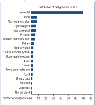
According to our results CRC was the most common cancer type (49.6%, 70), while the most common nonCRC malig
nancies were nonmelanotic skincancer (5.7%, 8), haema
tological and pulmonary cancer (5.75.7%, 88) (Fig. 1.). The Table 1. Demographic data of patients suffering from colorectal cancer (CRC) and non-colorectal cancer (non-CRC)
CRC patients
(N=70) non-CRC
patients (N=71) P value
Type of disease (N, %) 0.015*
• Ulcerative colitis 51 (72.9%) 35 (49.3 %)
• Crohn’s disease 18 (25.7%) 33 (46.5%)
• Indeterminate colitis 1 (1.4%) 3 (4.2%)
Gender (N, %) 0.090
• Male 50 (71.4%) 30 (42.3%)
• Female 20 (28.6%) 41 (57.7%)
Age at the time of diagnosis of IBD (mean ± SD, years) 33.9±16.7 42.2±18.4 0.010*
Age at the time of diagnosis of malignancy
(mean ± SD, years) 51.4±13.7 54.8±5.8 0.274
Disease duration at the time of diagnosis of
malignancy (mean ± SD, years) 17.9±10.7 12.6±9.7 0.001*
Age at the time of death (mean ± SD, years) 49.3±9.4+ 64.25±16.4++ 0.011*
Survival time after the diagnosis of malignancy
(mean ± SE, years) 0.73±1.01+ 1.19±0.83++ 0.208
*P<0.05 was considered significant, +N=11 patients, ++N=16 patients Colorectal
Non-melanotic skin Lung
0 80
Distribution of malignancies in IBD
10 20 30 40 50 60 70
Gynecological Prostate Haematological Pancreas and biliary tract Oropharyngeal Kidney Central nervous system Liver Upper gastriintestinal Breast Tonsil Melanoma malignum Urinary tract Appendix Sarcoma Thyroid gland Number of malignancies
Fig. 1. Distribution of malignancies in inflammatory bowel disease
proportion of UC patients is significantly higher in the CRC subgroup contrary to the nonCRC subgroup (P=0.015).
CRC patients were significantly younger at the time of diag
nosis of IBD (P=0.01), had a longer disease duration before the diagnosis of malignancy (P=0.001) and died younger (P=0.011) contrary to nonCRC patients. There was no statis
tical difference in gender and the age at the time of diagno
sis of malignancy. Table 1. shows the demographic data of the CRC and nonCRC patients in detail.
Colorectal cancer
As CRC can be a result of longterm IBD, it was analysed in detail. 70 patients were diagnosed with CRC. 72.9% (51 of 70) of CRC cases was associated with ulcerative colitis, more than 80% of them had extensive (50.9%, 26) and leftsided (31.2%, 16) colitis. Among CD patients ileocolo
nic and colonic localisation was observed in 44.4% (8/18) and 33.3% (6/18). In UC patients a male dominance was seen (80.4%). The mean age at the time of diagnosis of malignancy was 51.4±13.7 years. Mean disease duration of IBD was 17.9±10.7 years, but it was less than 8 years in 10 patients (70% of them had early stage CRC). In 5 cas
es CRC diagnosis was simultaneous with the diagnosis of IBD. Demographic data is detailed in Table 2. 27 patients (38.6%) had previous colonoscopies before the diagnosis of CRC, a mean 3.85±3.2 years. 21.4% (15 patients) of the patients were nonadherent to regular checkups. Family history was positive to CRC in 15.7% (11 patients).
The most common CRC localisation in UC patients was the rectosigmoid colon (52.9%, 27), and the rectum in CD pa
tients (38.9%, 7) (Figure 2.). There was no statistical difference between the tumour localisation and the nature of the dis
ease. Three patients had multiplex tumours: one ulcerative colitis (E2) patient had simultaneous adenocarcinoma in co
ecum and rectum; the second ulcerative colitis patient (E3) had three adenocarcinomas in coecum, descending colon and rectum; the third ulcerative colitis patient (E3), had four adenocarcinomas in colon and one in rectum. Tumour locali
Table 2. Demographic data of inflammatory bowel disease patients suffering from colorectal cancer. *(descending, transverse, ascending colon and coecum)
Ulcerative colitis
(N=51, 72.9%) Crohn’s disease
(N=18, 25.7%) Indeterminate colitis (N=1, 1.4%) Localisation of the disease, N (%) E1: 4 (7.8%) L1: 4 (22.2%)
E2: 16 (31.4%) L2: 6 (33.3%) E3: 31 (60.8%) L3: 8 (44.5%)
Behaviour of the disease, N (%) B1: 10 (55.5%)
B2: 5 (27.8%) B3: 3 (16.7%) Gender, N (%)
• Female 10 (19.6%) 10 (66.6%) 0 (0.0%)
• Male 41 (80.4%) 8 (44.4%) 1 (100.0%)
CRC localisation, N (%) (P=0.670) 27 (52.9%) 9 (50.0%) 1 (100.0%)
• Rectosigmoid colon E1: 4 L1: 1
E2: 10 L2: 3
E3: 13 L3: 5
• Colonic * 21 (41.2%) 9 (50.0%) 0 (0.0%)
E1: 0 L1: 4
E2: 7 L2: 3
E3: 14 L3: 2
• Multiplex 3 (5.9%) 0 (0.0%) 0 (0.0%)
E1: 0 E2: 1 E3: 2
Crohn’s disease Ulcerative colitis Indeterminate colitis 25
Rectum
Localisation of colorectal cancer
20 15 10 5 0 n
Sigmoid colo n
Descending colonTransverse colo n Ascending colo
n
Coecum Multiplex Other
CD = Crohn’s disease; UC = ulcerative colitis;
IC = indeterminate colitis; N = number of patients
Fig. 2. Localisations of colorectal cancer in inflammatory bowel disease
176
175 Central European Journal of Gastroenterology and Hepatology
Volume 7, Issue 4 / December 2021 Central European Journal of Gastroenterology and Hepatology
Volume 7, Issue 4 / December 2021
Eredeti közlemény / Original paper Eredeti közlemény / Original paper
sation of UC and CD patients is detailed in Fig. 2. Two patients had IBDrelated operations previously where the malignancy developed: one UC patient had pouch adenocarcinoma, the other patient with Crohn’s disease had adenocarcinoma in the ileotransverse anastomosis.
The nature of the disease was also evaluated. Chronic continuous symptoms, chronic remission, relapse and remission, relapse after chronic remission was observed in 37.1% (26), 24.3% (17), 20.0% (14) and 2.9% (2) of the patients, respectively (15.7%, 11 patients had no data ava
ilable). Half of the patients with CD had nonstricturing, nonpenetrating disease, 27.8% had stenotisating and 16.7% had penetrating disease behaviour (5.5% had no data about the behaviour of the disease).
Only 28.7% (20 patients) of the malignancies were detec
ted in early stage (Stage 0 or Stage 1). One (5%) of them died in pneumonia and sepsis in the year of the tumour diagnosis. Stage 2, Stage 3 and Stage 4 tumour was detec
ted in 50 patients (71.3%), 10 (18%) of them died after a mean 0.55 year (Fig. 3.). There was no statistical difference in the previously used therapy (immunosuppressive, bio
logical therapy or both) and the stage of the CRC.
At the time of the diagnosis of malignancy 71.4% (50) of the patients received aminosalicylates, 37.1% (26), 28.6% (20) received steroid and/or immunosuppressive therapy, and
only 11.4% (8) received biological therapy. Previously 42.9%
(30) of the patients received immunosuppressive therapy a mean 63.3±58.1 months, 60% of them received azathiopri
ne (AZA) alone, and 40% received a combination therapy.
Non-colorectal cancers
The most frequently observed nonCRC malignancies in our cohort were nonmelanotic skincancer (5.7%, 8), hae
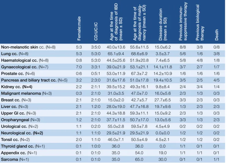
matological (5.7%, 8) and pulmonary cancer (5.7%, 8), then Table 3. Summary of non-colorectal cancers
Female:male CD:UC:IC Age at the time of diagnosis of IBD (mean ± SD) Age at the time of diagnosis of malig- nancy (mean ± SD) Disease duration (mean ± SD) Previous immuno- suppressive therapy Previous biological therapy Death
Non-melanotic skin cc. (N=8) 5:3 3:5:0 40.0±13.6 55.6±11.5 15.0±6.2 8/8 3/8 0/8
Lung cc. (N=8) 5:3 5:3:0 65.1±9.4 68.6±6.9 3.5±3.7 5/6 1/6 3/8
Haematological cc. (N=8) 0:8 5:3:0 44.5±25.6 51.9±20.8 7.4±6.5 5/8 4/8 1/8 Gynaecological cc. (N=7) 7:0 3:3:1 39.0±21.9 53.1±21.1 14.1±11.8 3/7 2/7 1/7
Prostate cc. (N=6) 0:6 0:5:1 53.0±11.9 67.3±7.2 14.2±10.9 1/6 1/6 1/6
Pancreas and biliary tract cc. (N=5) 3:2 2:3:0 31.6±17.6 51.0±17.8 19.4±10.5 3/5 2/5 4/5
Kidney cc. (N=4) 2:2 2:1:1 39.5±15.2 49.3±16.1 9.8±8.4 2/4 3/4 1/4
Malignant melanoma (N=3) 0:3 2:1:0 31.0±3.5 47.0±7.0 16.0±5.6 2/3 1/3 0/3
Breast cc. (N=3) 2:1 2:1:0 15.0±2.0 42.7±5.7 27.7±6.5 3/3 2/3 0/3
Liver cc. (N=3) 2:1 1:2:0 28.0±19.0 47.7±16.8 19.7±9.6 1/3 2/3 2/3
Upper GI cc. (N=3) 2:1 2:1:0 44.3±18.8 59.3±11.1 15.0±9.2 2/3 1/3 0/3
Oropharyngeal (N=3) 1:2 2:1:0 37.7±11.5 50.7±17.0 13.0±5.6 3/3 1/3 2/3
Urological cc. (N=2) 1:1 0:2:0 55.0±2.8 59.5±7.8 4.5±4.9 0/2 0/2 0/2
Neurological cc. (N=2) 1:1 1:1:0 29.5±21.9 29.5±21.9 0.0±0.0 1/2 1/2 0/2
Tonsil cc. (N=2) 2:0 1:1:0 46.0±7.1 50.5±4.9 4.5±2.1 1/2 1/2 0/2
Thyroid gland cc. (N=1) 0:1 1:0:0 36.0 36.0 0.0 1/1 0/1 0/1
Appendix cc. (N=1) 0:1 0:1:0 35.0 54.0 19.0 1/1 1/1 0/1
Sarcoma (N=1) 0:1 0:1:0 35.0 65.0 30.0 0/1 0/1 1/1
Cc. = cancer; GI = gastrointestinal; th = therapy
Stage 0 (in situ) 30.0
Stages of colorectal cancer
25.0
15.0 10.0 5.0 0 20.0
2.9%
25.7%
22.9%
27.1%
17.1%
4.3%
Stage 1 Stage 2 Stage 3 Stage 4 No data
Fig. 3. Stages of colorectal cancer at the time
of the diagnosis of malignancy gynaecological and prostate malignancies in 5% and 4.3% (7 and 6 patients). Noncolorectal cancers are detailed in Table 3.
Nonmelanotic skin cancers (NMSC) were the following: 5 cases had basal cell carcinoma and 3 cases had squamous cell carcinoma. All of the patients with NMSC had previ
ous immunosuppressive therapy for a mean 79.8±50.8 (12144) months. IBDrelated therapy of patients with skin cancer is detailed in Table 4.
8 patients had haematological malignancies, all of them were male. Three of them had ulcerative colitis with the following leukaemia: chronic myeloid leukaemia, acute myeloid leukae mia and chronic lymphoid leukaemia, and five of them had Crohn’s disease with the following dis eases: diffuse large Bcell lymphoma (2 cases), non
Hodgkin lymphoma, Waldenström macroglobulinemia and chronic myeloid leukaemia.
8 patients had lung cancer, 5 patients had Crohn’s dis ease, 3 had ulcerative colitis. 7 patients had gynaecological ma
lignancy. 6 patients had a prostate tumour, only one of them had longterm combination immunosuppressive treatment. Mean age at the time of diagnosis of prostate malignancy was 67.3 years (min: 54, max: 74); the 54 years old patient was that who had long term combination im
munosuppressive treatment, all the other patients were over 65 years old.
IBDrelated medical therapy was the following in the non
CRC subgroup: 76.1% (54) of the patients were on amino
salicylates, 15.5% (11) on steroids, 38.0% (27) on immuno
suppressive therapy and 18.3% (13) on biological therapy at the time of the diagnosis of malignancy. Previously, 56.3%
(40) received azathioprine (68.5±56.9 years): 23.9% (17) re
ceived azathioprine alone, 32.4% (23) received combination therapy and 5.6% (4) received biological therapy alone.
Comorbidities
Primary sclerosing cholangitis (PSC) and primary biliary cholangitis (PBC) was analysed in detail. 10 cases were as
sociated with PSC, from them 6 had CRC, 2 had liver ma
lignancy and 2 patients had biliary adenocarcinoma. 2 pa tients with colorectal cancer had PBC. Mean age at the time of diagnosis of CRC in patients with PSC or PBC was lower contrary to patients without comorbidities (45.5±7.4 versus 52.2±14.2 years).
Survival
19.3% (27/140) patients died after the diagnosis of ma
lignancy after a mean 1.0±0.92 years, 86% of the patients were adherent to regular checkups. 15.7% (11 of 70) of CRC patients and 22.5% (16 of 71) of nonCRC patients died during the followup period. Male dominancy was observed (19:8). CRC patients died significantly younger than nonCRC patients (49.3±9.4 versus 64.3±16.4 years).
There was no statistical difference between the survival of CRC (8.44±0.434 years) and nonCRC (7.91±0.459 years) patients (P=0.458) (Fig. 4.) and the survival of early or late stage colorectal cancers (P=0.758).
Discussion
In our nationwide prospective study 140 IBD patients were reported with malignancy over a time course of 4 years (2015–2019).
Based on our findings the most common malignan cies were colorectal cancers (49.6%), nonmelanotic skin cancers (5.8%), haematological neoplasms (5.8%) and malignant neoplasms of the lung (5.8%). These data are in concordance with the recently published Hungarian
Table 4. IBD-related therapy of patients with skin cancer
Squamous cell
carcinoma (N=3) Basal cell
carcinoma (N=5) Malignant melanoma (N=2) Therapy at the time of diagnosis of malignancy
• Aminosalicylate 3 5 2
• Steroid 0 0 0
• Immunosuppressive 3 3 2
• Biological therapy 1 0 1
Previously received therapy
• Immunosuppressive 3 5 2
• Biological therapy 3 1 1
0 1.0
Time (years) 0.9
0.7
0.6 0.8
2 4 6 8 10
Survival probability
CRC non CRC
Fig. 4. Survival after the diagnosis of colorectal cancer and non-colorectal cancer
177 Central European Journal of Gastroenterology and Hepatology Volume 7, Issue 4 / December 2021
Eredeti közlemény / Original paper
populationbased data, where 8.5% of the incident UC population was diagnosed with CRC (20). Similar data were presented by a Danish populationbased study from 1962 to 1987, where increased risk of colorectal cancer and melanoma was found in UC patients, but for other cancers they couldn’t verify this (21). A Scandina
vian population basedstudy also found an in creased risk of developing CRC both in UC and CD patients (HR [hazard ratio]: 1.66 and 1.40) (2, 3). Other studies sugg
est that the risk of CRC is declining over time (1, 22, 23). A recently published Swiss IBD cohort study found controversial results concerning CRC: they found an increased risk for lymphoma and biliary cancer, but not for colorectal cancer (22).
Colorectal cancers were more frequent in UC patients and developed after a significantly longer disease duration contrary to nonCRC patients (17.9 versus 12.6 years). In UC patients with colorectal cancer a male dominance was seen. NonCRC patients were older at the time of death (64.3 versus 49.3 years); and survival time was longer (1.2 versus 0.73 years) after the diagnosis of nonCRC malig
nancy than CRC. These findings are in concordance with the previously published findings, which showed that ulcerative colitis increases the risk of CRC and male gen
der, younger age at diagnosis of UC (disease duration), extensive colitis and geography were independent risk factors (9, 24).
Based on our results the most common CRC localisation in UC patients was the rectosigmoid part of the colon (54.9%), and the rectum in CD patients (38.9%). In line with our data, increased risk for CRC and hepatobiliary cancers among IBD patients was found, and rectal can
cer was more frequent among UC patients (25), but not among CD patients (6). 85.7% of the patients had long
standing disease (17.9 years) in our cohort, it should be noted that it was less than 8 years in 10 patients (70% of them had early stage CRC), which can be a result of diag
nostic delay. Primary sclerosing cholangitis can increase the risk of CRC in IBD patients, especially in ulcerative colitis patients (26). In our cohort IBD patients with PSC were younger at the time of CRC onset, contrary to pa
tients with IBD alone.
Colorectal cancer stages in our cohort are in concordan
ce with the findings of the Hungarian populationbased colorectal cancer screening pilot study’s result (27). In this pilot study stage 0 and 1 tumour was diagnosed in 26.7%, stage 2 in 27.2%, stage 3 in 22.6% and stage 4 in 23.5%, while in our cohort the data was 28.6%, 22.9%, 27.1% and 17.1%, respectively.
NonCRC patients were predominantly female. The most common nonCRC malignancies were nonmelanotic skin cancers, haematological neoplasms and malignant neoplasms of the lung. In our cohort all the patients with leukaemia suffered from UC. Although the overall risk of extraintestinal malignancies is not increased in IBD, the individual cancer types depending on the background po
pulation, CD patients have an especially increased risk to develop cancers (10, 13, 28). Contrary to our results, some studies found increased risk of lymphoma in CD patients,
especially in males. This risk was independent of thiopu
rine use (6, 25, 28). Pedersen et al found an increased risk of cancer of the upper gastrointestinal tract (SIR [standar
dised incidence ratio]: 2.85), lung (SIR: 1.82), urinary blad
der (SIR: 2.03) and skin (SIR: 2.35) among CD patients and of leukaemia (SIR: 2.0) and liverbiliary cancer (SIR: 2.58) among UC patients. A Spanish retrospective, multicentre, 5year follow up, observational study found an increased risk for nonmelanoma skincancer and for smallbowel cancer (RR [relative risk] = 3.85 and 3.70) (23). This differ
ence in the distribution of extraintestinal malignancies may be a result of different smoking habits and extrain
testinal manifestations (13).
All the patients with nonmelanotic skin cancer was on longstanding immunosuppressive therapy for a mean 79.8 months. All the patients had squamous cell carcino
ma and basal cell carcinoma, which is in range with previ
ous study’s results (13). Patients on thiopurine therapy has increased risk of nonmelanoma skin cancer (12), haema
tologic cancer, nonHodgkin lymphoma, skin squamous cell carcinoma and overall cancer (17).
This study has some limitations. However, this study was a prospective study there is still some incomplete data (medication, cancer stage, adherence). It could happen because not all the patients were initially diagnosed with IBD in the referral centre/physician, and some patients haven’t returned to the referral physician during the study period. This database is not representative of the Hungarian population, since participation was voluntary.
The CRC cases can be overestimated, due to the fact that patients with CRC visit gastroenterological centres more frequently, while patients with extraintestinal malignan
cies may be out of sight after the diagnosis of malignan
cy for years.
Conclusion
The most frequently observed IBDrelated malignancy in our cohort was colorectal cancer. Colorectal cancer pre
sented typically in the distal part of the colon by male ulcerative colitis patients with pancolitis or leftsided co
litis with a longstanding disease course of IBD. The most common noncolorectal cancer malignancies were non
melanotic skin cancer, haematological cancer and lung cancer. Patients with noncolorectal cancer malignancies were typically female and older than colorectal cancer patients at the time of the diagnosis of malignancy had a shorter diseasecourse of IBD and a longer survival time.
Acknowledgments
We thank the participants of the Hungarian IBD Study Group – Ágota Kovács, Patrícia Sarlós, Márta Varga, András Gelley, Zsuzsanna Kürti, Péter Lakatos, Lilla Lakner, Károly Palatka, Tamás Szamosi, Áron Vincze, Mihály Balog, Zsolt Barta, Ákos Iliás, János Novák, Tünde Pandur, Nóra Szigeti, János Banai, Ildikó Kovács, Márta Kovács, Pál Miheller, Ágnes Salamon, Valéria Sipos, Gyula Tolvaj, Gábor Veres, Tibor Wittmann, Alexandra Zádori-Born, Edit Balla, Anita Gaál, Krisztián Pepa – for their active participation in the study.
Eredeti közlemény / Original paper
Irodalom
1. Loo S, Vutcovici M, Bitton A, Lakatos P, Azoulay L, Suissa S, Brassard P. Risk of Malignant Cancers in Inflammatory Bowel Disease. J Crohns Colitis. 2019; 13(10): 1302–1310.
https://doi.org/10.1093/ecco-jcc/jjz058.
2. Olén O, Erichsen R, Sachs MC, Pedersen L, Halfvarson J, Askling J, Ekbom A, Sørensen HT, Ludvigsson JF. Colorectal cancer in Crohn’s disease: a Scandinavian population-based cohort study. Lancet Gastro- enterol Hepatol. 2020 May 1; 5(5): 475–484.
https://doi.org/10.1016/S2468-1253(20)30005-4.
3. Olén O, Erichsen R, Sachs MC, Pedersen L, Halfvarson J, Askling J, Ekbom A, Sørensen HT, Ludvigsson JF. Colorectal cancer in ulcerative colitis: a Scandinavian population-based cohort study. Lancet. 2020 Jan 11; 395(10218): 123–131.
https://doi.org/10.1016/S0140-6736(19)32545-0.
4. Lutgens MW, van Oijen MG, van der Heijden GJ, Vleggaar FP, Sierse- ma PD, Oldenburg B. Declining risk of colorectal cancer in inflammatory bowel disease: an updated meta-analysis of population-based cohort studies. Inflamm Bowel Dis. 2013; 19(4): 789–799.
https://doi.org/10.1097/MIB.0b013e31828029c0.
5. Taborelli M, Sozzi M, Del Zotto S, Toffolutti F, Montico M, Zanier L, Serraino D. Risk of intestinal and extra-intestinal cancers in patients with inflammatory bowel diseases: A population-based cohort study in northeastern Italy. PLoS One. 2020 Jun 23; 15(6): e0235142.
https://doi.org/10.1371/journal.pone.0235142
6. von Roon AC, Reese G, Teare J, Constantinides V, Darzi AW, Tekkis PP. The Risk of Cancer in Patients with Crohn's Disease. Dis Colon Rec- tum. 2007 Jun; 50(6): 839–855.
https://doi.org/10.1007/s10350-006-0848-z.
7. Rutter MD, Saunders BP, Wilkinson KH, Rumbles S, Schofield G, Kamm MA, Williams CB, Price AB, Talbot IC, Forbes A. Thirty-Year Analysis of a Colonoscopic Surveillance Program for Neoplasia in Ulce- rative Colitis. Gastroenterology. 2006; 130(4): 1030–1038.
https://doi.org/10.1053/j.gastro.2005.12.035
8. Herszényi L, Barabás L, Miheller P, Tulassay Z. Colorectal Cancer in Patients with Inflammatory Bowel Disease: The True Impact of the Risk.
Dig Dis. 2015; 33(1): 52–57. https://doi.org/10.1159/000368447.
9. tients With Ulcerative Colitis: A Meta-analysis of Population-Based Cohort Studies. Clin Gastroenterol Hepatol. 2012; 10(6): 639–645.
https://doi.org/10.1016/j.cgh.2012.01.010.
10. Annese V, Beaugerie L, Egan L, Biancone L, Bolling C, Brandts C, Dierickx D, Dummer R, Fiorino G, Gornet JM, Higgins P, Katsanos KH, Nissen L, Pellino G, Rogler G, Scaldaferri F, Szymanska E, Eliakim R;
ECCO. European Evidence-based Consensus: Inflammatory Bowel Dis- ease and Malignancies. Journal of Crohn’s and Colitis. 2015 Nov; 9(11):
945–965. https://doi.org/10.1093/ecco-jcc/jjv141.
11. Beaugerie L, Itzkowitz SH. Cancers complicating inflammatory bo- wel disease. Vol. 372, New England Journal of Medicine. 2015; 372:
1441–1452. https://doi.org/10.1056/NEJMra1403718
12. Chang M, Chang L, Chang HM, Chang F. Intestinal and Extraintesti- nal Cancers Associated With Inflammatory Bowel Disease. Clinical Co- lorectal Cancer. 2018; 17(1): e29–37.
https://doi.org/10.1016/j.clcc.2017.06.009
13. Pedersen N, Duricova D, Elkjaer M, Gamborg M, Munkholm P, Jess T. Risk of extra-intestinal cancer in inflammatory bowel disease: Meta- analysis of population-based cohort studies. Am J Gastroenterol. 2010 Jul; 105(7): 1480–1487. https://doi.org/10.1038/ajg.2009.760.
14. Hovde Ø, Høivik ML, Henriksen M, Solberg IC, Småstuen MC, Moum BA. Malignancies in patients with inflammatory bowel disease: Results from 20 years of follow-up in the IBSEN study. J Crohns Colitis. 2017;
11(5): 571–577. https://doi.org/10.1093/ecco-jcc/jjw193.
15. Armstrong RG, West J, Card TR. Risk of cancer in inflammatory bowel disease treated with azathioprine: A UK population-based case- control study. Am J Gastroenterol. 2010 Jul; 105(7): 1604–1609.
https://doi.org/10.1038/ajg.2009.745.
16. Pasternak B, Svanström H, Schmiegelow K, Jess T, Hviid A. Use of azathioprine and the risk of cancer in inflammatory bowel disease. Am J Epidemiol. 2013 Jun 1; 177(11): 1296–1305.
https://doi.org/10.1093/aje/kws375.
17. van den Heuvel TRA, Wintjens DSJ, Jeuring SFG, Wassink MHH, Romberg-Camps MJL, Oostenbrug LE, Sanduleanu S, Hameeteman WH, Zeegers MP, Masclee AA, Jonkers DM, Pierik MJ. Inflammatory bo- wel disease, cancer and medication: Cancer risk in the Dutch popula- tion-based IBDSL cohort. Int J Cancer. 2016 Sep 15; 139(6): 1270–1280.
https://doi.org/10.1002/ijc.30183.
18. Williams CJM, Peyrin-Biroulet L, Ford AC. Systematic review with meta-analysis: Malignancies with anti-tumour necrosis factor-a therapy in inflammatory bowel disease. Vol. 39, Aliment Pharmacol Ther. 2014.
Mar; 39(5): 447–458. https://doi.org/10.1111/apt.12624.
The rest of the references can be found in the editorial office and on the website http://www.gastronews.hu.