© European Crohn’s and Colitis Organisation (ECCO) 2017. 489 doi:10.1093/ecco-jcc/jjx162
Advance Access publication December 6, 2017 Review paper
This is an Open Access article distributed under the terms of the Creative Commons Attribution Non-Commercial License (http://creativecommons.org/
Review Article
Steroid but not Biological Therapy Elevates the risk of Venous Thromboembolic Events in Inflammatory Bowel Disease: A Meta-Analysis
Patricia Sarlos
a,b, Kata Szemes
a, Peter Hegyi
a,b,c, Andras Garami
b,
Imre Szabo
a, Anita Illes
a, Margit Solymar
b, Erika Petervari
b, Aron Vincze
a, Gabriella Par
a, Judit Bajor
a, Jozsef Czimmer
a, Orsolya Huszar
d,
Peter Varju
b,e, Nelli Farkas
b,e,faDivision of Gastroenterology, First Department of Medicine, University of Pécs, Pécs, Hungary bInstitute for Translational Medicine, University of Pécs, Pécs, Hungary cHungarian Academy of Sciences-University of Szeged, Momentum Gastroenterology Multidisciplinary Research Group, Szeged, Hungary dFirst Department of Surgery, Semmelweis University, Budapest, Hungary eSzentágothai Research Centre, University of Pécs, Pécs, Hungary
fInstitute of Bioanalysis, University of Pécs, Pécs, Hungary
Corresponding author: Patrícia Sarlós, MD, PhD, Division of Gastroenterology, First Department of Medicine, University of Pécs, 13 Ifjúság Street, 7624 Pécs, Hungary. Tel: 3672536145; fax: 3672536146, email: sarlos.patricia@pte.hu
Abstract
Background and Aims: Inflammatory bowel disease [IBD] is associated with a 1.5- to 3-fold increased risk of venous thromboembolism [VTE] events. The aim of this study was to determine the risk of VTE in IBD as a complication of systemic corticosteroids and anti-tumour necrosis factor alpha [TNFα] therapies.
Methods: A systematic review and meta-analysis was conducted, which conforms to the Preferred Reporting Items for Systematic Reviews and Meta-analyses [PRISMA] statement. PubMed, EMBASE, Cochrane Library and Web of Science were searched for English-language studies published from inception inclusive of 15 April 2017. The population-intervention-comparison- outcome [PICO] format and statistically the random-effects and fixed-effect models were used to compare VTE risk during steroid and anti-TNFα treatment. Quality of the included studies was assessed using the Newcastle–Ottawa scale. The PROSPERO registration number is 42017070084.
Results: We identified 817 records, of which eight observational studies, involving 58 518 IBD patients, were eligible for quantitative synthesis. In total, 3260 thromboembolic events occurred.
Systemic corticosteroids were associated with a significantly higher rate of VTE complication in IBD patients as compared to IBD patients without steroid medication (odds ratio [OR]: 2.202;
95% confidence interval [CI]: 1.698–2.856, p < 0.001). In contrast, treatment with anti-TNFα agents resulted in a 5-fold decreased risk of VTE compared to steroid medication [OR: 0.267; 95% CI:
0.106–0.674, p = 0.005].
Conclusion: VTE risk should be carefully assessed and considered when deciding between anti- TNFα and steroids in the management of severe flare-ups. Thromboprophylaxis guidelines should be followed, no matter the therapy choice.
Key Words: inflammatory bowel diseases; venous thromboembolism; therapy; anti-TNFα; corticosteroids
1. Introduction
Venous thromboembolism [VTE], mainly deep vein thrombosis [DVT] and pulmonary embolism [PE], is one of the most common types of cardiovascular disorders. VTE is associated with several ad- verse consequences including increased morbidity and mortality.1 The incidence rate of the first VTE event is approximately 1:1000/
year in the United States.2,3 Despite adequate anticoagulant therapy, recurrent VTE occurs frequently in the first few months.2,3 The long-term complications of VTE, such as post-thrombotic syndrome or chronic thromboembolic pulmonary hypertension, also signifi- cantly reduce the patient’s quality of life.4
The risk of VTE has been widely examined in inflammatory bowel disease [IBD] patients, including ulcerative colitis [UC] and Crohn’s disease [CD].5 Several population-based cohort studies have shown that IBD patients have a 1.5- to 3-fold higher risk of develop- ing VTE compared to non-IBD controls.6–10 In a recent meta-analysis, Yuhara et al. estimated the relative risk [RR] for DVT and PE among IBD patients to be 2.20 (95% confidence interval [CI]: 1.83–2.65).8 The risk of VTE in IBD patients is increased regardless of the diag- nosis [UC: RR = 2.57, 95% CI: 2.02–3.28; CD: RR = 2.12, 95% CI:
1.40–3.20]8 and sex.5,8,10
The pathogenesis of VTE in IBD patients is multifactorial and not completely understood. Inherited risk factors for VTE, such as factor V Leiden, factor II prothrombin, factor XIII, plasminogen ac- tivator inhibitor type 1, methylenetetrahydrofolate reductase gene polymorphism, antithrombin deficiency, protein C/protein S defi- ciencies, hyperhomocysteinaemia and dysfibrinogenaemia, also play a role in IBD-VTE. However, these hereditary risk factors contribute equally to VTE in patients with IBD compared to VTE in the general population.11,12
Indeed, acquired risk factors appear the most relevant ones in the development of VTE in IBD patients.13 The majority of VTE occurs during the acute flare-up of the disease [hazard risk (HR) = 8.4, 95% CI: 5.5–12.8], compared with periods of clinical remission [HR = 2.1, 95% CI: 1.6–2.9].14,15 Moreover, hospitalized IBD patients have higher rates of VTE than non-IBD hospitalized patients.6,14,16–20 The well-established, non-specific acquired VTE risk factors (e.g. smoking, oral contraceptives, immobilization, central venous catheters, pregnancy and dehydration) might also provoke the development of VTE in IBD patients.13,15
To date, most studies have suggested that IBD is associated with prothrombotic abnormalities, including activation of the coagula- tion cascade, downregulation of its natural inhibitors and impair- ment of fibrinolysis.21 Increased platelet count and dysfunction of the endothelium also contribute to VTE in IBD patients. Inflammatory mediators, such as tumour necrosis factor alpha [TNFα], CD40 ligand [CD40L], interleukin-6 [IL-6], interleukin-1 [IL-1] and C-reactive protein [CRP], have been shown to lead to activation of coagulation.13
Some drugs used in IBD treatment were found to influence the haemostatic system. Among them, glucocorticoids are potent anti-inflammatory drugs widely used for the induction of remission during the acute phase of IBD. Experimental studies have shown that glucocorticoid treatment alone increases the levels of clotting factors and fibrinogen.22
However, conflicting data exist regarding the association between coagulation and anti-TNFα treatment. Anti-TNFα agents can be used for induction and maintenance of remission in IBD. Increased TNFα levels in IBD seem to be a disease-specific risk factor for accel- erated thrombus formation, and thus therapies antagonizing TNFα potentially decrease the risk for thromboembolic complications.
In a prospective observational cohort study in rheumatoid arth- ritis patients, use of anti-TNFα therapy was not associated with an increased risk of VTE.23 In contrast to these findings, retrospective data from the French adverse drug reporting system suggested that VTE could be favoured by TNFα blockers.24 Moreover, the forma- tion of antiphospholipid antibodies during anti-TNFα treatment could promote hypercoagulation state in IBD patients.25
Therapy-specific risk factors for IBD, such as the effects of steroid treatment and anti-TNFα treatment on VTE complications, have not been compared in previous meta-analyses. Therefore, our primary objective was to determine whether corticosteroid and anti-TNFα treatment carry an increased risk of venous thromboembolic com- plication in IBD flare-ups.
2. Methods
The protocol of this systematic review and meta-analysis is in accord- ance with the Preferred Reporting Items for Systematic Reviews and Meta-analyses [PRISMA] statement26 and the Meta-analysis Of Observational Studies in Epidemiology [MOOSE] Statement27 [Supplementary Tables S1 and S2, respectively].
Details of the protocol for this systematic review were registered on the International Prospective Register of Systematic Reviews [PROSPERO] and can be accessed at www.crd.york.ac.uk/
PROSPERO/display_record.asp?ID=CRD42017070084.
2.1. Eligibility criteria
Observational studies evaluating venous thromboembolic complica- tions in patients with CD or UC requiring systemic corticosteroid or anti-TNFα therapy for flare-up of their disease were eligible for inclusion [Table 1]. In our meta-analysis, patients were divided into two subgroups, as follows: (A) steroid therapy vs treatment without steroids and (B) anti-TNFα vs steroid treatment. First, we selected studies that reported data on steroid vs no steroid treatment in IBD patients during hospitalization, within 3 months after discharge28 or in a postoperative setting.20,29,30 Publications for which data were retrieved from national registries31 or inception cohorts32 were also included. In the second part of the analysis we examined correlations between studies investigating VTE complications on anti-TNFα treatment or steroid therapy for active disease.28,33,34 Studies evaluat- ing only paediatric patients [age < 18 years] were excluded.
2.2. Search strategy
An electronic literature search was conducted until 15 April 2017 using PubMed [http://www.ncbi.nlm.nih.gov/pubmed], EMBASE [https://www.embase.com], Cochrane Library [http://www.
Table 1. Eligibility criteria
Eligibility criteria Observational studies Adults (aged ≥ 18 years) English language papers
Inflammatory bowel disease patients: Crohn’s disease, ulcerative colitis Patients with venous/pulmonary thromboembolism
Compared steroid therapy* with no steroid treatmenta Compared anti-TNFα therapy with steroid treatmentb
aFor the analysis of steroid effect on venous thromboembolism.
bFor the analysis of anti-TNFα effect on venous thromboembolism.
*Systemic prednisolone, methylprednisolone.
cochranelibrary.com] and Web of Science [www.webofknowledge.
com]. Key questions were formulated according to the ‘PICO’
method. P [population]: patients diagnosed with IBD; I [interven- tion]: drugs used for IBD treatment: systemic steroid usage in sub- group A; TNFα inhibitor therapy in subgroup B; C [comparator]: no steroid treatment compared with steroid therapy, steroid therapy as a comparator for TNFα inhibitors; O [outcome]: venous thrombo- embolism. To the best of our knowledge, no study assessing the risk of thromboembolism during anti-TNFα treatment compared to pla- cebo exists.
The following medical subject headings [MeSH] and/or free text terms were searched: ‘venous thromboembolism’, ‘deep vein throm- bosis’, ‘pulmonary embolism’ combined with ‘inflammatory bowel disease’, ‘ulcerative colitis’, ‘Crohn’s disease’ and ‘systemic corticos- teroids’ and ‘steroids’. Data were collected from inception up to 15 April 2017. In addition, the reference lists of relevant articles were scanned. Duplicate publications were excluded. The literature search was limited to English-language papers.
2.3. Study selection and data collection process After database searches, one author [KSz] removed duplicates using reference manager software [EndNote X8, Clarivate Analytics]. The titles of articles and abstracts were initially screened by two authors [PS and KSz] independently and relevant articles were shortlisted.
Full texts of the remaining articles were screened by two authors [PS and KSz] against the inclusion criteria [Table 1]. Discrepancies were resolved by consensus. A third person [PH] was involved when necessary. Case reports, letters and reviews were excluded from the quantitative synthesis. Additionally, studies lacking adequate data (without calculated odds ratio [OR]) or without extractable data were eliminated.
The following data were extracted from each included study: first author, year of publication, study design [prospective/retrospective and single-centre/multi-centre study], inclusion period, type of dis- ease [CD or UC], country/geographical region of origin, study popu- lation, age range of study subjects, number of total IBD patients [subdivided into CD and UC groups], number of VTE events in IBD cases, and number of VTE events in IBD cases [controls]. The number of patients on steroid or TNFα inhibitor therapy was also recorded when it was available.
2.4. Study quality and risk of bias
The quality and biases of the studies included in the analysis were assessed using a modified Newcastle–Ottawa scale [NOS] for obser- vational studies in meta-analyses.35 Two reviewers [NF and PS] inde- pendently evaluated the quality of each included study, discrepancies were discussed and if consensus was not reached, a third reviewer was consulted [PH]. The NOS scale for cohort studies contains eight items covering three main domains [selection, comparability and outcome]. A study can be awarded a maximum of one star for each numbered item; on the other hand, a maximum of two stars can be given for comparability. Each item was rated as ‘high risk’
[zero stars], ‘low risk’ [one star] or ‘unclear risk’ [zero stars] cor- responding to the definitions [Supplementary Table S3]. The item
‘Demonstration that outcome of interest was not present at start of study’ was not applicable because patients not exhibiting symptoms for VTE are not routinely tested in daily clinical practice. The last item in the outcome domain [‘Adequacy of follow-up of cohorts’]
was also removed from the tool because it is uninterpretable in retro- spective studies.
2.5. Outcome assessment
The main outcome studied in this meta-analysis was the chance of VTE occurrence in two patient groups: (A) steroid vs no steroid treatment group, (B) anti-TNFα vs steroid treatment group. If there were not sufficient data published to assess the risk for VTE in IBD patients [e.g. no OR calculated or insufficient data for calculating OR], the article was excluded from the meta-analysis.
2.6. Statistical data analysis
For our outcome data of VTE events from the individual studies we extracted the OR and its 95% CI. OR or CI were calculated from the original data, when these parameters were not specified.28,31 In the study of Ananthakrishnan et al.,33 we used HR rather than convert- ing it to OR. In the literature, there are methods for the conversion of HR to OR,36,37 but this transaction carries bias. Only three articles were identified where the TNFα inhibitor and the steroid treatment were compared. A meta-analysis including few studies bears greater publication bias and we did not want to increase the chance for errors with this conversion. The follow-up times in the studies were nearly the same, and in these cases, the HR can be handled as OR in a forest plot according to Cochran’s Handbook [Chapter 9].38
The meta-analytic calculations were performed by Comprehensive MetaAnalysis software [Version3, Biostat Inc.]. Heterogeneity was tested by using Cochrane’s Q and I2 statistics, where I2 = 100% × (Q−df)/Q and represents the magnitude of the heterogeneity [mod- erate: 30–60%, substantial: 50–90%, considerable: 75–100%].
In our study, as seen in Figure 2A, Q = 18.166, p = 0.006 and I2 = 66.971, and thus we applied the random-effects model, using the DerSimonian–Laird method.39 In Figure 2B, Peto’s methods [fixed- effects model] were used, because the results of the tests [Q = 1.486, p = 0.476, I2 = 0.000%] indicated a homogenous dataset.40
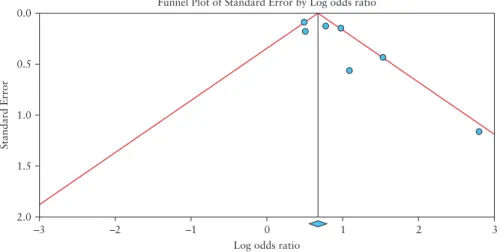
Publication bias was evaluated by visual assessment of the fun- nel plot because only six studies were included in our meta-analysis [subgroup A]. Tests for funnel plot asymmetry should be used only when there are at least ten studies included in the meta-analysis, be- cause the power of the test is too low to distinguish chance from real asymmetry when the number of studies is less than ten.38
3. Results
3.1. Literature search results
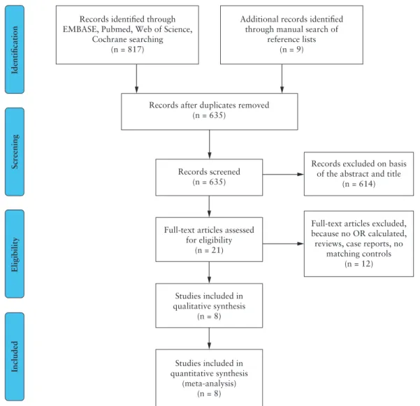
Our systematic literature search strategy identified 817 potentially relevant records [PRISMA flow diagram; Figure 1]. After removal of duplicates and screening of title and abstract, 21 papers remained for full-text evaluation. Following the revision of the eligible studies, 12 additional ones were excluded: studies without OR or extractable data [n = 3], case reports [n = 3], reviews [n = 4], no matching con- trol group [n = 2, e.g. non-IBD VTE controls]. One additional study was excluded from the A-subgroup analysis because of the lack of matching controls in the ‘no steroid’ group, although the data of the study were eligible for B-subgroup assessment.34 Finally, the remain- ing eight studies, published between 2012 and 2016, fulfilled the selection criteria [Table 1].
3.2. Study characteristics
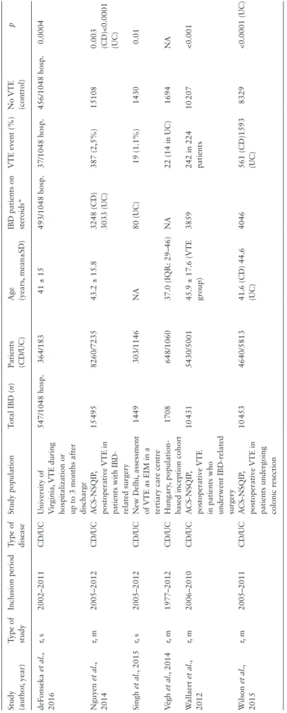
Eight studies were included in this meta-analysis. Of these, six reported on VTE complication in IBD patients during ster- oid treatment [A-group]20,28–32 and three on anti-TNFα therapy [B-group].28,33,34 OR was calculated in CD in one study,29 and in UC in two studies,30,31 while the others reported combined CD + UC
results28,32–34; only the publication from Nguyen et al.20assessed VTE risk in CD and UC separately. Since the risk for VTE in IBD patients is equally increased in both CD and UC, the results of the different studies can be evaluated together.8
Statistical analysis was carried out on 40 083 [A-subgroup] and 18 435 [B-subgroup] IBD patients. In the A-subgroup, 19 645 patients had CD and 20 438 had UC. There were 2861 [A-group] and 399 [B-group] VTE events identified. The mean age ranged from 37.0 to 45.9 years. Most of the patients included in the studies underwent an IBD-related surgery, were hospitalized and were followed during hospitalization or up to 3 months after discharge.20,28–30,33 Only a few articles reported outcomes retrieved from registries.31,32,34 All of the studies were retrospective, including two single-centre and six multi- centre analyses. The individual study characteristics are provided in Tables 2 and 3.
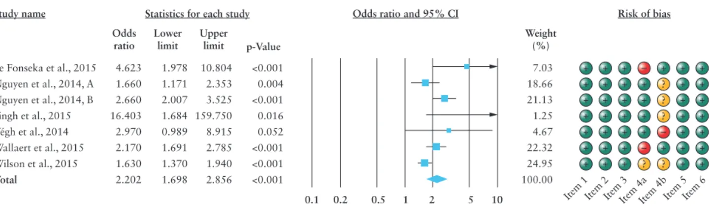
3.3. Risk of VTE in IBD patients treated with corticosteroids
Six studies assessed VTE risk in CD and UC patients treated or not with systemic corticosteroids.20,28–32 A total of 40 083 IBD patients were analysed and 2861 [7.13%] VTE events were identified as a complication. All individual studies had an OR > 0.989 reaching statistical significance [p < 0.05], except the study of Vegh et al. 32
[p = 0.052]. There was a significantly higher rate of VTE compli- cations in steroid-treated IBD patients compared to IBD patients without steroid medication [OR: 2.202; 95% CI: 1.698–2.856, p < 0.001] [Figure 2]. Evidence of heterogeneity was observed [I2 = 66.97; p = 0.006].
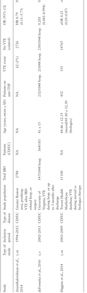
3.4. Risk of VTE in IBD patients treated with anti-TNFα
Only three studies assessed VTE risk in IBD patients treated with anti-TNFα compared to IBD patients treated with systemic corticos- teroids.28,33,34 A total of 18 435 IBD patients were analysed and 399 [2.16%] VTE events were reported. Two of three individual stud- ies had an OR < 1.0 attaining statistical significance [p < 0.05].28,34 There was a significantly lower rate of VTE complications in anti- TNFα-treated IBD patients compared to IBD patients with steroid medication [OR: 0.267; 95% CI: 0.106–0.674, p = 0.005] [Figure 3], with no statistically significant heterogeneity detected across studies [I2 = 0.000%, p = 0.476].
3.5. Risk of bias assessment
Overall NOS scores of the included studies in our meta-analysis ranged from 5 to 6 [Supplementary Table S4]. Since all studies were large IBD cohorts with a corresponding non-exposed control group, Records after duplicates removed
(n = 635) Records identified through
EMBASE, Pubmed, Web of Science, Cochrane searching
(n = 817)
Identification
Additional records identified through manual search of
reference lists (n = 9)
Records excluded on basis of the abstract and title
(n = 614)
Full-text articles excluded, because no OR calculated, reviews, case reports, no
matching controls (n = 12) Records screened
(n = 635)
Full-text articles assessed for eligibility
(n = 21)
Studies included in qualitative synthesis
(n = 8)
Studies included in quantitative synthesis
(meta-analysis) (n = 8)
ScreeningEligibilityIncluded
Figure 1. Flow diagram of the study selection process.
Table 2. Characteristics of studies evaluating thromboembolic complications during corticosteroid treatment Study (author, year)Type of studyInclusion periodType of diseaseStudy populationTotal IBD (n)Patients (CD/UC)Age (years, mean±SD)IBD patients on steroids*VTE event (%)No VTE (control)p deFonseka et al., 2016r, s2002–2011CD/UCUniversity of Virginia, VTE during hospitalization or up to 3 months after discharge
547/1048 hosp.364/18341 ± 15493/1048 hosp.37/1048 hosp.456/1048 hosp.0.0004 Nguyen et al., 2014r, m2005–2012CD/UCACS-NSQIP, postoperative VTE in patients with IBD- related surgery 15 4958260/723543.2 ± 15.83248 (CD) 3033 (UC)387 (2,5%)151080.003 (CD)<0.0001 (UC) Singh et al., 2015r, s2005–2012CD/UCNew Delhi, assessment of VTE as EIM in a tertiary care centre
1449303/1146NA80 (UC)19 (1.1%)14300.01 Végh et al., 2014r, m1977–2012CD/UCHungary, population- based inception cohort1708648/106037.0 (IQR: 29–46)NA22 (14 in UC)1694NA Wallaert et al., 2012r, m2006–2010CD/UCACS-NSQIP, postoperative VTE in patients who underwent IBD-related surgery
10 4315430/500145.9 ± 17.6 (VTE group)3859242 in 224 patients10 207<0.001 Wilson et al., 2015r, m2005–2011CD/UCACS-NSQIP, postoperative VTE in patients undergoing colonic resection 10 4534640/581341.6 (CD) 44.6 (UC)4046561 (CD)1593 (UC)8329<0.0001 (UC) r: retrospective, s: single-centre, m: multi-centre, CD: Crohn’s disease, UC: ulcerative colitis, IBD: inflammatory bowel disease, EIM: extraintestinal manifestation, hosp.: hospitalizations, VTE: venous thromboembolic event, ACS-NSQIP: The American College of Surgeon’s National Surgical Quality Improvement Program, IQR: interquartile range, NA: non available. *Systemic corticosteroid.
Table 3. Characteristics of studies evaluating thromboembolic complications during TNFα inhibitor treatment StudyType of studyInclusion periodType of diseaseStudy populationTotal IBDPatients (CD/UC)Age (years, mean ± SD)Patients on anti-TNFVTE eventNo VTE (control)OR (95% CI)p Ananthakrishnan et al., 2014r, m1994–2012CD/UCGreater Boston area, posthosp. VTE after IBD- related hosp. or surgery
2788NANANA62 (2%)2726HR 0.79 (0.11–5.73)NA deFonseka et al., 2016r, s2002–2011CD/UCUniversity of Virginia, VTE during hosp. or up to 3 months after discharge
547/1048 hosp.364/18341 ± 15232/1048 hosp.2/1048 hosp.230/1048 hosp.0.201 (0.041–0.994)0.0491 Higgins et al., 2014r, m2003–2009CD/UCTruven Health MarketScan database, VTE during steroid vs biologics therapy 15 100NA44.46 ± 12.55 (steroid)41.40 ± 12.30 (biologics)
45233514765aOR 0.21 (0.05–0.87)<0.05 r: retrospective, s: single-centre, m: multi-centre, CD: Crohn’s disease, UC: ulcerative colitis, IBD: inflammatory bowel disease, hosp.: hospitalizations, VTE: venous thromboembolic event, NA: not available, HR: hazard ratio, aOR: adjusted odds ratio.
they carried a low risk of bias in terms of representativeness, and also in terms of ascertainment of exposure. Comparability received the lowest count of stars: only 22% of the studies were rated as hav- ing low risk. All studies provided a clear definition of the diagnosis of VTE, including the details of the confirmation based on imaging techniques. Some studies used the international disease codes for
VTE diagnosis. All patients were followed up for at least 3 months, thereby fulfilling the corresponding NOS item. Risk of bias of the included studies is summarized in Figures 4 and 5.
The funnel plot in Figure 6 shows slight bias potentially as a consequence of a ‘small-study effect’, suggesting that smaller trials with no or moderate treatment effect may have remained unpub- lished. In our meta-analysis, the included articles are large cohort studies, except for that of Singh et al.,31 where the steroid-user group
Study name Statistics for each study Odds ratio and 95% CI Risk of bias
Ananthakrishnan et al., 2014 deFonseka et al., 2015 Higgins et al., 2014 Total
0.790 0.201 0.193 0.267
0.109 0.041 0.048 0.106
5.702 0.990 0.779 0.674
–0.234 –1.973 –2.312 –2.794
0.815 0.049 0.021 0.005 Odds
ratio Lower limit Upper
limit Z-Value p-Value Weight
(%) 21.99 33.80 44.21 100.00
0.01 0.1 1 10 100 Item
1 Item
2 Item
3
Item 4aItem 4bItem 5
Item 6 +
+ +
+ + +
+
+ +
+
+ + +
+ + + –
– – ?
?
Figure 3. Forest plot of studies evaluating thromboembolic complications during TNFα treatment with risk of bias assessment. Size of squares for risk ratio reflects weight of trial in pooled analysis. Horizontal bars represent 95% confidence intervals.
Ananthakrishnan et al., 2014 De Fonseka et al., 2016 Higgins et al., 2014 Nguyen et al., 2014 Singh et al., 2015 Végh et al., 2014 Wallaert et al., 2016 Wilson et al., 2015
Representativeness of the exposed Selection of non- exposed cohort Ascertainment of exposure Comparability of cohorts (age) Comparability of cohorts (smoking) Assessment of outcome Time for outcome to occur
+ + + – ? + +
+ + + + + +
+ + + + +
+ + + + +
+ + + + +
+ + +
+ +
+ + +
+ + + + + +
+ + + + +
– –
– –
?
?
?
?
?
Figure 4. Methodological quality of eligible studies using the Newcastle–
Ottawa scale [NOS] criteria.
100%
Low risk of bias Unclear risk of bias High risk of bias
90%
80%
70%
60%
50%
40%
30%
20%
10%
0%
Representativeness of the exposed Selection of non- exposed cohort Ascertainment of exposure Comparability of cohorts (age) Comparability of cohorts (smoking) Assessment of outcome Time for outcome to occur
Figure 5. Risk of bias.
Study name
de Fonseka et al., 2015 Nguyen et al., 2014, A Nguyen et al., 2014, B Singh et al., 2015 Végh et al., 2014 Wallaert et al., 2015 Wilson et al., 2015
4.623 1.660 2.660 16.403 2.970 2.170 1.630 2.202
1.978 1.171 2.007 1.684 0.989 1.691 1.370 1.698
10.804 2.353 3.525 159.750 8.915 2.785 1.940 2.856
<0.001 0.004
<0.001 0.016 0.052
<0.001
<0.001
<0.001
0.1 0.2 0.5 1 2 5 10
Total
Statistics for each study Odds ratio and 95% CI Risk of bias
Odds ratio Lower
limit Upper
limit p-Value
7.03 18.66 21.13 1.25 4.67 22.32 24.95
+ + + + + + +
+ + + + + + +
+ + + + + + +
–
– – + + + +
? +
+
?
?
?
? + + + + + + +
+ + + + + + + 100.00
Item 1
Item 2
Item 3
Item 4aItem 4bItem 5
Item 6 Weight
(%)
Figure 2. Forest plot of studies evaluating thromboembolic complications during corticosteroid treatment with risk of bias assessment. Size of squares for risk ratio reflects weight of trial in pooled analysis. Horizontal bars represent 95% confidence intervals.
included 80 patients. This study may have overestimated the risk of VTE, as is well known for small trials.
4. Discussion
Our meta-analysis evaluated pooled data from all currently available observational studies evaluating the risk of VTE in IBD as a com- plication of steroid and anti-TNFα therapies. Our results indicated that treatment with anti-TNFα was associated with a significantly lower risk of thromboembolic events in IBD patients [OR: 0.267;
95% CI: 0.106–0.674, p = 0.005] compared to steroid therapy. In contrast, VTE in IBD patients using systemic corticosteroids occurs twice as often as in the control group of IBD patients without steroid medication [OR: 2.202; 95% CI: 1.698–2.856, p < 0.001]. To our knowledge, no meta-analysis has evaluated the effect of steroid and anti-TNFα treatment in IBD patients. The major strength of our meta-analysis is the inclusion of large numbers of IBD patients with VTE events analysed at once, which would imply difficulties in ran- domized controlled trials.
Thromboembolism is one of the extraintestinal manifestations in IBD. The prevalence and incidence rate of VTE in IBD are 1.2–6.7% and 6.3 per 1000 person-years in clinical studies, respectively.15,41,42 DVT and PE are the most common types of VTE in IBD, but thromboses may also occur at unusual sites such as in cerebrovascular, retinal, portal, mesen- teric, splenic or internal jugular veins.42,43 The association between VTE and disease activity has been well established; at the time of a flare-up, the increase in VTE risk is more prominent.14 Other disease-specific risk factors, which have been identified in several studies, include colonic disease, fistulizing or stenotizing behaviour and surgical treatment.16,44 Recent literature indicates that drugs used in IBD treatment could also be considered as potential contributors to VTE pathogenesis. Systemic corticosteroids and anti-TNFα agents are both potent anti-inflammatory drugs used in moderately and severely active IBD flare-ups.
Steroids, however, have an independent thrombogenic effect from inflammatory processes. It has been shown that exogenous, systemic glucocorticoids and also excess endogenous cortisol [e.g. Cushing’s syndrome] are linked to VTE risk.45,46 Hypercoagulability in Cushing’s syndrome is due to increased production of procoagulant factors with activation of the coagulation cascade and an impaired fibrinolytic capacity.46 Additionally, the risk of VTE is increased among glucocorticoid users in non-IBD populations [for systemic glucocorticoids, adjusted incidence rate ratio (IRR) is 2.31, 95%
CI: 2.18–2.45], and this becomes more prominent at high doses.45,47 Corticosteroids administered to healthy volunteers also increase clotting factor levels and fibrinogen, which may also be related to the increased risk of thrombosis in the absence of inflammation.48
TNFα is a proinflammatory cytokine which has been reported to link inflammation and thrombosis in IBD. The findings of Yoshida et al. implicate TNFα in the enhanced microvascular thrombosis in dextran sodium sulphate-induced colitis model, and suggest that the action of TNFα accounts for most of the colitis-enhanced throm- botic response.49 Therefore, patients receiving anti-TNFα agents po- tentially have a decreased risk for thromboembolic complications. In addition to neutralizing TNFα, infliximab treatment in CD signifi- cantly reduced plasma soluble CD40L inflammatory cytokine levels, which might also decrease the risk of VTE.50
During our meta-analysis search, only three studies were found assessing VTE risk in IBD patients treated with anti-TNFα agents.
deFonseka et al. reported that the VTE risk is significantly lower when using TNFα inhibitor therapy in hospitalized IBD patients,28 whereas systemic corticosteroids were associated with an increased risk. In addition, use of anti-TNFα medication lowered the risk of post-hospitalization VTE in a greater Boston cohort.33 Higgins et al.
showed that biologics monotherapy resulted in a 5-fold reduction in VTE risk compared with patients receiving corticosteroid treat- ment.34 Moreover, this association between steroids and VTE risk seems to be dose-dependent.34 The study of Higgins et al. showed not only that the inflammatory effect itself is responsible for the elevated VTE risk in IBD but also that the corticosteroid drugs themselves increase VTE risk.34
Several limitations of this meta-analysis must be considered.
The first is the quality of data. All the included studies have a retrospective design, the data were extracted from medical records so patients were not randomized, and unrecognized confounding factors could bias our results and weaken the conclusions. At least four relevant studies were excluded due to unavailable ORs of VTE risk.42,51,52 In the study of Scoville et al.,53 there were non-IBD VTE controls as comparators for VTE risk in IBD, which did not match the control group criteria in our meta-analysis. Additionally, we can- not rule out that we lost relevant articles by having imposed English language, as a filter, on the search.
Secondly, there was substantial heterogeneity across the studies selected in subgroup A [I2 = 66.97]. Very different types of studies were included; some studies are population-based cohorts, while 0.0
0.5
1.0
Standard Error
1.5
2.0
–3 –2 –1 0
Log odds ratio
Funnel Plot of Standard Error by Log odds ratio
1 2 3
Figure 6. Funnel plot of corticosteroid studies with pseudo 95% confidence limits. Each circle indicates one study with its standard error indicating the weight of the study and its relative risk. The dotted lines represent 95% confidence interval to visualize the symmetry around the pooled estimate.
other data were derived from postoperative settings or hospitalized patients. However, the duration of follow-up was almost equal in the majority of the included studies. Likewise, there may be variability in disease phenotype of IBD among patients. Although colonic disease and disease extension correlate with VTE risk,16 the articles included no information on disease location or disease characteristics, so we were unable to analyse this parameter.
Thirdly, there were no quantitative activity indices for both UC and CD available in the publications, although the use of steroids and biologics indicates moderate to severe disease course.
Thus, we could not evaluate the effect of the exact severity of the disease and the effect of consecutive immobilization on thromboembolic risk.
In addition, we do not know how many IBD patients were given pharmacological thromboprophylaxis in the studies. Only two of the included studies report on the use of VTE prophylaxis, in which the use of thromboembolic prophylaxis based on hospital protocols was associated with reduced VTE risk.28,33 However, it is of note that the publication year of all trials is mainly before the implementation of VTE guidelines and consensus statements.41,54
Finally, if pharmacokinetic measurements had been used in the studies, it would have been possible to determine the plausible effect of drug levels and anti-drug antibodies on VTE risk.
In summary, the present meta-analysis confirms that cortico- steroid use in IBD patients carries a 2-fold risk of VTE. In contrast, thromboembolic risk decreases when using anti-TNFα agents for active disease instead of steroids. These associations highlight the importance of steroid-sparing therapy in IBD, especially in patients with additional risk factors for VTE. The choice between glucocorti- coids and anti-TNFα therapy in the management of severe flare-ups should take this into consideration, especially in patients with pre- vious VTE or a family history of thrombotic events.
According to recent guidelines, anticoagulant prophylaxis should be considered in all hospitalized and outpatients with se- vere IBD.41,54,55 Anticoagulant thromboprophylaxis is recommended with low-molecular-weight heparin, low-dose unfractioned heparin or fondaparinux.41 Recently, a risk assessment algorithm for IBD patients at risk of developing VTE has been proposed.56 Patients with IBD should be stratified according to their general and IBD- specific risk factors for VTE into high-risk and intermediate/low- risk patients. In addition to a detailed patient history focused on well-known general and disease-specific VTE risk factors, clot lysis parameters should be included in the risk assessment.56 We recognize the importance of our findings in the promotion of the appropriate thromboprophylaxis during steroid treatment. Further research is needed to establish the exact weight of these factors and large pro- spective cohort studies are awaited.
Funding
This work was supported by the Economic Development and Innovation Operative Programme Grant of the National Research, Development and Innovation Office [GINOP 2.3.2-15-2016-00048] STAY ALIVE grant.
Conflict of Interest
The authors have no conflicts of interest to declare.
Acknowledgments
The present scientific contribution is dedicated to the 650th anniversary of the foundation of the University of Pécs, Hungary.
Author Contributions
PS, PH and NF designed the research; PS, NF and KS performed the research and statistical analyses, and analysed and interpreted the data;
PS and NF wrote the article; AG, IS, AI, MS, EP, GB, JB, JC, OH, PV and AV made critical revisions related to important intellectual content of the manuscript; PS, AV and PH gave final approval of the version of the article to be published.
Supplementary Data
Supplementary data are available at ECCO-JCC online.
References
1. Søgaard KK, Schmidt M, Pedersen L, Horváth-Puhó E, Sørensen HT. 30- year mortality after venous thromboembolism: a population-based cohort study. Circulation 2014;130:829–36.
2. White RH. The epidemiology of venous thromboembolism. Circulation 2003;107:I4–8.
3. Tagalakis V, Patenaude V, Kahn SR, Suissa S. Incidence of and mortality from venous thromboembolism in a real-world population: the Q-VTE Study Cohort. Am J Med 2013;126:832.e13–21.
4. Piran S, Schulman S. Management of venous thromboembolism: an update. Thromb J 2016;14:23.
5. Bernstein CN, Blanchard JF, Houston DS, Wajda A. The incidence of deep venous thrombosis and pulmonary embolism among patients with inflam- matory bowel disease: a population-based cohort study. Thromb Haemost 2001;85:430–4.
6. Bernstein CN, Nabalamba A. Hospitalization-based major comorbid- ity of inflammatory bowel disease in Canada. Can J Gastroenterol 2007;21:507–11.
7. Kappelman MD, Horvath-Puho E, Sandler RS, et al. Thromboembolic risk among Danish children and adults with inflammatory bowel diseases: a population-based nationwide study. Gut 2011;60:937–43.
8. Yuhara H, Steinmaus C, Corley D, et al. Meta-analysis: the risk of venous thromboembolism in patients with inflammatory bowel disease. Aliment Pharmacol Ther 2013;37:953–62.
9. Saleh T, Matta F, Yaekoub AY, Danescu S, Stein PD. Risk of venous thromboembolism with inflammatory bowel disease. Clin Appl Thromb Hemost 2011;17:254–8.
10. Fumery M, Xiaocang C, Dauchet L, Gower-Rousseau C, Peyrin-Biroulet L, Colombel JF. Thromboembolic events and cardiovascular mortality in inflammatory bowel diseases: a meta-analysis of observational studies. J Crohns Colitis 2014;8:469–79.
11. Wang TJ, Lee SC, Liang SY, Tung HH, Wu SF, Lin YP. Comparing the effi- cacy of aquatic exercises and land-based exercises for patients with knee osteoarthritis. J Clin Nurs 2011;20:2609–22.
12. Zhong M, Dong XW, Zheng Q, Tong JL, Ran ZH. Factor V Leiden and thrombosis in patients with inflammatory bowel disease (IBD): a meta- analysis. Thromb Res 2011;128:403–9.
13. Danese S, Papa A, Saibeni S, Repici A, Malesci A, Vecchi M. Inflammation and coagulation in inflammatory bowel disease: The clot thickens. Am J Gastroenterol 2007;102:174–86.
14. Grainge MJ, West J, Card TR. Venous thromboembolism during active dis- ease and remission in inflammatory bowel disease: a cohort study. Lancet 2010;375:657–63.
15. Miehsler W, Reinisch W, Valic E, et al. Is inflammatory bowel disease an independent and disease specific risk factor for thromboembolism? Gut 2004;53:542–8.
16. Nguyen GC, Sam J. Rising prevalence of venous thromboembolism and its impact on mortality among hospitalized inflammatory bowel disease patients. Am J Gastroenterol 2008;103:2272–80.
17. Harbord M, Annese V, Vavricka SR, et al.; European Crohn’s and Colitis Organisation. The first European evidence-based consensus on extra- intestinal manifestations in inflammatory bowel disease. J Crohns Colitis 2016;10:239–54.
18. Van Assche G, Dignass A, Reinisch W, et al.; European Crohn’s and Colitis Organisation (ECCO). The second European evidence-based Consensus on the diagnosis and management of Crohn’s disease: Special situations. J Crohns Colitis 2010;4:63–101.
19. Van Assche G, Dignass A, Bokemeyer B, et al.; European Crohn’s and Colitis Organisation. Second European evidence-based consensus on the diagnosis and management of ulcerative colitis part 3: Special situations. J Crohns Colitis 2013;7:1–33.
20. Nguyen GC, Elnahas A, Jackson TD. The impact of preoperative steroid use on short-term outcomes following surgery for inflammatory bowel disease. J Crohns Colitis 2014;8:1661–7.
21. Giannotta M, Tapete G, Emmi G, Silvestri E, Milla M. Thrombosis in inflammatory bowel diseases: what’s the link? Thromb J 2015;13:14.
22. van Zaane B, Nur E, Squizzato A, et al. Systematic review on the effect of glucocorticoid use on procoagulant, anti-coagulant and fibrinolytic fac- tors. J Thromb Haemost 2010;8:2483–93.
23. Davies R, Galloway JB, Watson KD, Lunt M, Symmons DP, Hyrich KL;
BSRBR Control Centre Consortium, British Society for Rheumatology Biologics Register. Venous thrombotic events are not increased in patients with rheumatoid arthritis treated with anti-TNF therapy: results from the British Society for Rheumatology Biologics Register. Ann Rheum Dis 2011;70:1831–4.
24. Petitpain N, Gambier N, Wahl D, Chary-Valckenaere I, Loeuille D, Gillet P; French Network of Pharmacovigilance Centers. Arterial and venous thromboembolic events during anti-TNF therapy: a study of 85 spontan- eous reports in the period 2000-2006. Biomed Mater Eng 2009;19:355–64.
25. Vereckei E, Kriván G, Réti M, Szodoray P, Poór G, Kiss E. Anti-TNF- alpha-induced anti-phospholipid syndrome manifested as necrotizing vas- culitis. Scand J Rheumatol 2010;39:175–7.
26. Moher D, Liberati A, Tetzlaff J, Altman DG; PRISMA Group. Preferred reporting items for systematic reviews and meta-analyses: the PRISMA statement. PLoS Med 2009;6:e1000097.
27. Stroup DF, Berlin JA, Morton SC, et al. Meta-analysis of observa- tional studies in epidemiology: a proposal for reporting. Meta-analysis Of Observational Studies in Epidemiology (MOOSE) group. JAMA 2000;283:2008–12.
28. deFonseka AM, Tuskey A, Conaway MR, Behm BW. Antitumor necro- sis factor-α therapy is associated with reduced risk of thromboembolic events in hospitalized patients with inflammatory bowel disease. J Clin Gastroenterol 2016;50:578–83.
29. Wallaert JB, De Martino RR, Marsicovetere PS, et al. Venous thrombo- embolism after surgery for inflammatory bowel disease: are there modifiable risk factors? Data from ACS NSQIP. Dis Colon Rectum 2012;55:1138–44.
30. Wilson MZ, Connelly TM, Tinsley A, et al. Ulcerative colitis is associ- ated with an increased risk of venous thromboembolism in the postopera- tive period: The results of a matched cohort analysis. Annals of Surgery 2015;261:1160–6.
31. Singh B, Kedia S, Konijeti G, et al. Extraintestinal manifestations of inflam- matory bowel disease and intestinal tuberculosis: Frequency and relation with disease phenotype. Indian J Gastroenterol 2015;34:43–50.
32. Vegh Z, Golovics PA, Lovasz BD, et al. Low incidence of venous thrombo- embolism in inflammatory bowel diseases: prevalence and predic- tors from a population-based inception cohort. Scand J Gastroenterol 2015;50:306–11.
33. Ananthakrishnan AN, Cagan A, Gainer VS, et al. Thromboprophylaxis is associated with reduced post-hospitalization venous thromboembolic events in patients with inflammatory bowel diseases. Clin Gastroenterol Hepatol 2014;12:1905–10.
34. Higgins PD, Skup M, Mulani PM, Lin J, Chao J. Increased risk of venous thromboembolic events with corticosteroid vs biologic therapy for inflam- matory bowel disease. Clin Gastroenterol Hepatol 2015;13:316–21.
35. Wells GA, Shea B, O’Connell D, Peterson J, Welch V, Losos M, Tugwell P. The Newcastle-Ottawa Scale (NOS) for assessing the quality if non- randomized studies in meta-analyses. 2011. http://www.ohri.ca/programs/
clinical_epidemiology/oxford.asp. Accessed 2011.
36. Tierney JF, Stewart LA, Ghersi D, Burdett S, Sydes MR. Practical methods for incorporating summary time-to-event data into meta-analysis. Trials 2007;8:16.
37. Higgins JPT, Deeks JJ. Chapter 7: Selecting studies and collecting data.
Cochrane Handbook for Systematic Reviews of Interventions: Version 5.1.0 (updated March 2011). Wiley-Blackwell, 2011.
38. Deeks JJ, Higgins JPT, Altman DG. Chapter 9: Analysing data and under- taking meta-analyses. Cochrane Handbook for Systematic Reviews of Interventions: Version 5.1.0 (updated March 2011). Wiley-Blackwell, 2011.
39. DerSimonian R, Laird N. Meta-analysis in clinical trials. Control Clin Trials 1986;7:177–88.
40. Peto R. Clinical trial methodology. Biomedicine 1978;28:24–36.
41. Nguyen GC, Bernstein CN, Bitton A, et al. Consensus statements on the risk, prevention, and treatment of venous thromboembolism in inflam- matory bowel disease: Canadian Association of Gastroenterology.
Gastroenterology 2014;146:835–848.e6.
42. Papay P, Miehsler W, Tilg H, et al. Clinical presentation of venous thrombo- embolism in inflammatory bowel disease. J Crohns Colitis 2013;7:723–9.
43. Danese S, Semeraro S, Papa A, et al. Extraintestinal manifestations in inflammatory bowel disease. World J Gastroenterol 2005;11:7227–36.
44. Koutroumpakis EI, Tsiolakidou G, Koutroubakis IE. Risk of venous thromboembolism in patients with inflammatory bowel disease. Semin Thromb Hemost 2013;39:461–8.
45. Johannesdottir SA, Horváth-Puhó E, Dekkers OM, et al. Use of glucocor- ticoids and risk of venous thromboembolism: a nationwide population- based case-control study. JAMA Intern Med 2013;173:743–52.
46. van der Pas R, Leebeek FW, Hofland LJ, de Herder WW, Feelders RA.
Hypercoagulability in Cushing’s syndrome: prevalence, pathogenesis and treatment. Clin Endocrinol (Oxf) 2013;78:481–8.
47. Huerta C, Johansson S, Wallander MA, García Rodríguez LA. Risk fac- tors and short-term mortality of venous thromboembolism diagnosed in the primary care setting in the United Kingdom. Arch Intern Med 2007;167:935–43.
48. Brotman DJ, Girod JP, Posch A, et al. Effects of short-term gluco- corticoids on hemostatic factors in healthy volunteers. Thromb Res 2006;118:247–52.
49. Yoshida H, Yilmaz CE, Granger DN. Role of tumor necrosis factor-alpha in the extraintestinal thrombosis associated with colonic inflammation.
Inflammatory Bowel Diseases 2011;17:2217–23.
50. Danese S, Sans M, Scaldaferri F, et al. TNF-alpha blockade down-regu- lates the CD40/CD40L pathway in the mucosal microcirculation: a novel anti-inflammatory mechanism of infliximab in Crohn’s disease. J Immunol 2006;176:2617–24.
51. Shapiro R, Vogel JD, Kiran RP. Risk of postoperative venous thrombo- embolism after laparoscopic and open colorectal surgery: an add- itional benefit of the minimally invasive approach? Dis Colon Rectum 2011;54:1496–502.
52. Bollen L, Vande Casteele N, Ballet V, et al. Thromboembolism as an import- ant complication of inflammatory bowel disease. Eur J Gastroenterol Hepatol 2016;28:1–7.
53. Scoville EA, Konijeti GG, Nguyen DD, Sauk J, Yajnik V, Ananthakrishnan AN. Venous thromboembolism in patients with inflammatory bowel diseases: a case-control study of risk factors. Inflamm Bowel Dis 2014;20:631–6.
54. Gionchetti P, Dignass A, Danese S, et al.; ECCO. 3rd European evidence- based consensus on the diagnosis and management of Crohn’s disease 2016: part 2: surgical management and special situations. J Crohns Colitis 2017;11:135–49.
55. Guyatt GH, Eikelboom JW, Gould MK, et al. Approach to outcome meas- urement in the prevention of thrombosis in surgical and medical patients:
Antithrombotic therapy and prevention of thrombosis, 9th ed: American College of Chest Physicians evidence-based clinical practice guidelines.
Chest 2012;141:e185S–94S.
56. Bollen L, Vande Casteele N, Peeters M, et al. The occurrence of thrombosis in inflammatory bowel disease is reflected in the clot lysis profile. Inflamm Bowel Dis 2015;21:2540–8.