Accepted: 2015.04.21 Published: 2015.05.08
1856 1 3 26
Expression of VEGF in Neonatal Urinary Obstruction: Does Expression of VEGF Predict Hydronephrosis?
A 1 Zsófia Magyar
D 2 Julianna Schönleber
E 3 Miklós Romics
F 1 Ervin Hruby
CF 1 Bálint Nagy
B 4 Bálint Sulya
F 1 Artúr Beke
G 1 Ágnes Harmath
A 1 Judit Jeager
G 1 János Rigó jr.
AG 1 Éva Görbe
Corresponding Author: Éva Görbe, e-mail: gorbeeva@gmail.com Source of support: Departmental sources
Background: In animal studies, the inhibition of VEGF activity results in high mortality and impaired renal and glomerular development. Mechanical stimuli, like mechanical stretch in respiratory and circulatory systems, results in an elevated expression of VEGF. In animal models, the experimental urinary obstruction is associated with stretch- ing of tubular cells and activations of the renin-angiotensin system. This results in the upregulation of vascu- lar endothelial growth factor (VEGF) and TNF-alfa.
Material/Methods: Tissue samples from urinary tract obstruction were collected and immunohistochemistry was performed in 14 patients (average age: 7.1±4.1 years). The control histology group consisted of ureteropelvic junction tissue from 10 fetuses after midtrimester artificial abortion. The fetuses did not have any failure at ultrasound screen- ing and pathological examination. The mean gestational age was 20.6 weeks of gestation (±2.2SD). Expression of VEGF was detected with immunohistochemistry method.
Results: Expression of VEGF was found in varying intensity in the submucosa and subserosa layers, but only in the test tissue (placental tissue). The tissue of the patients with urinary obstruction and the tissue of the fetal uretero- pelvic junction without urinary obstruction were negative for expression of VEGF. The repeated examination showed negative cells and no color staining.
Conclusions: The pressure due to congenital urogenital obstruction resulting in mechanical stress in cells did not increase the expression of VEGF in young children in our study. To find a correlation between urogenital tract obstruc- tion and increased expression of VEGF, we need to perform more examinations because the connection may be of therapeutic significance.
MeSH Keywords: Endothelial Growth Factors • Hydronephrosis • Urinary Tract Full-text PDF: http://www.medscimonit.com/abstract/index/idArt/894133
Authors’ Contribution:
Study Design A Data Collection B Statistical Analysis C Data Interpretation D Manuscript Preparation E Literature Search F Funds Collection G
1 1st Department of Obstetrics and Gynecology, Semmelweis University, Faculty of Medicine, Budapest, Hungary
2 Department of Pathology, Jahn Ferenc Hospital, Budapest, Hungary 3 Department of Urology, Semmelweis University, Faculty of Medicine, Budapest,
Hungary
4 Department of Urology, Heim Pál Childrens Hospital, Budapest, Hungary
Background
VEGF is a major angiogenic factor and regulator of endotheli- al cell proliferation [1]. It has a crucial role in vasculogenesis and vascular permeability, and in the normal development of the glomerulus [2].
Its expression is tightly regulated. The VEGF gene has a 14-kb coding region with 8 exons and 7 introns. VEGF is produced by many cell types and its expression is induced by hypoxia [3].
Effects of mechanical stretch on cells in the respiratory and cir- culatory system were also found to be a stimulating factor [4].
In animal studies the inhibition of the vascular endothelial growth factor results in growth arrest and death. Histological changes lead to distinct liver and renal failure [5]. Regulation of VEGF plays an important role in development of renal dis- ease. VEGF-A expression in podocytes leads to glomerular dis- ease in mice [6].
In animal research, podocyte VEGF overexpression caused albu- minuria and congenital nephrotic syndrome [7]. Experimental urinary obstruction was associated with mechanical stretch- ing of tubular epithelial cells and activation of the renin-an- giotensin system, leading to the upregulation of cytokines and growth factors (VEGF and TNF-a). Tubulointerstitial nephritis (TIN) was induced in an experimental model of obstructive uropathy [8]. Effects of mechanical stretch on cells were found in respiratory and circulatory systems [9].
Material and Methods
Patients
We investigated the expression of VEGF in 14 different cas- es of urinary tract obstruction in children after surgical treat- ment. The mean age at surgery was 7.1 years (±SD 4.0) and the male/female ratio was 10/4.
The ureteropelvic obstruction was the most common malfor- mation, (11/14 patients), and in 7 cases the obstruction was on the left side. The treatment in these cases was Hynes- Anderson pyeloplasty.
In 1 case the surgical treatment was ureter-neoimplantation because of uretero-vesical stenosis. In another case the patient showed vesico-ureteral reflux and grade IV hydronephrosis, treated by uretero-nephrectomy. Hydronephrosis and hydro- ureter affecting both sides caused by posterior urethral valve was diagnosed in 1 case and the treatment was vesicostomy and bilateral ureter-neoimplantation. In 4 cases the diagnosis was confirmed by ultrasound examination following successful
previous prenatal diagnosis. In 5 cases the patients presented with abdominal pain, and in 3 patients urinary tract infection occurred before the diagnosis. In 2 cases the malformations were discovered during routine ultrasound in the newborn.
The control group consisted of 10 healthy fetuses, mean age 20.6 weeks of gestation (±2.2 SD), that did not show any mal- formations on autopsy examinations. Artificial abortion was performed before 23 weeks of gestation, as requested by the parents.
The expression of VEGF was measured in the lower third of the ureter in 3 cases and in the ureteropelvic junction tissue in 11 cases. The survey was completed using immunohisto- chemistry methods.
Immunohistochemistry
Monoclonal rabbit anti-human VEGF clone VG1 antibody (Dako, Denmark) was used for immunohistochemistry. Briefly, paraf- fin-embedded specimens were sliced (4 µm), dewaxed, and hydrated, incubated in citrate buffer solution for antigen re- covery (56 min at 91Cº), and were treated with primary anti- body for 48 min at 37Cº in a 1:30 dilution. Finally, the slides were incubated with Ventana UltraView Universal HRP (DAB) secondary antibody. We used a BenchMark Ultra automatic staining machine.
The immunostaining results were evaluated and scored by 2 pathologists independently who lacked knowledge of the clin- icopathological outcomes of the patients. Digital images were manually scored according to staining intensity and morpholo- gy. Positive expression of VEGF was identified by the presence of brown-yellow granules in the cytoplasm. In 5 random high- power fields, a positive slide had ³30% positive cells, and a neg- ative slide had <30% positive cells and no color staining [10].
The diagnosis of hydronephrosis was performed with assess- ment of the renal pelvic diameter [11,12].
Ethics permission was obtained from the authorities (ETT TUKEB permit 387/2013, 21300-4/2013/EKU), and the patients were informed and signed a consent.
Results
The characteristics of patients and clinical data are summa- rized in Table 1.
In 4 infants after prenatal diagnosis, there was a postnatal evaluation of hydronephrosis. In 5 cases the patients had ab- dominal pain and in 3 patients urinary tract infection occurred
before the diagnosis. In 2 newborn infants, the routine ul- trasound examination found congenital urinary obstruction.
The grade of urinary retention in the kidney was grade IV–V in cases of ureteropelvic obstruction and the surgical treatment was pyeloplasty (by Anderson-Hynes).
The expression of VEGF was measured in the tissue of the ure- teropelvic junction.
Only 1 patient had vesicoureteral obstruction on the left side;
he had a grade IV hydronephrosis. The surgical treatment was ureter neo-implantation. In this case the expression of VEGF was measured in the lower part of the ureter tissue.
In 1 patient vesico-ureteral reflux was present on the right side and hydronephrosis (grade V) was treated by uretero-nephrec- tomy. Hydronephrosis in grade IV-V and hydroureter on both
side caused by posterior urethral valve was the diagnosis in 1 patient and the treatment was vesicostomy and bilateral ure- ter-neoimplantation. The expression of VEGF was measured in these cases in the subserosal tissue of the ureter.
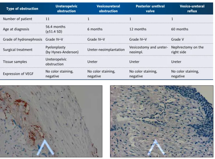
The expression patterns of VEGF staining are depicted in the test tissue (placental tissue). Figure 1 shows positive expression of VEGF identified by the presence of brown-yellow granules in the cytoplasm. A positive slide had ≥30% positive cells, and a negative slide had <30% positive cells and no color staining.
The transitional mucosal surface in the ureteropelvic junction tissue in a 5-year-old boy after a surgical treatment of uretero- pelvic obstruction by Hynes-Anderson pyeloplasty. The slide is negative for brown-yellow granules in the cytoplasm and had no positive cells and no color staining (Figure 2.).
Type of obstruction Ureteropelvic obstruction
Vesicoureteral obstruction
Posterior urethral valve
Vesico-ureteral reflux
Number of patient 11 1 1 1
Age at diagnosis 56.4 months
(±51.4 SD) 6 months 12 months 60 months
Grade of hydronephrosis Grade IV–V Grade IV–V Grade IV–V Grade V
Surgical treatment Pyelonplasty
(by Hynes-Anderson) Ureter-neoimplantation Vesicostomy and ureter- neoimpl.
Nephrectomy on the right side
Tissue samples Ureteropelvic
obstruction Ureter Ureter Ureter
Expression of VEGF No color staining, negative
No color staining, negative
No color staining, negative
No color staining, negative
Table 1. The mean demographic and clinical data of the surgical treatment and expression of VEGF on urinary obstruction in children.
Figure 1. Positive expression of VEGF identified by the presence of brown-yellow granules in the cytoplasm of endothelial cells in placental tissue (20×).
Figure 2. Mucosal surface on the ureteropelvic junction tissue in a 5 years old boy. The slide is negative for the brown- yellow granules in the cytoplasm, the slide has no positive cells and no color staining.
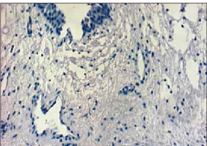
Our control group showed no increase in expression of VEGF in the ureteropelvic junction tissue of human fetuses. Our con- trol tissue sample had no positive cells in ureteropelvic slides in fetal tissue (without urinary obstruction) (Figure 3.)
Discussion
VEGF-A is a major angiogenic factor and a regulator of endo- thelial cell proliferation. It has a crucial role in vasculogenesis and vascular permeability and in the normal development of the glomerulus [13,14].
The expression is tightly regulated. VEGF is produced by many cell types, and the expression is induced by hypoxia [15]. Serum concentration of VEGF is high in bronchial asthma and in dia- betes mellitus [16]. The normal function of VEGF is the medi- ation of angiogenesis (formation of new blood vessels during embryonic development) and neoangiogenesis (the forma- tion of new blood vessels after injury or physical exercise) in vessels providing collateral circulation for hypoxemic areas.
Hypoxia affects cells producing hypoxia-inducible factor (HIF), a transcription factor. HIF then stimulates the release of VEGF-A, which binds to VEGF receptors on endothelial cells, triggering a tyrosine kinase pathway leading to angiogenesis. In the kidney, increased expression of VEGF-A in glomeruli directly causes the glomerular hypertrophy that is associated with proteinuria [17].
VEGF is a highly specific factor for promoting vascular endo- thelial cell division and inducing angiogenesis. In addition to VEGF being a potent angiogenic factor, it also acts as a mul- tifunctional cytokine. VEGF is found in normal human tissues such as heart, lung, kidney, and bladder. However, VEGF expres- sion in normal tissue occurs only at the low levels necessary to maintain normal blood vessel density and basic penetration
functions for facilitating the transport of nutrients. Since VEGF is secreted by tumor cells, a variety of malignant tumors, in- cluding bladder can cer, show significant VEGF expression.
The effects of stretching stimuli on molecules, including col- lagen 1a, lysyl oxidase, vascular endothelial growth factor-A, have been investigated in lung and blood vessels [18]. In the respiratory and circulatory system, the mechanical stretch re- sults in alteration of cell functions and an increase in surfac- tant secretion [19].
Urinary tract anomalies can be diagnosed during the prena- tal period in the second or third trimester of pregnancy. The most common malformation is hydronephrosis, which occurs in 1/500–700 live births. Hydronephrosis is more common in males, and in 20–40% of cases the malformation is bilateral [20–22]. The primary parameter for prenatal assessment and screening of hydronephrosis is the measurement of renal pel- vic diameter (RPD) by ultrasound [11,12].
The most common causes of hydronephrosis are the uretero- pelvic/ ureterovesical obstruction, posterior urethral valve, Eagle-Barrett syndrome, and vesicoureteral reflux. In 15% of patients the prenatally detected pelvis dilatation cannot be visualized after birth. The grade of pyelectasia is a predictive value for kidney injury [23,24].
In severe cases of bilateral hydronephrosis, prenatal percu- taneous vesicoamniotic shunt or drainage can be performed.
The intervention may serve as a solution for oligohydramnios and pulmonary hypoplasia and may also prevent later compli- cations with renal disease. Multicentric studies are necessary to compare both prenatal surgical methods of treatment and postnatal assessment of lower urinary tract obstruction [25].
The production of VEGF in the human kidney is localized to the podocytes and in the proximal and distal tubular system.
The receptor VEGFR-1 (Flt-1) and VEGFR-2 (Flk-1) are expressed in glomerular endothelial and in pre- and postglomerular ves- sels. The measured levels of circulating VEGF detected in pa- renchymal kidney diseases in the blood and in the urine may be of therapeutic significance. Another example of the signifi- cance of VEGF is in polycystic kidney disease, where angiogen- esis plays an important role of cyst formation and the struc- tural changes in normal renal parenchyma surrounding the cysts, which may eventually lead to end-stage renal failure.
The pivotal PKD1 gene and PKD2 genes are also expressed in the smooth muscle cells and endothelial cells of blood vessels.
Their effects are potentially augmented by the VEGF-A, having an important role in survival migration, permeability, and dil- atation of vessels. VEGF has been also detected with immu- nohistochemical method in the wall of cysts. The size of the cysts correlated with VEGF levels [2].
Figure 3. Subserosa in a fetal ureteropelvic tissue. The slide has no positive cells in the fetal tissue (without urinary obstruction).
In the early phase of the disease, consisting of cyst forma- tion, a high VEGF concentration is present, but later, when end-stage injury of renal blood vessels is developed, the VEGF concentration is low.
In experimental studies the glomerular-selective deletion or overexpression of VEGF leads to kidney disease in mice. The endogenous renal VEGF levels are increased in experimen- tal urogenital obstruction in animal studies. The tubular in- jury caused by obstructive nephropathy results in increased expression of VEGF. Urethral healing is associated with high levels of VEGF in animal studies [6,26].
In contrast with publications regarding the role of VEGF in uri- nary tract obstruction based on experimental animal models, our study is the first evaluation of VEGF expression in humans.
Limitations may be attributed to the sample size, which ex- plain the lack of a significant correlation between urinary tract obstruction and VEGF patterns.
Conclusions
Expression of VEGF shows a connection with glomerular de- velopment and with the regulation of permeability of kidney vessels. Experimental urinary obstruction was associated with mechanical stretching of tubular epithelial cells and activation of the renin-angiotensin system, leading to upregulation of cy- tokines and growth factors (VEGF and TNF-a). Tubulointerstitial nephritis (TIN) has been induced in an experimental model of obstructive uropathy. We investigated the expression of VEGF in different cases of urinary obstruction in children. Although our sample size was insufficient to investigate the associa- tion between mechanical stretch and increased expression of VEGF in congenital urinary obstruction, we believe that our pi- lot study can be applied to further studies and the assessing the expression of VEGF in the urinary obstruction may be of therapeutic significance.
References:
1. Schrijvers BF, Flyvbjerg A, De Vriese AS: The role of vascular endothelial growth factor (VEGF) in renal pathophysiology. Kidney Int, 2004;65: 2003–17 2. Simon M, Gröne HJ, Jöhren O et al: Expression of vascular endothelial growth factor and its receptors in human renal ontogenesis and in adult kidney.
Am J Physiol, 1995; 268: 240–50
3. Torres VE, Boletta A, Chapman A et al: Prospects for mTOR inhibitor use in patients with polycystic kidney disease and hamartomatous diseases. Clin J Am Soc Nephrol, 2010; 5: 1312–29
4. Breen EC: Mechanical strain increases type I collagen expression in pulmo- nary fibroblasts in vitro. J Appl Physiol, 2000; 88: 203–9
5. Gerber HP, Hillan KJ, Ryan AM et al: VEGF is required for growth and sur- vival in neonatal mice. Development, 1999; 126: 1149–59
6. Eremina V, Sood M, Haigh J et al: Glomerular-specific alterations of VEGF-A expression leaf to distinct congenital and acquired renal diseases. J Clin Invest, 2003; 111: 707–16
7. Veron D, Reidy K, Marlier A et al: Induction of podocyte VEGF164 overex- pression at different stages of development causes congenital nephrosis or steroid resistant nephrotic syndrome. Am J Pathol, 2010; 177: 2225–33 8. Truong LD, Gaber L, Eknoyan G: Obstructive uropathy. Contrib Nephrol,
2011; 169: 311–26
9. Breen EC: Mechanical strain increases type I collagen expression in pulmo- nary fibroblasts in vitro. J Appl Physiol, 2000; 88: 203–9
10. Sun YW, Xuan Q, Shu QA et al: Correlation of tumor relapse and elevated expression of survivin and vascular endothelial growth factor in superfi- cial bladder transitional cell carcinoma. Genet Mol Res, 2013; 12: 1045–53 11. Dighe M, Moshiri M, Phillips G et al: Fetal genitourinary anomalies – a pic-
torial review with postnatal correlation. Ultrasound Q, 2011; 27: 7–21 12. Vemulakonda V, YieeJ, Wilcox DT: Prenatal hydronephrosis: postnatal eval-
uation and management. Curr Urol Rep, 2014; 15: 430–35
13. Ferrara N, Gerber HP, LeCouter J: The biology of VEGF and its receptors. Nat Med, 2003; 9: 669–76
14. Gao H, Qian J, Chen B: Probing mechanical principles of focal contacts in cell-matrix adhesion with a coupled stochastic-elastic modelling frame- work. J R Soc Interface, 2011; 8: 1217–32
15. Holmes K, Roberts O, Angharad M et al: Vascular endothelial growth fac- tor receptor-2: Structure, function, intracellular signalling and therapeutic inhibition. Cellular Signalling, 2007;19: 2003–12
16. Cooper M, Dimitria V, Sherif Y et al: Increased renal expression of vascular endothelial growth factor (VEGF) and its receptor VEGFR-2 in experimen- tal diabetes. Diabetes, 1999; 48: 229
17. Liu E, Morimoto M, Kitajima S et al: Increased expression of vascular en- dothelial growth factor in kidney leads to progressive impairment of glo- merular functions. J Am Soc Nephrol, 2007; 8: 2094–104
18. Imsirovic J, Derricks K, Buczek-Thomas J et al: A novel device to stretch mul- tiple tissue samples with variable patterns: Application for mRNA regula- tion in tissue-engineered constructs. Biomatter, 2013; 3: e24650 19. Arold SP, Bartolák-Suki E, Suki B: Variable stretch pattern enhances sur-
factant secretion in alveolar type II cells in culture. Am J Physiol Lung Cell Mol Physiol, 2009; 296: 574–78
20. Ryckewaert-D’ Hallvin A, Le Bouar G, Odent S: Diagnosis of fetal urinary tract malformations: prenatal management and postnatal outcome. Prenat Diagn, 2011; 31: 1013–20
21. Fanaroff and Martin’s: Neonatal and Perinatal Medicine. Diseases of The Fetus and Infants, 8th Edition, 2009; Volume Two, Chapter 49: 1675–83 22. Gonzalez R, Schimke CM: Ureteropelvic junction obstruction in infants and
children. Pediatr Clin North Am, 2001; 48: 1505–8
23. Brown C, Morris RK, Daniels J et al: Effectiveness of percutaneous vesico- amniotic shunting in congenital lower urinary tract obstruction: divergence in prior beliefs among specialist groups. Eur J Obstet Gynecol Reprod Biol, 2010; 152: 25–29
24. Morris RK, Kilby MD: Long term renal and neurodevelopmental outcome in infants with LUTO, with and without fetal intervention. Early Hum Development, 2011; 87: 607–10
25. Ruano R: Fetal surgery for severe lower urinary tract obstruction. Prenat Diagn, 2011; 31: 667–74
26. Hofer MD, Cheng EY, Bury MI: Analysis of primary urethral wound healing in the rat. Urology, 2014; 84: 246–47