OBSTRUCTIVE SLEEP APNEA SYNDROME IN CHILDHOOD
Doctoral theses
Pálma Edina Benedek MD
Semmelweis University PhD School of Clinical Medicine
Supervisor: Prof. Gábor Katona MD
Opponents: Márta Novák MD, PhD
Péter Móricz MD, PhD
Head of doctoral committee: Prof. László Szabó MD Members of doctoral committee: Ágnes Szirmai MD, PhD
Zsolt Bella MD, PhD
Budapest 2014
1. Introduction
The sleep related disorders of breathing (SDB) can be observed in 10% of the pediatric population, which includes the habitual snoring, upper airway resistance syndrome, obstructive hypoventilation and the obstructive sleep apnea syndrome (OSAS). The major factor in childhood is the adenotonsillar hypertrophy but there is a lack of knowledge about the etiology, pathophysiology, diagnosis and therapy.
2. Objectives
The obstructive sleep apnea syndrome is one of the most researched topic in the field of pediatric otolaryngology.
However many questions are still unanswered. The hereby presented research was dedicated to elucidate some of these topics.
1. To compare exhaled biomarker pattern in children with OSAS and habitual snorers.
2. To elaborate a recommendation for the postoperative monitoring related to the severity of the OSAS to diminish the incidence of the postoperative respiratory complications.
3. To determine the level of the obstruction and identify the most appropriate therapeutic plan with sleep endoscopy in the cases when the site of the obstruction was not clear or the patients retained persistent OSAS after tonsilloadenotomy.
4. To study the effects of sleep disorders on different aspects of memory functions by testing children with sleep-disordered breathing (SDB)
3. Materials and Methods
3.1. To compare the exhaled biomarker profile in children with OSAS and habitual snorers.
Eighteen patients with diagnosed OSAS and ten habitually snoring control subjects were recruited amongst those who had undergone polysomnographic examination at the Sleep Disorders Laboratory of Heim Pál Children’s Hospital, Budapest Hungary from 1 November 2009 till 30 November 2011.
Tonsillary status was evaluated by physical examination and classified according to Brodsky and the status of the epy-meso and hypopharynx was evaluated by laryngofiberoscopy.
An 8 hour polysomnographic study was performed and the apnea-hypapnoe index (AHI), the desaturation index, the arrhythmia index were calculated. The leg movements and body positions were analyzed. We classified the severity of OSAS established by Mafcus as mild: AHI<5;moderate: AHI 5-10; severe AHI > 10.
Electronic nose
Electronic noses (E-noses) represent an innovative and noninvasive method of volatile compound analysis via composite sensor arrays allowing the online recognition of complex VOC (volatile organic compounds) mixtures using learning algorithms.
Exhaled breath tests
After inspiring VOC-filtered air by a single deep inspiratory capacity manoeuvre, subjects exhaled at a controlled flow rate (50 ml/sec) against resistance (15-20 cmH2O). The dead space was discarded and breath representing the alveolar region was collected in a Teflon-coated Mylar bag. Breath samples were examined with an electronic nose containing 32 carbon black polymer sensors (Cyranose 320). Autoscale normalization was
done and sensor responses were calculated as dR=(RS-R)/R where RS is the response to the sample and R is the response to the background signal i.e. VOC-filtered ambient room air.
Breathprints were analyzed using the signals from 28 sensors (water-sensitive sensors were excluded). Breath collections were performed in the same room in all cases.
3.2. To elaborate a recommendation for the postoperative monitoring related to the severity of the OSAS to diminish the incidence of the postoperative respiratory complications.
Between 2010-2012 142 patients were involved in the study.
After recording patient’s history a questionnaire with 25 questions was filled out by the parents. The physical examination included the laryngofiberoscopy and the tonsillary satus by Brodsky was recorded. Polysomnography was carried out. After midazolam premedication the operation was performed under intratracheal narcosis (with iv. propofol or sevoflurane/isoflurane inhalation, fentanyl, mivacurium).The wakening was uncomplicated.
Pulzoxymetry was utilized for the postoperative monitoring.
The respiratory rate, oxygen saturation, heart rate, the number and length of apneic and hypaneic events were monitored.
Patients’ data was recorded in the nursing documentations.
3.3. To determine the level of the obstruction and identify the most appropriate therapeutic plan with sleep endoscopy in the cases when the site of the obstruction was not clear or the patients retained persistent OSAS after tonsilloadenotomy.
Data from sleep endoscopic examination were collected between 2009 and 2011 in children with previously diagnosed OSAS. Out of the 213 patients 21 were then checked by sleep
endoscopy. The location of the obstruction was unclear in 18 patients and 3 patients retained complaints after surgery. All patients underwent a detailed patient’s history and questionnaire procedure (25 questions), physical examination including laryngofiberoscopy, Brodsky tonsillary status and Mallampati score followed by a polysomnography in the sleep laboratory of the Heim Pál Children’s Hospital. We classified the severity of OSAS established by Marcus as mild:
AHI<5;moderate: AHI 5-10; severe AHI > 10.
This method was introduced in children in whom adenotonsillectomy was unsuccessful or the size of the tonsils did not exceed stadium G2+ and suffered from OSAS. Sleep endoscopy was performed using a Fuji naso-pharyngo- laryngoscope with digital photographic and video documentation in the operating room. After a mucosa depletion with oxymethazoline the patient was given inhalation anaesthesia with sevofluran combined when necessary iv propophol at 0,5-1 mg/kg without loading dose to maintain the spontaneous breathing.
3.4. To study the effects of sleep disorders on different aspects of memory functions by testing children with sleep- disordered breathing
Ten children with SDB and 10 healthy controls participated in this study. Out of the patients in the SDB group 4 children had OSA and 6 of them were diagnosed with primary snoring. We administered the psychological tests in one session before the overnight polysomnographic study.
The following psychological tests were used:
Alternating Serial Reaction Time (ASRT)
We used a modified version of the original ASRT task in order to assess nondeclarative/implicit learning performance. In the
original version of this task, four open circles were displayed in the middle of the computer screen, and subjects had to press the corresponding button when the circles were filled in with black. In our version, a dog’s head appeared in one of the four empty circles on the screen, and the participants had to press the corresponding button. The computer was equipped with a special keyboard, which had four marked keys (Y, C, B, and M on a Hungarian keyboard), each corresponding to the circles.
Before beginning the task, detailed instructions were read to the participants. We emphasized that the aim was to try to respond as quickly and as correctly as possible.
The War of the Ghost test
Declarative memory performance was measured by “The War of the Ghosts” test. This is a story recall test, which is widely used to measure episodic memory performance. In this test, children are required to listen to a short story and then recall it immediately. The story consisted of 36 sentences; based on the standardized scoring, each sentence is allocated 1 point for the verbatim recalled sentences and 0.5 points for partly correct responses.
3.5. Statistical analysis
Data with normal distribution were compared with unpaired t- test and shown as mean ± standard deviation; categorical data were compared with Chi square test. Data which did not present normal distribution were analyzed with Mann-Whitney and Wilcoxon signed rank tests, Spearman correlation and expressed as median /min-max/. A p<0.05 value was considered significant.
Principal component analysis, an exploratory technique used to investigate how the data cluster in the multi-sensor space, was used to analyze the pattern of the sensor response of the E-nose
(SPSS 16.0). The spectra of sensor responses underwent data reduction, and the principal components (PC) were sorted by Initial Eigenvalue sizes, after which the highest principal components (capturing 99% of the variances within the dataset) were used for further analyses. Two statistical methods were utilized to assess the potential of PCs to discriminate between exhaled biomarker pattern of patients and controls. First, the Mahalanobis method, a stepwise classification technique was used to classify cases into categorical division; an F test (Wilks' lambda) was used to test if the discriminant model was significant. Second, PCs were included in logistic regression in order to develop a classification model and predicted probabilities were used to set up a receiver operating characteristic (ROC) curve analysis. to derive the optimum cut point for discrimination. During the examination of memory functions in the case of the “War of the Ghosts” task, we used one-sample t tests to determine whether participants could recall significantly more sentences than zero, separately for the SDB and the control group. Then, the performances of the two groups were compared using an independent samples t test.
A mixed-design analysis of variance (ANOVA) was conducted where the TRIPLET and the EPOCH was the within-subjects factors, and GROUP (SDB vs. control) was the between- subjects factor.
4. Results
4.1. To compare the exhaled biomarker profile in children with OSAS and habitual snorers.
Fifteen patients had mild and three had moderate OSAS based on the polysomnography.
The spectra of sensor responses underwent data reduction, and the principal components (PC) were sorted by Initial Eigenvalue sizes, after which the highest principal components
(capturing 99% of the variances within the dataset) were used for further analyses.
Four principal components were investigated to compare the pattern between patients with OSAS and healthy controls.
Using discriminant analysis it has been found that the exhaled breath pattern can be distinguished in OSAS from non-OSAS habitual snorers (Wilks' lambda=0.833, p=0.03). A ROC curve was designed on the four principal components using logistic regression (p=0.003, area under the curve: 0.84). The sensitivity and specificity of the model was 78% and 70%, respectively. The positive and negative predictive values were found to be 82% and 64%, respectively.
4.2. To elaborate a recommendation for the postoperative monitoring related to the severity of the OSAS to diminish the incidence of the postoperative respiratory complications Out of our study groups 45 mild, 50 moderate and 47 severe cases were diagnosed. There was no complication in the mild group. Complications were observed in the moderate group 6 times and 24 times in the severe group (desaturations, apnoea, stridor, stop breathing), which was significantly different (p<
0,001) No significant difference was noted between the severe group with and without complication in respect of figures of pre-operative apnoea-hypapnea index,(p=0,23), and levels of nadir oxygen saturation, (p=0,73) ( I. Table)
I. Table The occurrence of Postoperative complications was significantly higher in the sever OSAS group (p <0,001)
Mild Moderate
12%
Severe 51%
Desaturation<4 hour
0 2 9
Desaturation>4 hour
0 3 12
Dezaturation>24 hour
0 0 1
Apnea<4 hour 0 0 3
Apnea>4 hour 0 0 9
Stridor 0 1 7
Stop breathing 0 0 1
4.3. To determine the level of the obstruction and identify the most appropriate therapeutic plan with sleep endoscopy in the cases when the site of the obstruction was not clear or the patients retained persistent OSAS after tonsilloadenotomy.
From the total of 21 sleep endoscopy patients 15 were boys and 6 were girls. Grade III (severe) OSAS was diagnosed in 6, grade II (moderate) in 8 and grade I (mild) in 7 patients. (II.
Table)
II. Table Results
Causes Mild:
AHI 1-5
Moderate:
AHI 6-10
Severe:
AHI >
10
Solution Control After 1 Month
Laryngomalacia 4 2 1 supraglottoplasty
GERD therapy
4 Resolved 2 Improved Pharyngeal wall
Collapse
4 CPAP therapy Resolved Basal pole
Hypertrophy
7 3 RF tonsillotomy Resolved
Tongue retraction 1 Oral appliances Resolved
In 4 cases a complete collapse of the pharyngeal wall was observed during breathing. The treatment in those cases was CPAP /BIPAP therapy. Laryngomalatia was detected in 6 children. 1 patient needed supraglottoplasty, in 5 cases the conservative therapy of GERD was introduced. One tongue retraction was observed. During the sleep endoscopy Esmarch- Heiberg manover was applied, and with this the efficiency of the oral appliance became visible. In 10 cases the cause of OSAS was the hypertrophy of the basal pole of the tonsils in these cases the solution was the radiofrequency tonsillotomy.
4.4. The effects of sleep disorders on different aspects of memory functions by testing children with sleep- disordered breathing
1. Alternating Serial Reaction Time (ASRT)
To facilitate data processing, the blocks of ASRT were organized into epochs of five blocks. We calculated mean accuracy and reaction time (RT) medians for correct responses
only, separate for high- and low-frequency triplets and for each subject and each epoch.
A mixed-design analysis of variance (ANOVA) was conducted on the five epochs of the data shown in Figures 1a and 1b with triplet (2: high vs. low) and epoch (1–5) as within-subjects factors and group (SDB vs. control) as a between-subjects factor.
Figure 1. Results of accuracy for high and low triplets in (a) sleep-disordered breathing (SDB) and (b) control groups: Both groups showed significant sequence-specific learning, such that accuracy was greater on high- than on low-frequency triplets. There was a trend in general skill learning for accuracy to decrease across epochs in both groups. There were no differences between the groups: The pattern of learning was similar in the SDB and control groups.
The results of reaction time (RT) for high- and low-frequency triplets in (c) SDB and (d) control groups are also plotted: Both groups demonstrated significant sequence-specific and general skill learning, such that RT was faster on high- than on low-frequency triplets, and the RT decreased across epochs. There were no significant differences between the groups: The pattern of learning was similar in the SDB and control groups.
Similarly to the accuracy analysis, a mixed-design ANOVA was conducted on the five epochs of the data shown in Figures 1c and 1d with (triplet: high vs. low) and (epoch: 1–5) as within-subjects factors, and group (SDB vs. control) as a between-subjects factor.
War and the Ghost test
In the case of the “War of the Ghosts” task, we used one- sample t tests to determine whether participants could recall significantly more sentences than zero, separately for the SDB and the control group. Then, the performances of the two groups were compared using an independent-samples t test.
The result can be seen in Figure 3.
Figure 3. Declarative memory performance in sleep-disordered breathing (SDB) and control groups: The declarative memory performance of the SDB group was significantly lower than that of the control group. The dependent variable was the number of correctly recalled sentences.
5. Conclusions
5.1. To compare the exhaled biomarker profile in children with OSAS and habitual snorers.
The morning exhaled biomarker pattern is altered in untreated OSAS children compared to controls with habitual snoring.
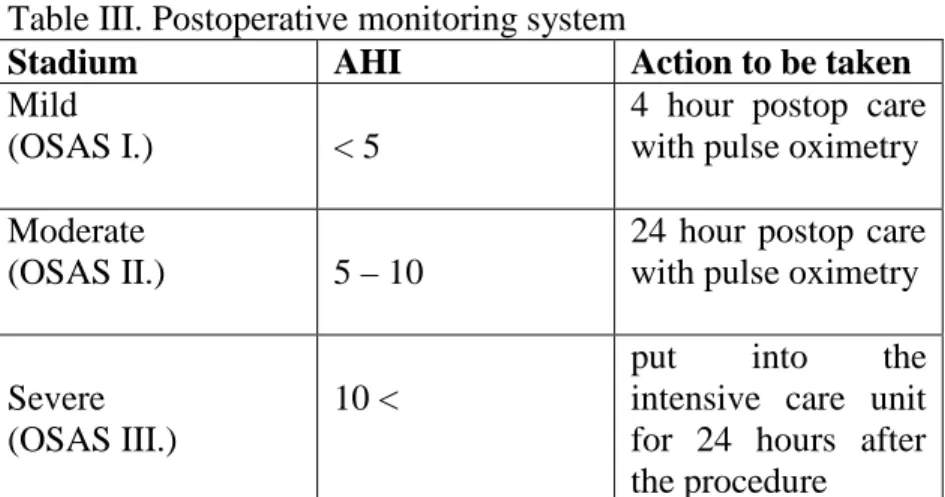
5.2. To elaborate a recommendation for the postoperative monitoring related to the severity of the OSAS to diminish the incidence of the postoperative respiratory complications The incidence of postoperative respiratory complications in this population is 16-27% compared with patients without OSAS. During my work I elaborated a recommendation for the postoperative monitoring related to the severity of the OSAS to diminish the incidence of the postoperative respiratory complications. (Table III.)
Table III. Postoperative monitoring system
Stadium AHI Action to be taken
Mild
(OSAS I.) < 5
4 hour postop care with pulse oximetry
Moderate
(OSAS II.) 5 – 10
24 hour postop care with pulse oximetry
Severe (OSAS III.)
10 <
put into the intensive care unit for 24 hours after the procedure
5.3.To determine the level of the obstruction and identify the most appropriate therapeutic plan with sleep endoscopy in the cases when the site of the obstruction was not clear or the patients retained persistent OSAS after tonsilloadenotomy.
Assessment of the site of obstruction is essential for the successful therapy of OSAS in children. Sleep endoscopy
provides accurate data on the location of obstruction. Together with the “gold standard” diagnostic method (polysomnography), sleep endoscopy can enable the physician to achieve the correct diagnosis and identify the most appropriate individual therapeutic plan.
5.4. The effects of sleep disorders on different aspects of memory functions by testing children with sleep- disordered breathing
Memory deficits lead to behavior problems, decreased learning abilities in patients suffering from OSAS. Declarative memory functions deteriorated due to the sleep disruption and desaturations while the nondeclarative form of learning was preserved, in spite of permanent sleep disruption in SDB.
7. List of publications
7.1. Publications related to the thesis
1. Benedek, P. (2009). A gyermekkori obstruktív alvási apnoe szindróma. Gyermekgyógyászat 60, 103–105.
2. Benedek, P., and Katona, G. (2011). A sleep
endoscopia helye a gyermekkori obstruktív alvási apnoe szindróma diagnosztikájában. Fül-Orr-Gégegyógyászat 57, 84–88.
3. Benedek, P., Sáringer, A., Katona, G., and Czinner, A.
(2002). A gyermekkori obstruktív alvási apnoe syndromás gyermekek kivizsgálása és kezelése kórházunkban. Fül-orr-gégegyógy 48, 156–159.
4. Benedek, P., Lázár, Z., Bikov, A., Kunos, L., Katona, G., and Horváth, I. (2013). Exhaled biomarker pattern is altered in children with obstructive sleep apnoea syndrome. Int. J. Pediatr. Otorhinolaryngol. 77, 1244–
1247. IF: 1,35
5. Benedek, P., Kiss, G., Csábi, E., and Katona, G. (2014).
Obstruktív alvási apnoe szindrómában szenvedő
gyermekek posztoperatív monitorozása. Orvosi Hetilap 155, 703–707.
6. Csábi, E., Benedek, P., Janacsek, K., Katona, G., and Nemeth, D. (2013). Sleep disorder in childhood impairs declarative but not nondeclarative forms of learning. J Clin Exp Neuropsychol 35, 677–685. IF: 1,862 7. Ricz, Z., Benedek, P., and Axmann (2002). Az
obstructiv alvási apnoe szindróma (OSAS) áttekintése egy súlyos esetünk kapcsán. Gyermekgyógyászat 53, 153–167.
1. 7.2. Other publications
Farkas, Z., Katona, G., Késmárszky, R., Benedek, P., Majoros, T., Pataki, L., and Csákányi, Z. (2001). A gyermekkori otitis media acuta purulenta (AOM) és a sinusitis maxillaris acuta purulenta (SM) kezelésének megváltozása. Medicus Universalis 34, 309–311.
2. Farkas, Z., Lőrincz, A., Csákányi, Z., Sultész, M., Majoros, T., Benedek, P., Dános, K., and Katona, G.
(2013). Idegentest (IT) eltávolítás gyermekkorban.
Gyermekorvos Továbbképzés 12, 9–11.
3. Katona, G., Benedek, P., Csákányi, Z., Farkas, Z., Majoros, T., and Pataki, L. (2002a). A radiofrekvenciás sebészet gyermek-fül-orr-gégészeti alkalmazása. Fül- Orr-Gégegyógyászat 48, 39–42.
4. Katona, G., Benedek, P., Csákányi, Z., Farkas, Z., Majoros, T., and Pataki, L. (2002b).
Aryepiglottoplasztika : a laryngomalacia műtéti kezelése. Fül-Orr-Gégegyógyászat 48, 79–83.
5. Kunos, L., Bikov, A., Lazar, Z., Korosi, B.Z., Benedek, P., Losonczy, G., and Horvath, I. (2014). Evening and morning exhaled volatile compound patterns are different in obstructive sleep apnoea assessed with electronic nose. Sleep Breath. IF: 2,256