Evaluation of commonly used tear sampling methods and their relevance in subsequent biochemical analysis
Aniko Rentka
1,*, Krisztina Koroskenyi
2,*, Jolan Harsfalvi
3, Zoltan Szekanecz
4, Gabriella Szucs
4, Peter Szodoray
5and Adam Kemeny-Beke
1Abstract
The human precorneal tear film is a special body fluid, since it is a complex mixture of proteins, lipids, small bioactive molecules, and their concentrations and relative distribution represent not only the metabolic state of the ocular surface but also the systemic and local homeostasis of the outer eye and the human body. This suggests that biochemical analysis of the precorneal tear film composition may provide a non-invasive tool for diagnosis and monitoring of disease pro- gression or treatment efficacy in human medicine. However, collecting tears is demanding, and obtaining reproducible and unaltered samples is challenging because of the small sample volumes of tears. Several methods are available for tear collection as a preparatory step of precorneal tear film analysis, and the collection method used has to be assessed since it has a critical impact on the effectiveness of the assays and on the quality of the results. Each sampling method has advantages and disadvantages; therefore, it is not easy to choose the appropriate collecting method for tear collection.
To overcome these limitations various methods have been recommended by different authors for special aspects of specific tests. The aim of our review was to evaluate tear sampling methods with regard to our ongoing biochemical analysis.
Keywords
Precorneal tear film, tear sampling, tear collection, bioactive components, tear diagnostics
Accepted: 2nd February 2017
Introduction
Precorneal tear film (PCTF) as a biological fluid is very easily accessible with non- or very low-invasive meth- ods at a relatively low cost. It not only lubricates the ocular surface carrying secreted molecules from corneal epithelial cells and tissues producing tear components but can also represent the physiological status of the body. Due to the very limited number of samples and the relative instability of the components, sample col- lection is a critical step in tear research and diagnostics.
In the present review, we summarize the most com- monly used tear sampling methods, emphasizing their advantages and disadvantages based particularly on the subsequent analysis.
1Department of Ophthalmology, Faculty of Medicine, University of Debrecen, Debrecen, Hungary
2Department of Biochemistry and Molecular Biology, Signaling and Apoptosis Research Group, Hungarian Academy of Sciences, Research Center of Molecular Medicine, University of Debrecen, Debrecen, Hungary
3Department of Biophysics and Radiation Biology, Semmelweis University, Budapest, Hungary
4Department of Rheumatology, Institute of Medicine, Faculty of Medicine, University of Debrecen, Debrecen, Hungary
5Institute of Immunology, Rikshospitalet, Oslo University Hospital, Oslo, Norway
*Contributed equally.
Corresponding author:
Adam Kemeny-Beke, Department of Ophthalmology, Faculty of Medicine, University of Debrecen, Nagyerdei krt. 98, Debrecen H-4012, Hungary.
Email: kemenyba@med.unideb.hu
Annals of Clinical Biochemistry 2017, Vol. 54(5) 521–529
!The Author(s) 2017 Reprints and permissions:
sagepub.co.uk/journalsPermissions.nav DOI: 10.1177/0004563217695843 journals.sagepub.com/home/acb
Tear biology: Functions and pathological relations of the human tear
The tear film covering the ocular surface has several functions including protecting the external surface of the eyeball thus constituting a mechanical and anti- microbial barrier. It lubricates the eye surface and nourishes also the avascular tissues of the cornea.
Since the tear film is also an optical refractive medium, its stability is pivotal in achieving appropriate vision.1–3
The human tear film is composed of two layers: there is a lipid layer and an aqueous layer that include soluble proteins and mucins and also membrane-bound mucins.3The lipid component, which originates from the Meibomian glands of the tarsus, forms the superfi- cial layer of the tear film. The aqueous component is primarily secreted by the lacrimal gland. This is the thickest layer, which contains water, electrolytes and diverse proteins, peptides and glycoproteins. Mucins, which are glycoproteins expressed by epithelial tissues of mucous surfaces, protect tissues since they are anti- oxidants, provide lubrication and inhibit bacterial adherence.4–7
Under normal conditions, the PCTF flow in humans is around 0.5–2.2lL/min with a turnover rate of approximately 16% per minute.8PCTF volume present in the human eye is 7–10lL.9 A continuous cycle of tear production, evaporation, drainage and absorption results in a dynamic equilibrium of the ocular surface.
Osmolarity is the index of tear dynamics and is mainly determined by the electrolytes of the aqueous phase of the PCTF. Under normal conditions the expected range is 3028 mOsm/L. The dry eye workshop in 2007 identified increased tear osmolarity and tear film instability as ‘core mechanisms’ of DE, regardless of the oetiology.4,6 DE studies using the TearLab Osmolarity System have found that the mean tear osmolarities of the mild-to-moderate and severe DE patients were 315.011.4 and 336.422.3 mOsm/L, respectively.5
Furthermore, tears have to be viscous enough to protect and lubricate the surface, but not too viscous so as to avoid ocular surface damages of high shear forces caused by blinking. The normal tear fluid has a viscosity of 1.3–5.9 cP.7
The approximate properties of normal tear fluid are outlined in Table 1.
Irritating stimuli like environmental fluctuations, diurnal patterns and physiological status indicate reflex tear secretion via activation of the corneal nerves.8–12 Open and closed eye PCTF differ in com- position and origin, hence eye closure also influences the result of tear analysis.13
Changes in tear composition are associated with many ocular diseases, such as dry eye syndrome,
Sjo¨gren’s syndrome, diabetic retinopathy, glaucoma, Meibomian gland dysfunction, autoimmune thyroid eye disease, pterygium, keratoconus, ocular rosacea, blepharitis, as well as various systemic diseases such as inflammatory diseases and infections, diabetes melli- tus, allergies, Parkinson’s disease and certain types of cancers (breast, lung, prostate).14–17 Despite the fact that some biochemical properties (e.g. pH or osmolal- ity) of tears and serum are similar, the protein compos- ition, as well as the relative and absolute amount of the components are different. For example, Vitamin A con- centration in tears is remarkably higher than in serum.
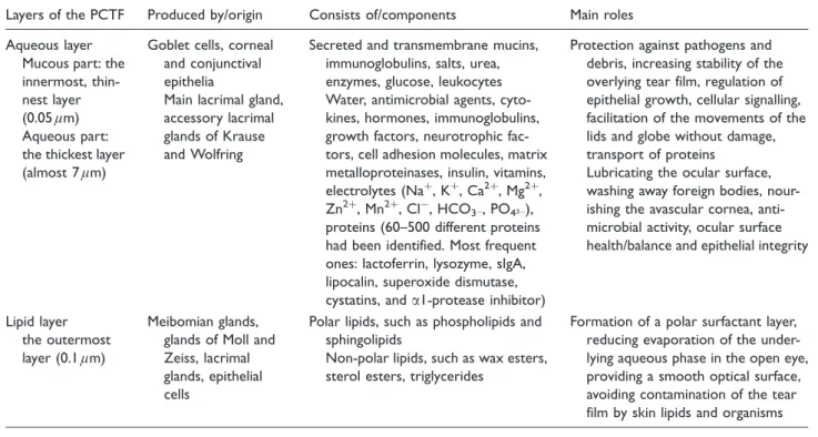
Similarly, the fibronectin content of PCTF is one order of magnitude higher than that of serum.18 Therefore, tears should be considered a unique body fluid. Table 2 summarizes data of tear film lipid layers, i.e. their origin, components and main roles (Table 2).
Major bioactive components of PCTF
Despite its small volume, tear film is a remarkably com- plex biological fluid consisting of peptides, electrolytes, lipids, carbohydrates, salts and small bioactive mol- ecules such as amino acids, nucleosides, vitamins, etc.10,19 Normal tears have a total protein concentra- tion of approximately 7 g/L and they contain hundreds of different proteins, though the method of tear collec- tion greatly influences the relative proportion of the proteins present in any individual tear sample, as clar- ified in the early 1980s.20–22Many of the tear proteins play an important role in corneal wound healing, inflammatory processes and corneal protection against various pathogens.23
The most frequent proteins detected in tear samples include lactoferrin, lysozyme, secretory immunoglobulin Table 1. Properties of precorneal tear film (PCTF).
Origin Lacrimal functional unit: the main and accessory lacrimal glands;
the ocular surface: cornea, con- junctiva and Meibomian glands, the eyelids, the interconnecting sensory and motor nerves
Volume 7–10lL
Flow (secretion velocity) 0.5–2.2lL/min
Osmolarity 4290 mOsmol/L
Turnover rate 16%/min
Layers (1) Aqueous, including soluble
proteins and mucins and membrane-bound mucins (2) Lipid/oily
Thickness 3–11lm
Total protein concentration 7 g/L
A (sIgA), lipocalin, superoxide dismutase, cystatins and a1-protease inhibitor. These proteins account for more than 90% of all tear proteins.2,24,25 In the early years, tear film protein profiles were characterized using gel electrophoresis and Edman degradation.26–28 Later, sensitive immunoassay-based methods identified other proteins in mammals’ tears, including growth factors, neurotrophic factors, cytokines and cell adhesion molecules, matrix metalloproteinases, immunoglobulins and insulin.29,30
Prospects for the future: Tear diagnostics
Body fluid analysis is a widely accepted, readily repeated, convenient and low cost method in diagnos- tics. Biomarker screening of various body fluids may have potential benefits not only for the examination of physiological processes but also for the early diag- nosis and effective therapy of several diseases. Fluid biomarkers include macromolecules such as lipids, proteins, RNA and DNA, as well as cells such as immune, endothelial or even cancer cells.31
Tears, a body fluid exposed to both internal and external environment, contain an amazing amount of molecular information, which is useful for the diagno- sis, prognosis and treatment of ocular surface diseases.
This may promote the development of personalized medicine and the utilization of biomarkers in certain diseases.10
Tear collection methods and their application in practice
Quantitative determination of tear proteins is of increasing interest in ophthalmology, but still there remains a technical problem due to small sample volumes available and the complexity of sample composition.32Tear sampling is definitely a major chal- lenge and has the greatest significant influence on the precision and reproducibility of the analytical results.
Direct sampling methods. Regarding the direct sampling method, microcapillary tubes (MCT) or micropipettes are used for sampling and this manner frequently requires previous stimulation or instillation of different volumes of saline (100–200lL) into the cul-de-sac and collecting after sufficient mixing. This procedure can cause dilution and may not permit collection of samples from specific sites of the ocular surface.33Kalsow et al.
investigated tear cytokine response to multipurpose solutions in contact lens wear. Prior to contact lens removal, non-stimulated tears (NST) were collected from each eye from the inferior tear meniscus between the 6 o’clock and lateral canthus positions using a 10lL flame-polished glass micropipette. Following collection, a 5.5lL volume of tear was immediately transferred to a sterile 0.2 mL tube containing 49.5lL of storage solution to produce a 1:10 tear dilution for immediate storage at80C.34
Table 2. Layers of tears: Origins, components and main roles.
Layers of the PCTF Produced by/origin Consists of/components Main roles Aqueous layer
Mucous part: the innermost, thin- nest layer (0.05lm) Aqueous part:
the thickest layer (almost 7lm)
Goblet cells, corneal and conjunctival epithelia
Main lacrimal gland, accessory lacrimal glands of Krause and Wolfring
Secreted and transmembrane mucins, immunoglobulins, salts, urea, enzymes, glucose, leukocytes Water, antimicrobial agents, cyto- kines, hormones, immunoglobulins, growth factors, neurotrophic fac- tors, cell adhesion molecules, matrix metalloproteinases, insulin, vitamins, electrolytes (Naþ, Kþ, Ca2þ, Mg2þ, Zn2þ, Mn2þ, Cl, HCO3, PO43), proteins (60–500 different proteins had been identified. Most frequent ones: lactoferrin, lysozyme, sIgA, lipocalin, superoxide dismutase, cystatins, anda1-protease inhibitor)
Protection against pathogens and debris, increasing stability of the overlying tear film, regulation of epithelial growth, cellular signalling, facilitation of the movements of the lids and globe without damage, transport of proteins
Lubricating the ocular surface, washing away foreign bodies, nour- ishing the avascular cornea, anti- microbial activity, ocular surface health/balance and epithelial integrity
Lipid layer the outermost layer (0.1lm)
Meibomian glands, glands of Moll and Zeiss, lacrimal glands, epithelial cells
Polar lipids, such as phospholipids and sphingolipids
Non-polar lipids, such as wax esters, sterol esters, triglycerides
Formation of a polar surfactant layer, reducing evaporation of the under- lying aqueous phase in the open eye, providing a smooth optical surface, avoiding contamination of the tear film by skin lipids and organisms PCTF: precorneal tear film.
In 2012, Guyette et al. compared low-abundance biomarker concentrations in capillary-collected NST and washout (WO) tears of aqueous-deficient and normal patients. Ten-microliter polished micropipettes were used to collect tears from the inferior marginal strip taking great care in minimizing ocular surface con- tact. Tear collection rate was continuously monitored.
Individual NST samples were collected in 10 min ali- quots and then immediately transferred to a sterile polymerase chain reaction (PCR) tube. An equal volume of assay buffer was added and the sample was stored at 86C. A total of at least 6.5lL NST was collected from each study participant; each 10 min ali- quot was stored without delay in a separate PCR tube.
Prior to WO tear sample collection 10lL sterile physio- logic saline solution was added to the lower conjunctiva by a digital pipette. The patient was instructed to gently close their eyes and avoid any eye movements for 1 min.
Tears were then collected using the same method as for NST samples, but a shorter collection time of 5 min per aliquot was used to make up the 6.5lL minimum volume requirement. Tear collection volume and time were continually monitored to measure the tear collec- tion rate.35
There have been several research projects in dry eye syndrome and today emphasis in dry eye research has shifted towards the role of inflammation in the anterior surface of the eye.36Since inflammatory mediators ori- ginating from various ocular surface sources and the main lacrimal gland do not constitute a totally homo- genous mix the way tears are collected can influence the resulting biomarker profile. NST from the inferior mar- ginal strip covers a broader spectrum of the sources, whereas stimulated tear (ST) samples contain a higher proportion of the lacrimal gland secretion.1 Explicit protein profile differences between NST and ST dem- onstrate that these two sample types are not equiva- lent.37,38 Although NST represents specifically the inflammatory status of the ocular surface, the volume of NST is limited, especially in aqueous deficient dry eye (ADDE). Even though tear sampling frequently makes use of capillaries as they are less irritating and the resulting sample is an exact representative concen- tration of molecules, the main limitation of the method is the volume of the sample (2–3lL) to be gained.8
One way to increase the available tear sample volume is adding fluid (e.g. sterile saline) to the eye prior to sample collection, effectively ‘washing out’
ocular surface molecules.39,40 Validity of the WO method depends on the extent to which it changes the NST biomarker profile. By determining tear sIgA, inducement of reflex tearing is easily detected because tear sIgA concentrations decrease with reflex tear flow rate. Markoulli et al. found equal tear sIgA–total tear ratios in WO and NST, which suggests that WO
samples do not significantly induce reflex tearing.
Guyette’s study evaluated WO tear collection as a replacement for capillary NST and applied this to com- pare biomarker concentrations between ADDE and non-ADDE patients.35,38,41,42
Indirect methods. Regarding indirect methods, collection of PCTF is carried out using absorbing supports such as Schirmer test strips (STS), filter paper disks, cellulose sponges and polyester rods. The most common method among them is STS collection.43
Inflammatory markers were analysed in the PCTF of patients with ocular surface disease. Ten microliters of tear was collected by a Weck-Cell Sponge. The concen- trations of interleukin (IL)-1, IL-6 and pro-MMP-9 were measured by enzyme-linked immunosorbent assay (ELISA), and the MMP-9 activity was evaluated with gelatine zymography.44
Ophthalmic sponges and extraction buffers were compared for quantifying cytokine profiles in tears using Luminex technology. Luminex detection of cyto- kine/chemokine profiles of tears collected with Merocel sponges was found to be useful in clinical studies, for instance to assess cytokine profiles evaluation in ocular surface diseases.45
Samples obtained from the Schirmer procedure have a higher mucus, lipid and cellular content than MCT samples.46 STS also suffers incomplete, non-uniform elution of proteins from the filter matrix.43 Although micropipette and STS collection provide different bio- marker profiles for a given donor, the correctly applied micropipette method is more consistent.47STS is widely accepted as the volume of sample collected with this method is larger compared to other methods, but it can cause reflexive tearing due to irritation, which increases the volume of the samples, therefore aggra- vates the detection of the investigated tear compo- nent(s), e.g. drug concentrations.
In comparative studies tears of one and the same patient are collected by several collection methods to determine the same biomarkers from different tear samples.
Green-Church et al. collected tears using small volume (1–5lL) Drummond glass MCT tubes with 1.6slit-lamp magnification. Non-reflex tears were col- lected from the inferior tear prism without contact with the lower lid until a total of 5lL were collected. During a separate visit, tear collection was performed by placing a STS over the lower lid. The lid was not anesthetized and the STSs were placed approximately 6 mm nasally from the lateral canthus. The subject was instructed to close their eyes for the 5 min test duration;
the wet length was not recorded but was observed to be within reference ranges in all cases. The STS was then placed in a 1.6 mL amber Eppendorf tube at 4C until
analysis. Their results suggest that the tear film collec- tion method does impact the proteins present in the sample, so care should be exercised in choosing a tear collection method in order to best correlate to the experiment being conducted or the hypothesis being tested.47
Lee et al. used two collection techniques for the com- parative analysis of PCR assay to detect a pathogen, namely herpes simplex virus 1.48 Tears were collected from the lower fornix using STSs for 5 min, a method adopted in a previous study of Satpathy et al.49 The other collection method was micro pipetting of tears, after irrigating 100lL saline in the lower fornix. This method was described in a previous study, where the
‘flush’ tear collection technique was validated as a viable alternative to basal and reflex tear collection.42 Based on their PCR results Lee et al. established that PCR positive rate was not dependent on the tear col- lection method or primers.48 Comparison of a direct and an indirect method was performed by Jones et al.
They collected tears from healthy volunteers with either porous polyester rods or glass capillary micropipettes.
Tear collection rate and recovery of two tear proteins, EGF and lactoferrin, were compared in samples col- lected with the two methods. Their results showed that polyester rods collected tears an average of 3.9- fold faster than glass capillary micropipettes, but this difference was not statistically significant. Lastly, they suggested that polyester rods may have greater clinical utility, facilitating routine analysis of the PCTF.50 Table 3 summarizes the different tear collection meth- ods published by various authors from the past few years. All studies performed involved human participants.
The main advantage of the direct sampling methods is the straight way to collect tears from the ocular sur- face, and the main disadvantages are the possible need of dilution and the impossibility of sample collection from detailed sites of tears. They may be difficult to perform in practice, but analytically they provide the most proper analyte concentration of tears. The indir- ect methods, however, are easy to implement, but ana- lytically they do not inevitably represent the biochemical characteristics of tears.
The critical aspects of tear sampling with respect to the subsequent analysis
In the last decade, advances in proteomics/metabolo- mics/lipidomics technologies have greatly expanded our knowledge of the biochemical composition of the ocular tear fluid. To date, a number of tear proteins and lipids have been identified as possible disease- related biomarkers.51The rapid development of various
‘omics’ methods facilitates the identification and
examination of tear-based biomarkers. In some cases, these techniques require specific sample collection, handling and storage procedures. Hereunder we would like to summarize the most commonly used methods in proteomics and lipidomics with special regard to their minimum sampling requirements.
Proteomics. Qualitative and quantitative tear protein examination methods include one- and two-dimensional gel electrophoresis, ELISA, high performance liquid chromatography (HPLC), mass spectrometry (MS) related techniques such as MS–MS, matrix-assisted laser desorption/ionization time-of-flight MS, surface- enhanced laser desorption/ionization time-of-flight MS, liquid chromatography–MS, various antibody arrays, multiplex bead analysis, Western blot analysis, etc.52,53 Using highly sensitive techniques – like isobaric tags for relative and absolute quantitation – more than 500 tear proteins have been identified so far.
Previous studies have indicated that sample handling variables such as sample collection conditions and time, storage temperature and time, storage tube, freeze/thaw cycles and protease inhibitors have significant effects on the results of protein analysis.54In general, sample col- lection should be scheduled at the same time of day (e.g. early morning) and samples should be transferred onto dry ice immediately after sampling to prevent pro- tein degradation. Addition of protective or stabilizing compounds (reducing agents, protease and peptidase inhibitors, etc.) would be desirable but inhibitor cock- tails may interfere with the subsequent MS analysis.
The collected samples should be aliquoted and stored with minimization of thaw/refreeze cycles, preferably at 80C.55 Theoretically, the frozen samples (at20 to 80C or in liquid nitrogen) can be stored for years protected from degradation.56
Another critical aspect of tear analysis is the limited volume of the samples and the relatively low number of proteins of interest. In addition, the stimulus conditions (NST versus ST) and collection technique can strongly affect the protein profile and volume of the tear sample.
Fullard and Snyder observed that the concentrations of eight proteins (IgA-SC, IgA1, IgA2, IgM, IgG, IgA, transferrin, serum albumin) from the 12 analysed tear proteins showed significantly higher concentrations in NST, and only four (lactoferrin, tear-specific prealbu- min, peroxidase, lysozyme) were in similar concentra- tions in both types of tears.38 On the other hand, the total tear protein content of NST samples decreased from 9.1 to 6.0 g/L in the ST ones.37These data indicate that the diluting effect of reflexive tearing has a decisive effect on the quantitative composition of the tear sample and highlights the importance of controlling tear flow rate during tear collection.55 Based on the above observation NST sampling seems to be more
Table3.Tearcollectionmethodsofsomeauthorsincludingstudiesfromthepastfewyearswithalargenumberofsamples.Allstudieswereperformedinhumans. Reference(s)StatusofindividualsAnalyteTearcollection method(s)TearvolumeAdvantage/DisadvantageBiochemicalmethod Kalsowetal.34 Contactlens wearersCytokinesNST(collectedwith 10lLglass micropipette) 5.5lLNSTcollectionwithglass micropipetteisafeasible methodfortearcytokine examinationwithcytokine assay
Multiplexcytokine beadassay Guyetteetal.35 ADDEandnon- ADDEpatientsLow-abundance biomarkersNSTandWO(both collectedwith10lL glassmicropipette)
Minimumof6.5lLSubstantiallyelevatedNSTcyto- kineconcentrationsmaynot beaccuratelyreflectedinWO tears.
PCR,multiplexcyto- kinebeadassay Markoullietal.42 HealthyindividualssIgANST,STandWO (collectedwith 10lLglasscapillary tubes)
100lLoftearsdiluted (1:3000)insample diluent
TearcollectionwiththeWO methodismuchfasterthan NSTsampling.Itreturns essentiallythesamespectrum ofproteinsinsimilarpropor- tions,buttearsecretionvel- ocitycannotbedetermined
ELISA,SDS-PAGE,MS Aceraetal.44 Patientswithocular surfacediseaseInflammatory cytokinesOphthalmicsponge10lLOnlyvalidatedforIL-betaIL-6 andMMP-9ELISA Inic-Kanadaetal.45 HealthyindividualsCytokinesDifferentophthalmic sponges(Merocel, Pro-ophta,Weck- Cell)
NotdefinableOphthalmicspongesarewell toleratedbythepatient, especiallychildren.Accurate tearvolumecannotbedefined
Multiplexcytokine beadassay Green-Church etal.47HealthyindividualsTearproteomeGlassmicrocapillary tubesandSchirmer strips
Minimumof5lL,3–16 pooledsamplesnot definable,Schirmer stripswereputinto 100lLbuffer Itisdifficulttodeterminesimilar proteinconcentrationson totalproteinquantitiesfroma Schirmerstripsincethe volumecollectedcannotbe measured
SDS-PAGE,2D-SDS- PAGE,and2D LC–MS/MSmultidi- mensionalprotein identification technology Leeetal.48 and Satpathyetal.49Patientswith herpeskeratitisHerpessimplex virus1SchirmerstripsWO (collectedwithglass capillary micropipette)
10lLThepositivityoftearPCR seemedtonotbedependent onthetearcollectionmethod ortheprimersused PCR ADDE:aqueousdeficientdryeye;ELISA:enzyme-linkedimmunosorbentassay;MS:massspectrometry;NST:non-stimulatedtear;PCR:polymerasechainreaction;SDS-PGE:sodiumdodecylsulphate polyacrylamidegelelectrophoresis;sIgA:secretoryimmunoglobulinA;ST:stimulatedtear;2DLC:two-dimensionalliquidchromatography;WO:washout.
beneficial unless the experimental goal is the investiga- tion of ST tears or reflexive tearing.
Lipidomics. While the protein composition of the human tear has been described in great detail, the lipidomic analysis of the tear is noticeably lagging behind due to the low lipid content of the tear fluid.57 Because of the various difficulties (lipid diversity and complexity, chemical stability or instability of different types of lipids) the qualitative and quantitative analysis of tear lipids is a difficult task.
Regarding the technical limitations (sensitivity and performance of the method; volatility and stability of intact lipids) the commonly used analytical methods – gas chromatography–mass spectroscopy and liquid chromatography–mass spectrometry (LC/MS) – are not sufficiently efficient and accurate for the lipidomic analysis of the human tear. HPLC and its newer and faster ‘relative’, ultra-high performance liquid chroma- tography (UPLC) mass spectrometry (HPLC/MS and UPLC/MS) and atmospheric pressure ionization MS are more suitable methods for the examination of intact lipids in tear fluid.58Nuclear magnetic resonance spectroscopy, Raman and infrared spectroscopy may also be used as alternative approaches, although their sensitivity and selectivity is far below that of MS.4,58
The requirements of sample collection are the same as in the case of protein determination. The samples should be stored frozen at 80C or lower, multiple melting of the samples should be avoided and the sam- ples should be handled in deeply frozen conditions (e.g.
at80C) until assessment. During the examination of lipids that are sensitive to light or the O2content of the air – such as retinoids – special attention is needed, e.g.
usage of amber-coloured Eppendorf tubes.58Basal tear collection using capillary tubes is recommended for tear lipid analysis as the concentration of several lipid classes is below the limit of detection in reflex and flush tears.59
Conclusions
Biomarker-based diagnostics, as well as personalized medicine utilizing its results are becoming more widely used in modern medicine. In addition to the
‘classical’ sampling methods (e.g. biopsy), there is a growing demand for fast, painless and non-invasive sampling procedures such as examination of various easily accessible body fluids. The identification and potential application of biomarkers carried by urine, sweat, amniotic fluid and last but not least precorneal tear are the subjects of intensive interest and research nowadays.
Similarly to other newly developed methods, precor- neal tear analysis has no standard methodology.
Sampling techniques used by different research groups are not exceptions to this rule. Both the investigation of
‘tear physiology’ and tear biomarker research are based on the biochemical characterization of so-called basal (or NST) tears, which differ from ST tears (also known as reflex tears) both in terms of quantitative and quali- tative biochemical characteristics. In practice, two quickly and easily adaptable sampling methods, the STS and the MCT technique are widely used for the collection of NST. Based on the previous statements the authors unequivocally recommend the capillary- collected NST sampling method for protein content determination since it represents the most accurate pro- tein concentration of tears. For lipid content determin- ation, NST is the most commendable sampling method as well, but in this case glass or polished micropipette is also available. There are no special considerations for mucin analysis as they consist of glycocalyx expressed by epithelial tissues of mucous surface and there is no direct simple method to evaluate ocular surface glycocalyx.
Since the STS method triggers more or less intense tearing, this technique is suitable for the isolation of a mixed sample containing NST and ST. Thus, the ana- lysis of samples collected by the STS method does not necessarily represent the biochemical properties of NS tears.
In the last decade, numerous studies have addressed the comparison of STS and MCT methods. It was observed in the late 1960s that the STS method may underestimate the actual protein concentrations of tears due to increased fluid flow. Some studies have since revealed that not only the diluting effect of reflex tear- ing, but also the protein binding and retention capacity – which is strongly associated with the molecular weight and hydrophobic surface area of the studied proteins – of STS paper is responsible for the lower protein content of STS samples. The STS method modifies both the quantitative and the qualitative char- acteristics of tear samples. In the late 1970s and 1980s, various research groups reported elevated concentra- tions of certain proteins – like albumin, IgG, transfer- rin, urokinase and plasmin as well as various intracellular enzymes involved in metabolism such as aspartate aminotransferase and alanine aminotransfer- ase, lactate dehydrogenase, aldolase, glucose-6- phosphate dehydrogenase – in the STS-collected tears compared to MCT samples. Comparing the proteome of tear samples collected by MCT and STS methods Green-Church et al. found that more than 50 different proteins were detected exclusively in the STS samples (MCT-specific proteins: 13; STS-specific proteins: 54;
overlapping proteins: 30; total: 97 proteins; determin- ation: in-gel tryptic digestion followed by liquid chromatography–tandem mass spectrometry and
multidimensional protein identification technology).
Several studies have confirmed that the STS method – which often triggers irritation in the lower cul-de-sac of the eye – changes the protein composition of tears by injuring the conjunctival surface and microvasculature.
In contrast to STS-based sampling, the MCT tech- nique is believed to be a less invasive procedure. If it is performed by a specialist who has practice and experi- ence in this collection method, the MCT technique does not induce reflex tearing, nor does it involve a potential risk of injury. Therefore, the MCT sampling method is more suitable for the collection of NST than the STS procedure. On the other hand, some researchers have highlighted the following disadvantages of the MCT method: the sampling is interrupted by blinking; STS could be more pleasant for the test subjects than the capillary tube; the investigator has to hold the capillary tube for the duration of the sampling procedure, which entails constant and prolonged work on the open eye.
For the compensation of low sample volume, several research groups use the so-called WO method, in which the tears are ‘flushed out’ through the addition of exogenous fluid (e.g. sterile physiol. saline). The dilut- ing effect of the WO method is reflected by the sup- pressed concentrations and the decreased variances of the most abundant cytokines (e.g. IL-8, IL-1 and vascular endothelial growth factor). In addition, the concentrations of ‘minor’ tear components drop below the detection limit and the slight differences between the samples become unrecognizable.
Therefore, the WO method can be used successfully only in those experiments which target proteins found in high concentration in tears.
Acknowledgements None.
Declaration of conflicting interests
The author(s) declared no potential conflicts of interest with respect to the research, authorship, and/or publication of this article.
Funding
The author(s) received no financial support for the research, authorship, and/or publication of this article.
Ethical approval
Local bioethics committee (DE KK RKEB/IKEB 4242-2014, and HBR/052/
01761-2/2014) and Declaration of Helsinki (1996).
Guarantor AKB.
Contributorship
All authors researched literature to perform the study. AR and KK wrote the first draft of the manuscript. All authors reviewed and edited the manuscript and approved the final version of the manuscript.
References
1. Tiffany JM. The normal tear film.Dev Ophthalmol2008; 41: 1–20.
2. Pflugfelder SC and Stern ME (eds)Dry Eye and Ocular Surface Disorders.
New York: Marcel Dekker, 2004, pp.44–49.
3. Lamberts DW. Punctal occlusion.Int Ophthalmol Clin1994; 34: 145–150.
4. The definition and classification of dry eye disease: report of the Definition and Classification Subcommittee of the International Dry Eye WorkShop (2007).Ocul Surf2007; 5: 75–92.
5. Sullivan BD, Whitmer D, Nichols KK, et al. An objective approach to dry eye disease severity.Invest Ophthalmol Vis Sci2010; 51: 6125–6130.
6. Potvin R, Makari S and Rapuano CJ. Tear film osmolarity and dry eye disease: a review of the literature.Clin Ophthalmol2015; 9: 2039–2047.
7. Coffey MJ, Decory HH and Lane SS. Development of a non-settling gel formulation of 0.5% loteprednol etabonate for anti-inflammatory use as an ophthalmic drop.Clin Ophthalmol2013; 7: 299–312.
8. Mishima S, Gasset A, Klyce SD Jr, et al. Determination of tear volume and tear flow.Invest Ophthalmol1966; 5: 264–276.
9. Carney LG and Hill RM. Human tear pH. Diurnal variations. Arch Ophthalmol1976; 94: 821–824.
10. Zhou L and Beuerman RW. Tear analysis in ocular surface diseases.Prog Retin Eye Res2012; 31: 527–550.
11. Walker PM, Lane KJ, Ousler GW, et al. Diurnal variation of visual func- tion and the signs and symptoms of dry eye.Cornea2010; 29: 607–612.
12. Benito MJ, Gonzalez-Garcia MJ, Teson M, et al. Intra- and inter-day variation of cytokines and chemokines in tears of healthy subjects.Exp Eye Res2014; 120: 43–49.
13. Sack RA, Tan KO and Tan A. Diurnal tear cycle: evidence for a nocturnal inflammatory constitutive tear fluid.Invest Ophthalmol Vis Sci1992; 33:
626–640.
14. Zhou L, Beuerman RW, Ang LP, et al. Elevation of human alpha-defensins and S100 calcium-binding proteins A8 and A9 in tear fluid of patients with pterygium.Invest Ophthalmol Vis Sci2009; 50: 2077–2086.
15. Wong TT, Zhou L, Li J, et al. Proteomic profiling of inflammatory signal- ing molecules in the tears of patients on chronic glaucoma medication.
Invest Ophthalmol Vis Sci2011; 52: 7385–7391.
16. Kim HJ, Kim PK, Yoo HS, et al. Comparison of tear proteins between healthy and early diabetic retinopathy patients.Clin Biochem2012; 45:
60–67.
17. Bohm D, Keller K, Pieter J, et al. Comparison of tear protein levels in breast cancer patients and healthy controls using a de novo proteomic approach.Oncol Rep2012; 28: 429–438.
18. Geerling G, Maclennan S and Hartwig D. Autologous serum eye drops for ocular surface disorders.Br J Ophthalmol2004; 88: 1467–1474.
19. Lam SM, Tong L, Duan X, et al. Extensive characterization of human tear fluid collected using different techniques unravels the presence of novel lipid amphiphiles.J Lipid Res2014; 55: 289–298.
20. Tiffany JM. Tears and conjunctiva. In: Harding JJ (ed.)Biochemistry of the eye. London: Chapman & Hall, 1997, pp.45–78.
21. Tuft SJ and Dart JK. The measurement of IgE in tear fluid: a comparison of collection by sponge or capillary.Acta Ophthalmol (Copenh)1989; 67:
301–305.
22. Copeland JR, Lamberts DW and Holly FJ. Investigation of the accuracy of tear lysozyme determination by the quantiplate method.Invest Ophthalmol Vis Sci1982; 22: 103–110.
23. de Souza GA, Godoy LM and Mann M. Identification of 491 proteins in the tear fluid proteome reveals a large number of proteases and protease inhibitors.Genome Biol2006; 7: 72.
24. Rantamaki AH, Seppanen-Laakso T, Oresic M, et al. Human tear fluid lipidome: from composition to function.PLoS One2011; 6: 19553.
25. Azzarolo AM, Brew K, Kota S, et al. Presence of tear lipocalin and other major proteins in lacrimal fluid of rabbits. Comp Biochem Physiol B Biochem Mol Biol2004; 138: 111–117.
26. Holopainen JM, Moilanen JA, Sorsa T, et al. Activation of matrix metal- loproteinase-8 by membrane type 1-MMP and their expression in human tears after photorefractive keratectomy.Invest Ophthalmol Vis Sci2003; 44:
2550–2556.
27. Dartt DA. Interaction of EGF family growth factors and neurotransmit- ters in regulating lacrimal gland secretion.Exp Eye Res2004; 78: 337–345.
28. Bours J, Reitz C, Strobel J, et al. Detection of secretory IgM in tears during rhino-conjunctivitis.Graefes Arch Clin Exp Ophthalmol2005; 243:
456–463.
29. Rocha EM, Cunha DA, Carneiro EM, et al. Identification of insulin in the tear film and insulin receptor and IGF-1 receptor on the human ocular surface.Invest Ophthalmol Vis Sci2002; 43: 963–967.
30. Barton K, Nava A, Monroy DC, et al. Cytokines and tear function in ocular surface disease.Adv Exp Med Biol1998; 438: 461–469.
31. Martin KJ, Fournier MV, Reddy GP, et al. A need for basic research on fluid-based early detection biomarkers.Cancer Res2010; 70: 5203–5206.
32. Li K, Chen Z, Duan F, et al. Quantification of tear proteins by SDS-PAGE with an internal standard protein: a new method with special reference to small volume tears.Graefes Arch Clin Exp Ophthalmol2010; 248: 853–862.
33. Small D, Hevy J and Tang-Liu D. Comparison of tear sampling techniques for pharmacokinetics analysis: ofloxacin concentrations in rabbit tears after sampling with Schirmer tear strips, capillary tubes, or surgical sponges.J Ocul Pharmacol Ther2000; 16: 439–446.
34. Kalsow CM, Reindel WT, Merchea MM, et al. Tear cytokine response to multipurpose solutions for contact lenses. Clin Ophthalmol 2013; 7:
1291–1302.
35. Guyette N, Williams L, Tran MT, et al. Comparison of low-abundance biomarker levels in capillary-collected nonstimulated tears and washout tears of aqueous-deficient and normal patients.Invest Ophthalmol Vis Sci 2013; 54: 3729–3737.
36. Report of the research subcommittee of the international dry eye work shop, 2007, pp.179–193.
37. Fullard RJ and Tucker D. Tear protein composition and the effects of stimulus.Adv Exp Med Biol1994; 350: 309–314.
38. Fullard RJ and Snyder C. Protein levels in nonstimulated and stimulated tears of normal human subjects. Invest Ophthalmol Vis Sci 1990; 31:
1119–1126.
39. Petznick A, Evans MD, Madigan MC, et al. A comparison of basal and eye-flush tears for the analysis of cat tear proteins.Acta Ophthalmol2011;
89: 75–81.
40. Bjerrum KB and Prause JU. Collection and concentration of tear proteins studied by SDS gel electrophoresis. Presentation of a new method with special reference to dry eye patients.Graefes Arch Clin Exp Ophthalmol 1994; 232: 402–425.
41. Senchyna M and Wax MB. Quantitative assessment of tear production: a review of methods and utility in dry eye drug discovery.J Ocul Biol Dis Infor2008; 1: 1–6.
42. Markoulli M, Papas E, Petznick A, et al. Validation of the flush method as an alternative to basal or reflex tear collection.Curr Eye Res2011; 36:
198–207.
43. VanDerMeid KR, Su SP, Krenzer KL, et al. A method to extract cytokines and matrix metalloproteinases from Schirmer strips and analyze using Luminex.Mol Vis2011; 17: 1056–1063.
44. Acera A, Rocha G, Vecino E, et al. Inflammatory markers in the tears of patients with ocular surface disease.Ophthalmic Res2008; 40: 315–321.
45. Inic-Kanada A, Nussbaumer A, Montanaro J, et al. Comparison of oph- thalmic sponges and extraction buffers for quantifying cytokine profiles in tears using Luminex technology.Mol Vis2012; 18: 2717–2725.
46. Choy CK, Cho P, Chung WY, et al. Water-soluble antioxidants in human tears: effect of the collection method.Invest Ophthalmol Vis Sci2001; 42:
3130–3134.
47. Green-Church KB, Nichols KK, Kleinholz NM, et al. Investigation of the human tear film proteome using multiple proteomic approaches.Mol Vis 2008; 14: 456–470.
48. Lee SY, Kim MJ, Kim MK, et al. Comparative analysis of polymerase chain reaction assay for herpes simplex virus 1 detection in tear.Korean J Ophthalmol2013; 27: 316–321.
49. Satpathy G, Mishra AK, Tandon R, et al. Evaluation of tear samples for Herpes Simplex Virus 1 (HSV) detection in suspected cases of viral keratitis using PCR assay and conventional laboratory diagnostic tools. Br J Ophthalmol2011; 95: 415–418.
50. Jones DT, Monroy D and Pflugfelder SC. A novel method of tear collec- tion: comparison of glass capillary micropipettes with porous polyester rods.Cornea1997; 16: 450–458.
51. Tsuji F and Kawazu K. Biomarker identification of tear fluid.
Metabolomics2012; 2: 105.
52. Sack RA, Conradi L, Krumholz D, et al. Membrane array characterization of 80 chemokines, cytokines, and growth factors in open- and closed-eye tears: angiogenin and other defense system constituents.Invest Ophthalmol Vis Sci2005; 46: 1228–1238.
53. Hagan S and Tomlinson A. Tear fluid biomarker profiling: a review of multiplex bead analysis.Ocul Surf2013; 11: 219–235.
54. Lygirou V, Makridakis M and Vlahou A. Biological sample collection for clinical proteomics: existing SOPs.Methods Mol Biol2015; 1243: 3–27.
55. Hu S, Loo JA and Wong DT. Human body fluid proteome analysis.
Proteomics2006; 6: 6326–6353.
56. de Jager W, Bourcier K, Rijkers GT, et al. Prerequisites for cytokine meas- urements in clinical trials with multiplex immunoassays.BMC Immunol 2009; 10: 52.
57. Lam SM, Tong L, Reux B, et al. Rapid and sensitive profiling of tear wax ester species using high performance liquid chromatography coupled with tandem mass spectrometry.J Chromatogr A2013; 1308: 166–171.
58. Butovich IA. Tear film lipids.Exp Eye Res2013; 117: 4–27.
59. Rohit A, Stapleton F, Brown SH, et al. Comparison of tear lipid profile among basal, reflex, and flush tear samples. Optom Vis Sci 2014; 91:
1391–1395.