Transcriptome analysis of an ochratoxin-A biodegrading bacteria
Mohammed AL-NUSSAIRAWI1– Balázs KRISZT1– Csilla KRIFATON1– Mátyás CSERHÁTI1 1: Department of Environmental Protection and Safety, Faculty of Agricultural and Environmental Sciences, Szent István University, 1 Páter Károly Street, Gödöll˝o
Corresponding author: Mátyás CSERHÁTI, E-mail: cserhati.matyas@mkk.szie.hu
Abstract: Fighting against and decreasing the effect of mycotoxins is an emerging problem. Among post- harvest methods are physical, chemical, and biological ones. This study is focusing on the biological tools for minimalizing the harmful effect of the ochratoxin-A (OTA) occurring on crops and fodders. The bac- teriaCupriavidus basilensis ÖR16 strain has very good ability to detoxify ochratoxin-A to phenylalanine and ochratoxin-alfa. In previous studies the degradation rate of the ÖR16 bacteria was over 98%. The whole genome sequencing was also performed by our group in 2012. During this research, the enzymes, and genes responsible for the OTA degradation were characterized via transcriptome analyses. 15 genes were identified, which could play role in the degradation of OTA. Testing and investigating these nominated genes and en- zymes could lead for a prepared fodder additive, which can help in the elimination of the negative effects of OTA in the future.
Keywords: OTA, genes, biodegradation
Received 23 May 2020, Revised 15 June 2020, Accepted 15 September 2020
Introduction
Ochratoxin-A (OTA) is one of the most im- portant mycotoxins, which is produced as toxic metabolites by genera belonging toAs- pergillusandPenicilliumspecies, mainlyAs- pergillus niger, A. ochraceus, A. carbonar- ius, and Penicillium verrucosum (De Bel- lis et al., 2015). The chemical structure of OTA is (N-[(3R)-5-chloro-8-hydroxy-3- methyl-1-oxo-3,4-dihydro-1H-isochromen- 7-yl]carbonyl-L-phenylalanine) includes a b- phenylalanine-dihydro isocoumarine deriva- tive, which is very constant at intense tem- perature and resistant to hydrolysis. The cur- rently used managing methods of raw ma- terials in feed and food production does not reduce the OTA, therefore the toxin persists in the final food and feed products (Ferenczi et al., 2014). OTA is considered to be one of the important contaminants which targets ce- real grains and crop products, peanuts, coffee beans, red wine and pork products (Bragu- lat et al., 2008). The occurrence of OTA can be detected in all regions due to inappro- priate storage of human foods and animal derived products and weather conditions.
The International Agency for Research on
Cancer (IARC) has been classified OTA as group 2B - possible carcinogenic to humans (IARC, 1993). OTA mainly target the kidney in humans as well as to its ability to induce porcine nephropathy (Duarte et al., 2012).
There are several strategies focusing on the reduction or elimination of OTA concen- tration in human food and animal feeds.
The physical adsorbents procedure, besides, has various disadvantages despite that its widely used method, such as nonspecific bindings of some important nutrients (vi- tamins, minerals, and therapeutic agents), high cost and limited efficiency. Accord- ing to the literature, the biological meth- ods are the most effective and promising methodology for controlling the contami- nation of OTA in food and animal feeds, thus diminish the risk on animals and hu- man health. More than ten species of bacte- ria, including our selected bacteria, have the ability to degrade OTA: Acinetobacter cal- coaceticus (De Bellis et al., 2015), Acine- tobacter sp. (Liuzzi et al., 2017), Alcali- genes faecalis(Zhang et al., 2017),Bacillus licheniformis (Petchkongkaew et al., 2008), Bacillus amyloliquefaciens (Chang et al.,
2015),Brevibacterium spp.(Rodríguez et al., 2011), Lactobacillus acidophilus (Fuchs et al., 2008),Pediococcus parvulus(Abrunhosa et al. 2014), Lactobacillus spp. (Luz et al., 2017).
In this study the Cupriavidus basilensis ÖR16 strain OTA degradation was inves- tigated in the level of RNA expression.
The genus Cupriavidus was identified in 2004 (Coenye et al., 2003). Members of this genus are Gram-negative, chemoorgan- otrophic and facultative chemolithotrophic bacteria that can be found in diverse habi- tats such as soil, root nodules and aquatic environment. The genus Cupriavidus be- longs to the family Burkholderiaceae and the class b-proteobacteria. The genus con- sists of nineteen type strains. Remarkable heavy metal tolerance of environmental iso- lates has been confirmed (Goris et al., 2001).
According to the literature in the case of 7 strains different xenobiotic biodegradation was observed. For example chlorinated aro- matic chemicals; halo benzoate and nitro- phenols were degraded byCupraividus neca- tor CCUG 52238T (Makkar and Casida, 1987) and some xenobiotic genes and en- zymes such as benzoate1,2-dioxygenase and chlorocatechol-degradative for this strain were reported (Ogawa and Miyashita, 1999).
Cupriavidus basilensis RK1 DSM 11853T strain was originally isolated as a 2,6- dichlorophenol degrading strain (Steinle et al., 1998). Other isolates of the species are also capable for degradation of various xeno- biotics such as furfural, 5-hydroxymethyl furfural, bisphenol-A, chlorophenols and atrazine (Stamper et al. 2003, Koopman et al., 2010).
In the case of Cupriavidus basilensis ÖR16 one study in mice demonstrated the effec- tiveness of the strain’s detoxification ability (Ferenczi et al., 2014). There was efficient degradation for the OTA by the 5th day of the experiment by the strain. The by-product group which arrived from OTA degradation
process of the C. basilensis ÖR16 was not toxic on the mice kidney cells. In 2012 the total genome project was preceded of theC.
basilensisÖR16 strain for making a founda- tion of future studies (Cserháti et al., 2012).
Materials and Methods The experiment reagents
Ochratoxin-A mycotoxin (OTA) (Sigma- Aldrich Co., USA). Luria-Bertani medium (LB) (100%: 10 g tryptone, 5 g yeast ex- tract, 9 g sodium-chloride). Minimal buffer medium (3.1 g of K2HPO4, 1.7 g of NaH2PO4 · 2H20, 4.0 g of (NH4)2SO4, 0.2 g of MgCl2 · 6H2O, 20 mg of EDTA, 4 mg of ZnSO4·7H20, 2 mg of CaCl2·2H20, 10 mg of FeSO4 ·7H20, 0.4 mg of Na2MoO4 · 2H20, 0.4 mg of CuSO4 · 5H2O, 0.8 mg of CoCl2·6H2O, 2 mg of MnCI2·2H2O).
Bacterial strain and culture conditions in the biodegradation experiment
The strainCupriavidus basilensisÖR16, was isolated from a Hungarian pristine soil sam- ple. It was deposited in the National Collec- tion of Agricultural and Industrial Microor- ganisms (NCAIM BO2487). It was grown on LB agar plates and incubated at 28 °C for 72 h. Single colonies were inoculated into 50 ml liquid LB medium and incubated at 170 rpm at 28 °C for 72 h. After resuspen- sion, the optical density (OD600) of the cul- ture was measured at 600 nm (OD 600) (IM- PLEN SpectroPhotometer, GENESIS 10S, Thermo Fischer Scientific) and adjusted to 0.6 (OD600 = 0.6) to prepare bacterial in- oculum, from this 10 ml was added to 45 ml minimal buffer, which contained 7 mg/l OTA in final concentration.
OTA degradation experiment for getting the RNA to transcriptome analysis
The C. basilensis ÖR16 strain was cultured in LB media for growing and getting the ex- act cell number. The OTA degradation was
carried out in a minimal buffer,C. basilensis ÖR16 was grown on LB agar plates and incu- bated at 28 °C for 72 h. Single colonies were inoculated into 50 ml liquid LB medium and incubated at 170 rpm at 28 °C for 72 h. Cul- tures then centrifuged and cleaned from LB media via minimal buffer.
10 ml of the ÖR16 was added to 45 ml minimal buffer and 45 ml 2% fructose (200 ml Demineralized Water, 4.0 g D-Fructose), only with fructose as carbon source, to acti- vate just those genes, which are responsible or act in the presents of OTA or OTA degra- dation and incubated for 11 hours (till reach- ing the log phase of ÖR16).
For control E. coli TOP10 was used in LB and in minimal buffer media, incubated in the same circumstance as ÖR16. After 11 hours incubation, OD was measured to reach 0.4-0.8 (to be suitable with the requirement of the RNA isolation kit). 7 ppm of OTA was added to the target groups (ÖR16 + OTA).
Samples were set in duplicates.
Remaining OTA concentrations in the su- pernatant and pellet were analysed by High Performance Liquid Chromatography (Szent Istvan University, Advanced Chemistry De- partment) and by Neogen Accuscan Gold ELISA equipment (Szent Istvan University, Environmental Protection and Safety Depart- ment).
RNA extraction, RNA quality test
In order to obtain good quality RNA, 100 ml of the matrix (45 ml of 2% fructose + 45 ml minimal buffer + 10 ml of culture of ÖR16 + 7 mg/l of OTA) was used for the biodegrada- tion experiment for the transcriptome anal- ysis. Samples were centrifuged at 4600 rpm at 4 °C for 30 minutes after reaching the log phase (11 h). Total RNA was extracted from the pellets using the Trizol Plus RNA Pu- rification Kit (Thermo Fisher Scientific Co., USA) at SZIU, Gödöll˝o, according to the manufacturer’s instructions. The quality and the quantity or RIN (RNA integrity number)
of the RNA sample were analysed by Agi- lent 2200 Technologies and using TapeSta- tion software (Seqomics Ltd, Hungary) (Ta- ble 1.).
Transcriptome analysis
Whole transcriptome sequencing was per- formed using TrueSeq RNA Library Prepa- ration Kit v2 (Illumina Co., USA) according to the manufacturer’s instructions. Briefly, RNA quality and quantity measurements were performed using RNA ScreenTape and Reagents on TapeStation (all from Agilent Co., USA) and Qubit (Thermo Fisher Sci- entific Co., USA); only high quality (RIN 7 and 8) total RNA samples were processed.
Next, 1 µg of RNA was treated with DNa- seI (Thermo Fisher Scientific Co., USA), the ribosomal RNA depleted using RiboZero Magnetic Kit for Gram-negative bacteria (Epicentre Co., USA) and the leftover was ethanol precipitated. The success of rRNA removal was determined by measurement on TapeStation using high-sense RNA Screen- Tape and Reagents (Agilent Co., USA).
RNA was purified and fragmented; first strand cDNA synthesis was performed us- ing SuperScript II (Thermo Fisher Scientific Co., USA) followed by second strand cDNA synthesis, end repair, 30-end adenylation, adapter ligation, and PCR amplification. All the purification steps were performed using AmPureXP Beads (Beckman Coulter Co., USA). Final libraries were quality checked using D1000 ScreenTape and Reagents on TapeStation (Agilent Co., USA). The con- centration of each library was determined using the KAPA Library Quantification Kit for Illumina (KAPA Biosystems Co., USA).
RNA quality control, RNA preparation and sequencing were performed by Seqomics, Ltd, Hungary on an Illumina NextSeq instru- ment using the NextSeq 500/550 High Out- put Kit v2 (300 cycles; Illumina Co., USA) generating ~10 million clusters for each sam- ple.
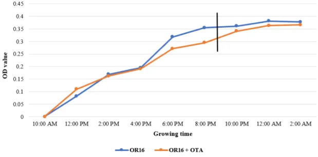
Figure 1. The log phase age ofC. basilensisÖR16 in the minimal buffer, black line means the end of the log phase.
Bioinformatics analysis of RNA-sequencing data
After sequencing, paired-end Illumina reads were quality trimmed in CLC Genomics Workbench Tool (v.11.0, Qiagen Bioinfor- matics Co., Denmark) applying an error probability threshold of 0.01. No ambiguous nucleotide was allowed in trimmed reads.
For filtering, reads were mapped on CLC with a length fraction of 0.9 and a se- quence identity threshold of 0.95. RNA-Seq analysis package from CLC was then used to map filtered reads on a custom-masked C. basilensis ÖR16 genome version. Only those reads were considered that displayed an alignment longer than 80% of the read length while showing at least 95% sequence identity against the reference genome. Next
“Total gene read” RNA-Seq count data was imported from CLC into R 3.3.2 for data normalization and differential gene expres- sion analysis. Function “calcNormFactors”
from package “edgeR” v.3.12.1 was ap- plied to perform data normalization based on the “trimmed mean of M-values” (TMM) method. Genes displaying at least one -fold gene expression change with an FDR (false
discovery rate) value below 0.05 were con- sidered as significant (Seqomics Ltd, Hun- gary).
Results
Log phase identification of Cupriavidus basilensis ÖR16 strain
Estimating the log phase of Cupriavidus basilensis ÖR16 was important, to find the correct time for extracting the best quality RNA from the inoculum. During the pre- experiments, the RNA extraction was ac- cording the peak of the OTA degradation process on the 3rd day, but at that time the RNA was already broken, not useful for tran- scriptome analysis. The 11th hour was the proper time for making the RNA extraction, getting good quality of RNA, which can be used for the analysis (Figure 1).
Results of the RNA isolation
The biodegradation in minimal buffer was stopped at the 11th hour, according the log phase peak for getting good quality RNA.
There were two parallel settings from each
Figure 2. RNA bands ofCupriavidus basilensisÖR16 with and without OTA from the OTA degradation experiment conducted in minimal buffer for transcriptome analysis, sampled after 11 hour of incubation.
Table 1. RNA quality results of the different setting from the OTA degradation matrix in minimal buffer from the Agilent 2200 Technologies (Seqomics Ltd, Mórahalom, Hungary), RIN =RNA integrity number.
Sample description 23S/16S (Area) Conc. [ng/µl] RIN
Electronic Ladder - 84.9 -
OR 16_1 0.8 99.2 8.2
OR 16_2 0.7 57.8 8.2
OTA OR 16_1 1.3 92.3 7.0
OTA OR 16_2 0.5 70.1 7.6
set: OTA+ÖR16 strain; ÖR16 strain; E.coli LB (in LB media) and E. coliMB (in mini- mal buffer media). RNA was extracted and the RNA integrity was confirmed in 1%
agarose gel electrophoresis (Figure 2). RNA quality was tested by Agilent 2200 Tech- nologies and using TapeStation software (Se- qomics Ltd, Mórahalom, Hungary) (Table 1).
Results of the transcriptome analysis
During bioinformatics analysis it turned out that 3500 genes were up regulated in the ÖR16 strain in the presents of OTA. A deci- sion system had to be developed for the iden- tification of the potential genes and enzymes,
which could play a role in the OTA degrada- tion. The decision system concluded the fol- lowing circumstances:
• At least 2-fold expression
• Playing role in any aromatic ring open- ing
• Should be a protease
• Could be connected to the hypothetical degradation pathways in literature
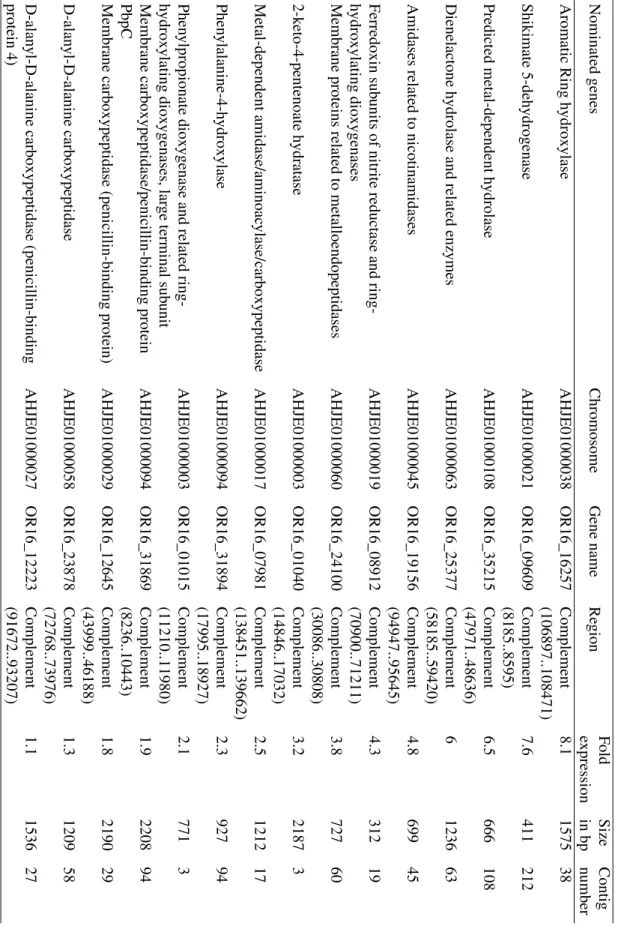
• There is any literature about the role in degradation of xenobiotics or OTA The ideal case was when all the circum- stances were standing. Of course there were exceptions like the low fold expression genes. Out of the 3500 gene, only 15 genes
could be enrolled into the criteria of the de- cision system (Table 2).
Discussion
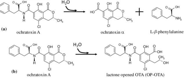
In the literature there are two hypothet- ical microbiological degradation pathways involved in the OTA degradation as illus- trated in Figure 3. The first pathway (a) is the hydrolysis occurred in the amide bond, which links the L-b-phenylalanine molecule to ochratoxin-alpha (OTa) moiety both of them are non-toxic (Abrunhosa et al., 2010).
The second pathway (b), the lactone ring hydrolysis can be considered a more hypo- thetical process in the OTA degradation and detoxification (Bruinink et al., 1998; Abrun- hosa et al., 2010).
According to the literature, there are sev- eral enzymes which might be involved in the biodegradation of OTA. There are two types of carboxypeptidases among mi- crobes, which may be involved in the OTA- biodegradation (Chang et al., 2015; Liuzzi et al., 2017). The first one is carboxypeptidase- A (CPA), where the “A” refers to aromatic compound, carboxypeptidases that have a stronger preference for those amino acids containing aromatic or branched hydrocar- bon chains. The CPY is the second enzyme, where the “Y” refers to yeast origin. A CPY was isolated fromSaccharomyces cerevisiae, it could degrade 52% of OTA and converted it to OTA-a after five days of incubation with pH 5.6 at 37 °C (Abrunhosa et al., 2010).
There are different enzymes besides the car- boxypeptidases, which can degrade OTA.
Aspergilus niger strains have a few enzy- matic tool for OTA degradation: a lipase en- zyme can hydrolyse OTA through the amide bond (Stander et al., 2000) and Protease-A have been reported to degrade around 87.3%
of 1µg OTA respectively with pH 7.5 in 25 hour-incubation period (Abrunhosa et al., 2010). At last, amidase 2, which is encoded by open reading frame (ORF) ofAspergillus
niger has the hydrolytic activity to degrade 83% of 50 µg/ mL of OTA (Loi et al., 2017).
Among the 15 identified genes, there are in- teresting members, which can degrade dif- ferent chemicals according to the literature.
For example, dienelactone hydrolase can degrade protoanemonin, which is a toxic metabolite, which may be formed during the degradation of some chloroaromatic com- pounds, such as polychlorinated biphenyls (Brückman et al. 1998). TheAromatic Ring hydroxylase can convert closed-ring struc- tures to non-aromatic cis-diols (Neidle et al., 1991) ThePredicted metal-dependent hydro- lase are acting on carbon – nitrogen bonds and 2-keto-4-pentenoate hydratase partici- pates in 9 metabolic pathways: phenylala- nine metabolism, benzoate degradation via hydroxylation, biphenyl degradation, toluene and xylene degradation, 1,4-dichlorobenzene degradation, fluorene degradation, carbazole degradation, ethylbenzene degradation and styrene degradation (Zhen et al., 2006). In 2017 Luizzi and colleagues cloned and in- vestigated CPA genes of the Acinetobac- ter sp. neg1 strain responsible for OTA biodegradation. In our transcriptome result the ÖR16 CPAs showed weak fold expres- sion, not matching by the criteria of the deci- sion system. Even so the future investigation of these CPAs is still important.
According to the results of the transcriptome analyses there is a chance to identify the enzymes via cloning and expression. Test- ing the expressed proteins in OTA degra- dation system the OTA degraders can be identified. The following genes should be investigated in the future for their OTA- biodegradation and detoxification poten- tial: OR16_12645 coded membrane CPA (penicillin-binding protein), OR16_24100 coded membrane proteins related to met- alloendopeptidases, OR16_31869 coded membrane CPA (penicillin-binding proteins PbpC), OR16_31894 coded phenylalanine- 4-hydroxylase and OR16_16257 coded aro-
Figure 3. The biodegradation pathways of OTA adapted from Abrunhosa et al., 2010).
matic ring hydroxylase.
Acknowledgements
1. Thank you for the Seqomics Ltd. for helping this research to realize.
2. Development and Innovation Fund (NKFIA); Grant Agree-
ment: NVKP_16-1-2016-0009 and the Higher Education Institu- tional Excellence Program (1783- 3/2018/FEKUTSTRAT) by the Min- istry of Human Capacities projects.
3. Tempus Public Foundation (TPF) / Stipendium Hungaricum Scholarships Program.
References
Abrunhosa L, Paterson RRM, Venâncio A. (2010) Biodegradation of ochratoxin a for food and feed decontamination. Toxins (Basel) 2 :1078–1099. DOI: https://doi.org/10.3390/toxins2051078
Abrunhosa, L., Inês, A., Rodrigues, A.I., Guimarães, A., Pereira, V.L., Parpot, P., Mendes- Faia, A., Venâncio, A. (2014): Biodegradation of ochratoxin A by Pediococcus parvulus isolated from Douro wines. Int. J. Food Microbiol. 188, 45–52. DOI: https://doi.org/10.1016/J.IJFOODMICRO.
2014.07.019
Bragulat, M.R., Martínez, E., Castellá, G., Cabañes, F.J. (2008): Ochratoxin A and citrinin producing species of the genusPenicilliumfrom feedstuffs. Int. J. Food Microbiol. 126, 43–48. DOI:
https://doi.org/10.1016/j.ijfoodmicro.2008.04.034
Brückmann M., Blasco R., Timmis N. K., Pieper H. D. (1998): Detoxification of Protoanemonin by Dienelactone Hydrolase, Journal of Bacteriolgy, 180, p. 400–402, DOI: https://doi.org/10.1128/
JB.180.2.400-402.1998
Chang, X., Wu, Z., Wu, S., Dai, Y., Sun, C. (2015): Degradation of ochratoxin A byBacillus amyloliquefaciensASAG1. Food Addit. Contam. - Part A Chem. Anal. Control. Expo. Risk Assess.
32, 564–571. DOI: https://doi.org/10.1080/19440049.2014.991948
Coenye, T., Vandamme, P., Lipuma, J.J. (2003):Ralstonia respiraculi sp . nov., isolated from the respiratory tract of cystic fibrosis patients. Int J Syst Evol Microbiol. 53(Pt 5):1339-1342. DOI:
https://doi.org/10.1099/ijs.0.02440-0
Table2.Fifteennominatedgenesoutof3500accordingthetranscriptomeanalysesoftheÖR16straininpresentofOTAinminimalbuffer. NominatedgenesChromosomeGenenameRegion FoldSizeContigexpressioninbpnumberAromaticRinghydroxylaseAHJE01000038OR16_16257Complement8.1157538(106897..108471)Shikimate5-dehydrogenaseAHJE01000021OR16_09609Complement7.6411212(8185..8595)Predictedmetal-dependenthydrolaseAHJE01000108OR16_35215Complement6.5666108(47971..48636)DienelactonehydrolaseandrelatedenzymesAHJE01000063OR16_25377Complement6123663(58185..59420)AmidasesrelatedtonicotinamidasesAHJE01000045OR16_19156Complement4.869945(94947..95645)Ferredoxinsubunitsofnitritereductaseandring-AHJE01000019OR16_08912Complement4.331219hydroxylatingdioxygenases(70900..71211)MembraneproteinsrelatedtometalloendopeptidasesAHJE01000060OR16_24100Complement3.872760(30086..30808)2-keto-4-pentenoatehydrataseAHJE01000003OR16_01040Complement3.221873(14846..17032)Metal-dependentamidase/aminoacylase/carboxypeptidaseAHJE01000017OR16_07981Complement2.5121217(138451..139662)Phenylalanine-4-hydroxylaseAHJE01000094OR16_31894Complement2.392794(17995..18927)Phenylpropionatedioxygenaseandrelatedring-AHJE01000003OR16_01015Complement2.17713hydroxylatingdioxygenases,largeterminalsubunit(11210..11980)Membranecarboxypeptidase/penicillin-bindingproteinAHJE01000094OR16_31869Complement1.9220894PbpC(8236..10443)Membranecarboxypeptidase(penicillin-bindingprotein)AHJE01000029OR16_12645Complement1.8219029(43999..46188)D-alanyl-D-alaninecarboxypeptidaseAHJE01000058OR16_23878Complement1.3120958(72768..73976)D-alanyl-D-alaninecarboxypeptidase(penicillin-bindingAHJE01000027OR16_12223Complement1.1153627protein4)(91672..93207)
Cserháti, M., Kriszt, B., Szoboszlay, S., Tóth, Á., Szabó, I., Táncsics, A., Nagy, I., Horváth, B., Nagy, I., Kukolya, J. (2012): De novo genome project ofCupriavidus basilensisOR16. J. Bacteriol.
194, 2109–2110. DOI: https://doi.org/10.1128/JB.06752-1
De Bellis, P., Tristezza, M., Haidukowski, M., Fanelli, F., Sisto, A., Mulè, G., Grieco, F.
(2015): Biodegradation of ochratoxin a by bacterial strains isolated from vineyard soils. Toxins (Basel). 7, 5079–5093. DOI: https://doi.org/10.3390/toxins7124864
Duarte, S.C., Lino, C.M., Pena, A. (2012): Food safety implications of ochratoxin A in animal- derived food products. Vet. J. 192, 286–292. DOI: https://doi.org/10.1016/j.tvjl.2011.11.002
Ferenczi, S., Cserháti, M., Krifaton, C., Szoboszlay, S., Kukolya, J., Szoke, Z., Koszegi, B., Albert, M., Barna, T., Mézes, M., Kovács, K.J., Kriszt, B. (2014): A new ochratoxin a biodegradation strategy usingCupriavidus basilensisOr16 strain. PLoS One 9. DOI: https://doi.org/10.1371/journal.
pone.0109817
Fuchs, S., Sontag, G., Stidl, R., Ehrlich, V., Kundi, M., Knasmüller, S. (2008): Detoxification of patulin and ochratoxin A, two abundant mycotoxins, by lactic acid bacteria. Food Chem. Toxicol.
46, 1398–1407. DOI: https://doi.org/10.1016/j.fct.2007.10.008
Goris, J., De Vos, P., Coenye, T., Hoste, B., Janssens, D., Brim, H., Diels, L., Mergeay, M., Kersters, K., Vandamme, P. (2001): Classification of metal-resistant bacteria from industrial biotopes asRalstonia campinensis sp. nov.,Ralstonia metallidurans sp. nov.andRalstonia basilensis Steinle et al. 1998 emend. Int. J. Syst. Evol. Microbiol. 51, 1773–1782. DOI: https://doi.org/10.1099/
00207713-51-5-1773
IARC, (1993): Some naturally occurring substances: food items and constituents, heterocyclic aromatic amines and mycotoxins. IARC Monogr. Eval. Carcinog. Risk Chem. to Humans 56, 1–521.
DOI: https://doi.org/10.1002/food.19940380335
Koopman, F., Wierckx, N., Winde, J.H. De, Ruijssenaars, H.J. (2010): Identification and char- acterization of the furfural and 5- ( hydroxymethyl ) furfural degradation pathways ofCupriavidus basilensisHMF14 Proc Natl Acad Sci U S A. 107. DOI: https://doi.org/10.1073/pnas.0913039107
Liuzzi, V.C., Fanelli, F., Tristezza, M., Haidukowski, M., Picardi, E., Manzari, C., Lionetti, C., Grieco, F., Logrieco, A.F., Thon, M.R., Pesole, G., Mulè, G. (2017): Transcriptional Analysis ofAcinetobactersp. neg1 Capable of Degrading Ochratoxin-A, Front. Microbiol. 7, 09 1–9. DOI:
https://doi.org/10.3389/fmicb.2016.02162
Luz, C., Ferrer, J., Mañes, J., Meca, G. (2018): Toxicity reduction of ochratoxin A by lactic acid bacteria. Food Chem. Toxicol. 112, 60–66. DOI: https://doi.org/10.1016/j.fct.2017.12.030
Makkar, N.S., Casida, L.E. (1987): Cupriavidus necator gen. nov., sp. nov.; a Nonobligate Bacterial Predator of Bacteria in Soil†. Int. J. Syst. Evol. Microbiol. 37, 323–326. DOI: https://doi.
org/10.1099/00207713-37-4-323
Neidle EL, Hartnett C, Ornston LN, Bairoch A, Rekik M, Harayama S. (1991): Nucleotide sequences of theAcinetobacter calcoaceticusgenes for benzoate 1,2-dioxygenase reveal evolution- ary relationships among multicomponent oxygenases. Journal of Bacteriology 173, 5385-95. DOI:
https://doi.org/10.1128/jb.173.17.5385-5395.1991
Ogawa, N., Miyashita, K., (1999): The Chlorocatechol-Catabolic Transposon Tn 5707 of Alcaligenes eutrophus NH9 , Carrying a Gene Cluster Highly Homologous to That in the 1,2,4- Trichlorobenzene-Degrading BacteriumPseudomonassp . Strain P51 , Confers the Ability To Grow on 3-Chloroben, Appl Environ Microbiol. 65(2): 724–731. PMCID: PMC91086
Petchkongkaew, A., Taillandier, P., Gasaluck, P., Lebrihi, A. (2008): Isolation ofBacillusspp.
from Thai fermented soybean (Thua-nao): screening for aflatoxin B1 and ochratoxin A detoxification.
J. Appl. Microbiol. 104, 1495–1502. DOI: https://doi.org/10.1111/j.1365-2672.2007.03700.x Stamper, D.M., Radosevich, M., Hallberg, K.B., Traina, S.J., Tuovinen, O.H., (2003):Ralsto- nia basilensisM91-3, a denitrifying soil bacterium capable of using s-triazines as nitrogen sources, Canadian Journal of Microbiology 48(12):1089-98. DOI: https://doi.org/10.1139/W02-113
Steinle, P., Stucki, G., Stettler, R., Hanselmann, K.W., (1998): Aerobic Mineralization of 2 , 6 Dichlorophenol byRalstoniasp . Strain RK1 64, Appl Environ Microbiol. 64(7):2566-71. PMCID:
PMC106427
Rodriguez, H., Reveron, I., Doria, F., Costantini, A., De Las Rivas, B., Muˇnoz, R., Garcia- Moruno, E. (2011): Degradation of Ochratoxin A byBrevibacteriumSpecies. J. Agric. Food Chem.
59, 10755–10760. DOI: https://doi.org/10.1021/jf203061p
Zhang, H.H., Wang, Y., Zhao, C., Wang, J., Zhang, X.L. (2017): Biodegradation of ochratoxin A byAlcaligenes faecalisisolated from soil. J. Appl. Microbiol. 123, 661–668. DOI: https://doi.org/
10.1111/jam.13537
Zhen D, Liu H, Wang SJ, Zhang JJ, Zhao F, Zhou NY. (2006): Plasmid-mediated degrada- tion of 4-chloronitrobenzene by newly isolatedPseudomonas putidastrain ZWL73. Appl Microbiol Biotechnol 72(4);797-803. PMID: 16583229