Effects of Zinc Oxide Nanoparticles (ZnO NPs) and some Plant Pathogens on the Growth and Nodulation of Lentil (Lens culinaris Medik.)
Z. A. SIDDIQUI1*, A. KHAN1, M. R. KHAN1 and E. F. ABD-ALLAH2
1Department of Botany, Aligarh Muslim University, Aligarh-202002(U.P.), India
2Department of Plant Production, Faculty of Food and Agricultural Sciences, King Saud University, Riyadh 11451, Saudi Arabia
(Received: 8 June 2018; accepted: 5 July 2018)
Effects of ZnO nanoparticles (NPs) were studied on lentil plants inoculated with Alternaria alternata, Fusarium oxysporum f. sp. lentis, Xanthomonas axonopodis pv. phaseoli, Pseudomonas syringae pv. syringae and Meloidogyne incognita. Plant growth, chlorophyll, carotenoid contents, nitrate reductase (NR) activity and nodulation of lentil both in the presence and absence of Rhizobium sp. were examined in a pot test. Inoculation of plants with A. alternata / F. oxysporum f. sp. lentis / X. axonopodis pv. phaseoli / P. syringae pv. syringae or M. incognita caused a significant reduction in plant growth, number of pods per plant, chlorophyll, carotenoids and NR activity over uninoculated control. Inoculation of plants with Rhizobium sp. with or without pathogen increased plant growth and number of pods per plant, chlorophyll, carotenoids and NR activity. When plants were grown without Rhizobium, a foliar spray of plants with 10 ml solution of 0.1 mg ml–1of ZnO NPs per plant caused a significant increase in plant growth and number of pods, chlorophyll, carotenoid contents and NR activity in both inoculated and uninoculated plants. Spray of ZnO NPs to plants inoculated with Rhizobium sp.
caused non significant increase in plant growth, number of pods per plant, chlorophyll, carotenoid contents and NR activity when plants were either uninoculated or inoculated with pathogens. Numbers of nodules per root system were high in plants treated with Rhizobium sp. but foliar spray of ZnO NPs had adverse effect on nod- ulation. Inoculation of plants with test pathogens also reduced nodulation. Spray of ZnO NPs to plants reduced galling, nematode multiplication, wilt, blight and leaf spot disease severity indices.
Keywords: Root nodulation Rhizobium sp., disease management, plant pathogens.
Lentil (Lens culinaris Medik.) is a bushy and annual shrub. Lentils are nutritious and provide a good source of minerals, containing about 25% proteins, 56% carbohy- drates, 1.0% fat in seeds. Lentil is one of the best and cheapest sources of vegetable pro- teins (Adsule et al, 1989). Lentils indulge in the nitrogen fixation process that helps the soil to revive its nitrogen content. The lentils are an important source of essential amino acids, fatty acids and trace mineral (Haq et al., 2011).
Unfortunately, there are some constraints of pests and pathogens in the successful cultivation of lentil (Taylor et al., 2007). Fungal diseases on lentil includes Alternaria blight (Alternaria alternata), Fusarium wilt (Fusarium oxysporum f. sp. lentis). Bacterial
* Corresponding author; e-mail: zaki_63@yahoo.co.in
diseases of lentil are bacterial leaf spot caused by Xanthomonas axonopodis pv. phaseoli and bacterial blight caused by Pseudomonas syringae pv. syringae. Root knot nematode Meloidogyne incognita is also a major pathogen.
Root nodulation is a complex symbiotic process between host plant and Rhizobium sp. Rhizobium is a genus of Gram-negative soil bacteria that fix nitrogen. It forms an en- dosymbiotic nitrogen fixing association with roots of legumes. The association of Rhizo- bia with plant pathogens in the rhizosphere is not always detrimental since it sometimes leads to stimulation of nodulation (Hussey and Barker, 1976). The presence of rhizobia in the rhizosphere may also protect the host root from damage caused by pathogens (Sid- diqui and Husain, 1992; Siddiqui and Mahmood, 1995).
Nanoparticles can offer green and eco-friendly alternatives for plant disease manage- ment (Alghuthaymi et al., 2015) because of increased antimicrobial activity and decreased ecological toxicity (Neal, 2008). Zinc oxide nanoparticles (ZnO NPs) have remarkable optical, physical, and antimicrobial properties (Sabir et al., 2014) and have potential to enhance agriculture production due to active oxygen species generated by these NPs.
During the course of survey of lentil fields of Aligarh district of U.P., we found frequent occurrence of Meloidogyne incognita (Kofoid and White) Chitwood, Alternaria alternata (Fr.) Keissler, Fusarium oxysporum f. sp. lentis Gordon, Xanthomonas axono
podis pv. phaseoli (E. F. Smith) Dowson and Pseudomonas syringae pv. syringae van Hall. Plants infected with these pathogens were poor in growth and showed typical dis- ease symptoms. These pathogens were highly destructive and considered as major con- straints in the successful cultivation of lentil.
In the present study an attempt was made to use ZnO NPs for the management of M. incognita, A. alternata, F. oxysporum f. sp. lentis, X. axonopodis pv. phaseoli and P. syringae pv. syringae on lentil both in the presence and absence of Rhizobium sp.
Materials and Methods
Root and soil samples were collected from lentil fields of Aligarh district, U.P., India. These samples were collected in polythene bags and stored in a refrigerator at 4 °C until processing began. The samples were examined for the presence of the root-knot nematode Meloidogyne spp., fungi and plant pathogenic bacteria.
Isolation and identification of rootknot nematode M. incognita
Lentil roots were examined for root-knot symptoms. Root galls were dissected to take out the females of root-knot nematodes. Identification of Meloidogyne sp. was made on the basis of perineal patterns (Taylor and Sasser, 1978). Soil samples were also pro- cessed for the nematodes isolation by Cobb’s sieving and decanting technique followed by a Baermann funnel (Southey, 1986). Identification of root-knot juveniles were also made from the nematode suspension.
Isolation of fungi from lentil
Roots and shoots with fungal disease symptoms were transferred to sterilized Petri dishes containing sterilized distilled water and gently freed of soil particles. These parts were transferred to other Petri dishes and the process was repeated until all adhering soil particles were removed. Later, these infected parts were cut into approximately 5 mm pieces and transferred to a Petri dish containing 0.1% sodium hypochlorite (NaOCl) solu- tion. After one minute the pieces were washed at least thrice in distilled water and dried on filter paper. Five of these pieces were then plated in Petri dishes containing PDA ( Potato dextrose agar) using sterilized forceps under aseptic conditions. Petri dishes were incu- bated at 25±2 °C for 10 days. The fungus that developed from infected root and shoot pieces were examined and identified. For confirmation of identity as Alternaria alter
nata and Fusarium oxysporum f. sp. lentis, pure cultures of these fungi were prepared.
Identification of A. alternata was made based on morphological characters (Simmons, 2007), while F. oxysporum was identified using the key proposed by Nelson et al. (1983).
Pathogenicity and characterization of the isolate as forma specialis lentis was tested by in- oculating the susceptible lentil cultivar P 651. The pure cultures were stored at 5 °C until used. The composition of PDA medium was as follows:
Potato infusion from 200 g
Dextrose 20 g
Agar agar 15 g
Distilled water 1000 ml Isolation of bacteria from lentil
Collected lentil plants showing symptoms of bacterial blight or leaf spot were used for the isolation of bacteria. Plant parts having rot or blight symptoms were transferred to a sterilized Petri dishes containing sterilized distilled water and gently freed of soil particles. These parts were transferred to another Petri-dish and process was repeated till all adhering soil particles were removed. Later, the plant parts were cut into 5 mm pieces and then surface sterilized with 0.1% sodium hypochlorite (NaOCl) solution, followed by three repeated washings with distilled water and dried on filter paper. Five of these pieces were then plated in each of the Petri dishes containing nutrient agar medium with the help of sterilized forceps under aseptic condition. Petri dishes were then incubated at 30 + 2 °C for 24–48 h. The bacteria that developed from infected pieces were examined and identified. Pseudomonas syringae pv. syringae was identified using biochemical and physiological tests (Ashorpour et al., 2008) while X. axonopodis pv. phaseoli was identi- fied according to Nunes et al. (2008). To confirm identity as Pseudomonas syringae pv.
syringae and Xanthomonas axonopodis pv. phaseoli, pure cultures of these bacteria were prepared and stored at 5 °C. The composition of nutrient agar medium was as follows:
Beef extract 3.0 g
Peptone 5.0 g
Agar agar 15.0 g
Distilled water 1.0 liter
Preparation of zinc dioxide nanoparticles solution
Zinc oxide, dispersion nanoparticles (ZnO NPs), <100 nm particle size (TEM),
≤40 nm average particle size, 20 wt. % in H2O, pH 7.5±1.5 was obtained from Sig- ma-Adrich (Product No. 721077-100G). Solution of ZnO NPs was prepared by dissolving 0.50 ml ZnONPs in one litre distilled water to obtain a final concentration of 0.1 mg ml-1 for the foliar spray. Ten ml suspension of ZnO NPs was used as foliar spray per plant.
Preparation and sterilization of soil mixture
Sandy loam soil is collected from a field belonging to the Department of Botany, A.M.U., Aligarh and passed through a 10 mesh sieve. The soil, river sand and organic manure were mixed in the ratio 3:1:1 and 15 cm diameter clay pots were each filled with 1 kg of the mixture. A little water was poured into each pot to just wet the soil surface before sterilization at 137.9 kPa for 20 minutes. Sterilized pots were allowed to cool at room temperature before use.
Raising and maintenance of test plants
Lentil (Lens culinaris L.) seeds of cultivar K-75 were surface sterilized with 0.1%
sodium hypochlorite (NaOCl) for 2 minutes and rinsed three times with sterile water.
Half of the seeds were treated with Rhizobium sp. while the other half remained untreated before sowing. To treat the seeds with Rhizobium sp., commercial culture of Rhizobium sp. was obtained from Quarsi Agriculture farm, Aligarh. For seed treatment, 100 g com- mercial culture of Rhizobium lentil strain was placed on butter paper and mixed with 50 g sucrose. Later, 50 ml distilled water was poured over culture and mixed. Hundred g seeds were mixed with commercial culture and left to dry at room temperature. Sowing of Rhizobium-treated and untreated seeds were done separately. Three seeds were sown in the 15 cm diameter clay pots and one week after germination thinning was done to re- tain single seedling per pot. Seedlings were subjected to the treatments and uninoculated plants served as a control and plants were kept in a glass house at 20±2 °C. Pots were ar- ranged in a randomize block design and each treatment was replicated 5 times. Pots were watered as needed and experiment was terminated 90 days after inoculation.
Inocula of fungi
For obtaining sufficient inocula, A. alternata and F. oxysporum f. sp. lentis were separately cultured on Richard’s liquid medium having following composition:
Potassium nitrate 10.0 g
Potassium dihydrogen phosphate 05.0 g
Magnesium sulphate 02.5 g
Ferric chloride 0.02 g
Sucrose 50.0 g
Distilled water 1000 ml
The medium was prepared and filtered through muslin cloth, sterilized in an auto- clave at 103.4 kPa for 15 minutes in 250 ml Erlenmeyer flasks each containing 80 ml
liquid medium. A. alternata and F. oxysporum f. sp. lentis were separately inoculated in each flask with the help of inoculation needle. Inoculated flasks were incubated at 25±1 °C for about 15 days to allow sufficient growth of each fungus. The mycelium of each fungus was collected on blotting papers separately to remove excess water and nu- trients. The inoculum of each fungus was prepared separately by mixing 50 g mycelium in 500 ml distilled water and blending it for 30 seconds in Waring blender. Ten ml of this suspension containing 1 g fungus was used as inoculum. Pure cultures of A. alternata and F. oxysporum f. sp. lentis were continuously maintained on PDA by re-inoculation after every 15 days.
Inocula of bacteria
For obtaining sufficient inocula, P. syringae and X. axonopodis were separately cultured on Nutrient agar medium. Medium was poured in sterilized Petri dishes and these dishes were later streaked separately with a pure colony of P. syringae / X. axonopodis and incubated at 30±1 °C for 24 h. Single colonies from a 24-h-old pure culture of P. sy
ringae / X. axonopodis were inoculated separately into nutrient broth flasks and incubated at 30±1 °C for 72 h. Cell density was determined following Sharma (2001) to achieve 1.2×10⁵ colony-forming units (CFU) / ml. Ten ml of this suspension was added to each pot around a lentil seedling.
Preparation of nematode inoculum
Meloidogyne incognita was collected from lentil roots and multiplied on the roots of egg plants (Solanum melongena L.) using single egg mass. Large numbers of egg masses from heavily infected eggplant roots were hand-picked with the help of sterilized forceps from the previously maintained pure culture of M. incognita. The egg masses were washed with distilled water and placed in a small sieve (9 cm diameter with 1-mm pore size) containing crossed layers of tissue paper. The sieve was placed in a Petri-dish containing distilled water deep enough to contact the egg masses. A number of these as- semblies were kept in an incubator running at 25±1 °C in order to obtain the required number of second-stage juveniles for inoculation. The hatched second-stage juveniles were collected from the Petri-dishes every 24 h, fresh water was added, and the process was repeated. For counting nematode juveniles, an average of 5 counts was made to de- termine the density of nematodes in the suspension. The inoculum was adjusted to 200±5 nematodes per ml. Ten ml of this suspension (i.e. 2000 freshly hatched juveniles) was added to each pot around a lentil seedling.
Inoculation techniques
Ten days after germination, inoculations were performed on well-established healthy seedlings. For inoculation of M. incognita, fungi and bacteria, the soil around the roots was carefully moved aside without damaging the roots. The inoculum suspen- sion was poured around the roots before the soil was replaced. As a control treatment, water was used. There were 2 sets of experiments 1. Plants with Rhizobium sp. 2. Plants without Rhizobium sp. Each set comprised of six treatments, i.e. (A) Control (without
pathogen); (B) M. incognita; (C) P. syringae pv. syringae; (D) X. axonopodis pv. phaseoli;
(E) A. alternata; (F) F. oxysporum f. sp. lentis (6×2=12 treatments). These 12 treat- ments were combined with (I) water (control, without ZnO NPs); (II) ZnO NPs 0.50 ml/L (12×2=24). Each treatment was replicated 5 times (24×5=120 pots). Experiment was conducted in 2017–2018.
Chlorophyll and carotenoid estimation
The chlorophyll and carotenoid contents in the fresh leaf was estimated 90 days after inoculation following Mackinney (1941). One g of freshly cut leaves was ground to fine pulp using a mortar and pestle after adding 20 cm³ of 80% acetone. The mixture was centrifuged at 5,000 rpm for 5 minutes. The supernatant was collected in 100 cm³ volumetric flask. The absorbance was read at 645 and 663 nm for chlorophyll and 480 and 510 nm for carotenoid against the blank (80% acetone) on spectrophotometer (Shimadzu UV-1700, Tokyo, Japan).
Estimation of Nitrate Reductase (NR) activity
Nitrate reductase (NR) activity (EC 1.7.1.1) was estimated 90 days after inocula- tion following Jaworski (1971). The amount of nitrite formed was determined spectro- photometrically. Fresh leaves (0.2 g) were chopped and transferred to a plastic vial; each vial contained 2.5 mL of phosphate buffer (pH 7.5), 0.5 mL of 0.2 M potassium nitrate solution, and 2.5 mL of 5% isopropanol. These vials were incubated for 2 h at 30 °C in dark. Then 0.4 mL of the incubated mixture was transferred to a test tube and 0.3 mL each of 1% sulfanilamide and 0.02% N-(1-naphthyl) ethylenediamine dihydrochloride solution was added. Finally, the vials were kept at room temperature for 20 min for colour devel- opment and the vial content was diluted to a volume of 5 mL with double distilled water.
The optical density (OD) of the content was recorded at 540 nm using the spectropho- tometer (UV-1700, Shimadzu, Japan). A blank was run simultaneously with each sample.
Standard curve was plotted by using known graded concentrations of NaNO2 (sodium nitrite) solution. NR activity was expressed as nano-moles of nitrite produced per gram fresh weight of leaf tissue per hour (nM NO2– g–1 FW h–1).
Observations
The plants were harvested 90 days after inoculation. Data on plant length, plant fresh weight, plant dry weight, no. of pods per plant and number of nodules per root system were recorded. The length of plant was recorded in centimeter from the top of the first leaf to the end of the root. Excess water was removed by blotting before weighing the plant. The plants were cut with knife above the base of the root emergence zone to separate shoot and root. Shoot and root were kept in envelope at 60 °C for 4 days before weighing for dry weight determination. Number of galls per root system were counted.
For nematode population, a 250 g subsample of well-mixed soil from each treatment was processed by Cobb’s sieving and decanting technique followed by Baermann funnel ex- traction (Southey, 1986). Nematode suspension was collected after 24 h and the numbers of nematodes were counted in five aliquots of 1 ml of suspension from each sample. The
means of five counts were used to calculate the population of nematodes per kg soil. To estimate the number of juveniles, eggs and females inside the roots, a 1 g subsample of roots was macerated in a Waring blender and counts were made from the suspension thus obtained. Numbers of nematodes present in roots were calculated by multiplying the number of nematodes present in 1 g of root by the total weight of root. Disease severity caused by fungal and bacterial pathogens were also recorded. Wilt, leaf blight, bacterial blight and leaf spot were determined by scoring the severity of disease. Disease rating was on a scale from 0 to 5 where 0=no disease (no wilt / leaf blight / leaf spot / bacterial blight symptoms observed); 1=wilt / leaf blight / leaf spot / bacterial blight symptoms up to 12.5% on leaf; 2=wilt / leaf blight / leaf spot / bacterial blight symptoms 12.6 to 25% on leaf; 3=wilt / leaf blight / leaf spot / bacterial blight symptoms 25.1 to 37.5% on leaf; 4=wilt / leaf blight / leaf spot / bacterial blight symptoms 37.6 to 50% on leaf, and 5>50% wilt / leaf blight / leaf spot / bacterial blight symptoms on leaf.
Microscopic examination of eggs and second stage juveniles of M. incognita
Effect of ZnO NPs was observed on eggs and second stage juveniles of M. incog
nita. Forty ml of ZnO NPs solution (0.1 mg ml-1)was placed in five Petri dishes and ten egg masses of M. incognita were added for hatching. Twenty four hours after incubation, juveniles and eggs were observed under microscope.
Statistical analysis
Plant length, plant fresh weight, plant dry weight, chlorophyll, carotenoid and ni- trate reductase activity were analysed statistically using analysis of variance in the sta- tistical software R (R development core team 2011; package library agricolae). Least significant differences (L.S.D.) were calculated at p=0.05. Duncan’s multiple range test (DMRT) was applied to denote the significant differences between the treatments.
Results
Analysis of variance revealed that Rhizobium, ZnO NPs and pathogen exposure significantly (P=0.001) affected plant length, plant fresh weight, plant dry weight, no.
of pods, no. of nodules, chlorophyll and carotenoid contents and nitrate reductase activ- ity (Table 1). Interaction of Rhizobium×NPs was significant at P=0.05 on plant length, plant fresh weight, no. of pods, no. of nodules, chlorophyll, carotenoid and nitrate re- ductase activity. Interactions of NPs×pathogens and Rhizobium×NPs×pathogens were found non-significant on all parameters studied (Table 1).
Effects of pathogens and ZnO NPs on plants growth in the absence of Rhizobium sp.
In plants sown without Rhizobium sp., inoculation with P. syringae pv. syringae / X. axonopodis pv. phaseoli / M. incognita / A. alternata or F. oxysporium f. sp. lentis caused a significant reduction in plant growth and number of pods / plant (Table 2).
Among these pathogens, M. incognita and F. oxysporium f. sp. lentis resulted in relatively
Table 1
Analysis of variance (ANOVA) showing the effect of Rhizobium, ZnO nanoparticles, pathogens and their interaction on the growth and physiological parameters of lentil
Parameters Treatments Df Sum of square Mean of
square F value Pr (>F)
Plant length Rhizobium 1 135.00 134.90 43.280 2.49e-09 ***
Nanoparticles (NPs) 1 93.00 92.80 29.777 3.78e-07 ***
Pathogens 5 6302.00 1260.40 404.272 < 2e-16 ***
Rhizobium × NPs 1 18.00 17.80 5.717 0.0187 *
Rhizobium × pathogens 5 9.00 1.80 0.564 0.7274ns
NPs × pathogens 5 1.00 0.20 0.060 0.9975ns
Rhizobium × NPs × pathogens 5 2.00 0.40 0.120 0.9876ns
Residuals 96 299.00 3.10
Plant fresh
weight Rhizobium 1 79.80 79.80 64.683 2.31e-12 ***
Nanoparticles (NPs) 1 47.20 47.20 38.255 1.51e-08 ***
Pathogens 5 3136.30 627.30 508.510 < 2e-16 ***
Rhizobium × NPs 1 3.60 3.60 2.881 0.0929*
Rhizobium × pathogens 5 3.30 0.70 0.532 0.7513ns
NPs × pathogens 5 0.60 0.10 0.104 0.9911ns
Rhizobium × NPs × pathogens 5 0.50 0.10 0.076 0.9957ns
Residuals 96 118.40 1.20
Plant dry
weight Rhizobium 1 20.10 20.13 31.115 2.24e-07 ***
Nanoparticles (NPs) 1 10.80 10.83 16.739 8.93e-05 ***
Pathogens 5 862.10 172.42 266.498 < 2e-16 ***
Rhizobium × NPs 1 1.70 1.67 2.579 0.112ns
Rhizobium × pathogens 5 0.40 0.08 0.116 0.989ns
NPs × pathogens 5 0.20 0.04 0.068 0.997ns
Rhizobium × NPs × pathogens 5 0.20 0.04 0.064 0.997ns
Residuals 96 62.10 0.65
No. of pods Rhizobium 1 649.00 648.70 61.586 5.96e-12 ***
Nanoparticles (NPs) 1 400.00 399.70 37.946 1.70e-08 ***
Pathogens 5 9037.00 1807.40 171.598 < 2e-16 ***
Rhizobium × NPs 1 52.00 52.00 4.938 0.0286 *
Rhizobium × pathogens 5 34.00 6.90 0.653 0.6601ns
NPs × pathogens 5 4.00 0.90 0.083 0.9947ns
Rhizobium × NPs × pathogens 5 4.00 0.80 0.077 0.9956ns
Residuals 96 1011.00 10.50
No. of nodules Rhizobium 1 51875.00 51875.00 62720.06 < 2e-16 ***
Nanoparticles (NPs) 1 500.00 500.00 60.479 8.40e-12 ***
Pathogens 5 3672.00 734.00 88.791 < 2e-16 ***
Rhizobium × NPs 1 317.00 317.00 38.312 1.48e-08 ***
Rhizobium × pathogens 5 2779.00 556.00 67.189 < 2e-16 ***
NPs × pathogens 5 24.00 5.00 0.569 0.723ns
Rhizobium × NPs × pathogens 5 22.00 4.00 0.529 0.754ns
Residuals 96 794.00 8.00
higher reduction in plant growth and number of pods / plant, than A. alternata. Inocula- tion of plants with P. syringae pv. syringae, X. axonopodis pv. phaseoli, M. incognita, A. alternata and F. oxysporium f. sp. lentis resulted in a 43.18, 45.23, 49.42, 38.69 and 48.91 % reductions in plant dry weight, respectively (Table 2).
In the absence of Rhizobium sp., a spray of ZnO NPs to plants inoculated with either test pathogens resulted in a significant increase in plant growth and number of pods / plant (Table 2). Spray of ZnO NPs to uninfected plants grown without Rhizobium sp. resulted in a 3.93% increase in plant dry weight. Similarly, ZnO NPs treatment caused 9.52, 9.62, 10.17, 8.60 and 13.43% increases in plant dry weight upon inoculation with P. syringae pv. syringae, X. axonopodis pv. phaseoli, M. incognita, A. alternata and F. oxy sporium f. sp. lentis, respectively (Table 2).
Nodulation was very poor (1 to 5 per root system) in plants without Rhizobium, irrespective of pathogen presence or ZnO NPs treatment (Table 2). Plants inoculated with P. syringae pv. syringae, X. axonopodis pv. phaseoli, A. alternata and F. oxysporium f. sp.
lentis showed severe symptoms of bacterial blight, bacterial leaf spot, fungal blight and wilt (with severity indices of 4, 4, 3 and 4, respectively) (Table 2). Spray of ZnO NPs to plants inoculated with P. syringae pv. syringae, X. axonopodis pv. phaseoli, A. alternata
Parameters Treatments Df Sum of square Mean of
square F value Pr (>F) Chlorophyll
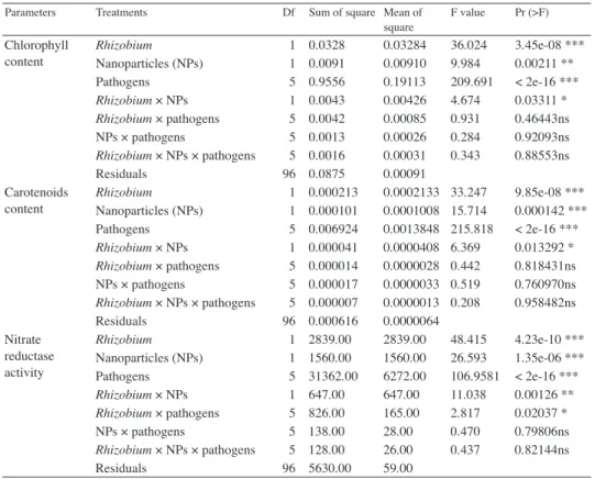
content Rhizobium 1 0.0328 0.03284 36.024 3.45e-08 ***
Nanoparticles (NPs) 1 0.0091 0.00910 9.984 0.00211 **
Pathogens 5 0.9556 0.19113 209.691 < 2e-16 ***
Rhizobium × NPs 1 0.0043 0.00426 4.674 0.03311 *
Rhizobium × pathogens 5 0.0042 0.00085 0.931 0.46443ns
NPs × pathogens 5 0.0013 0.00026 0.284 0.92093ns
Rhizobium × NPs × pathogens 5 0.0016 0.00031 0.343 0.88553ns
Residuals 96 0.0875 0.00091
Carotenoids
content Rhizobium 1 0.000213 0.0002133 33.247 9.85e-08 ***
Nanoparticles (NPs) 1 0.000101 0.0001008 15.714 0.000142 ***
Pathogens 5 0.006924 0.0013848 215.818 < 2e-16 ***
Rhizobium × NPs 1 0.000041 0.0000408 6.369 0.013292 *
Rhizobium × pathogens 5 0.000014 0.0000028 0.442 0.818431ns
NPs × pathogens 5 0.000017 0.0000033 0.519 0.760970ns
Rhizobium × NPs × pathogens 5 0.000007 0.0000013 0.208 0.958482ns
Residuals 96 0.000616 0.0000064
Nitrate reductase activity
Rhizobium 1 2839.00 2839.00 48.415 4.23e-10 ***
Nanoparticles (NPs) 1 1560.00 1560.00 26.593 1.35e-06 ***
Pathogens 5 31362.00 6272.00 106.9581 < 2e-16 ***
Rhizobium × NPs 1 647.00 647.00 11.038 0.00126 **
Rhizobium × pathogens 5 826.00 165.00 2.817 0.02037 *
NPs × pathogens 5 138.00 28.00 0.470 0.79806ns
Rhizobium × NPs × pathogens 5 128.00 26.00 0.437 0.82144ns
Residuals 96 5630.00 59.00
*=F values are significantly different at P=0.05, **=F values are significantly different at P=0.01,
***=F values are significantly different at P=0.001 and ns=F values are not significant.
Table 1 cont.
and F. oxysporium f. sp. lentis decreased disease severity indices to 3, 3, 2 and 3, respec- tively. Without Rhizobium sp., inoculation of plants with M. incognita caused 29 galls per root and high nematode population. Galling and nematode population was reduced by 37.93 and 38.10%, respectively, followed by a spray application of ZnO NPs (Table 2).
Effects of pathogens and ZnO NPs on plants growth in the presence of Rhizobium sp.
Inoculation of Rhizobium resulted in a significant increase in plant growth and number of pods / plant compared to plants without Rhizobium (Table 2). Inoculation of Rhizobium-treated plants with P. syringae pv. syringae, X. axonopodis pv. phaseoli, M. in
cognita, A. alternata and F. oxysporium f. sp. lentis resulted in a significant reduction in plant growth and number of pods / plant. Inoculation of plants with P. syringae pv.
syringae, X. axonopodis pv. phaseoli, M. incognita, A. alternata and F. oxysporium f. sp.
Table 2
Effect of ZnO NPs on M. incognita (M), A. alternata (A), F. oxysporum f. sp. lentis (F), P. syringae pv. syringae (P) and X. axonopodis pv. phaseoli (X)
on the growth and nodulation of lentil both in the presence and absence of Rhizobium sp.
Treatments Plant length
(Cm) Plant fresh
weight (g) Plant dry weight (g) No. of
pods No. of nodules No. of
galls Nematode population Disease
index Without
Rhizobium Without
ZnO NPs Control 63.24b 32.82c 15.74c 54c 5i – – –
P 46.90fgh 21.88ijkl 8.92kl 36hijk 3i – – 4
X 45.15hi 20.74klmn 8.62lm 35jkl 4i – – 4
M 41.90j 17.38p 7.96m 29m 2i 29a 8740a –
A 48.65def 23.18ghi 9.65ghij 40fghi 3i – – 3
F 44.52hi 18.52op 8.04m 31lm 2i – – 4
With ZnO
NPs Control 66.19a 34.26b 16.36bc 60b 4i – – –
P 49.26cdef 23.42fghi 9.77fghi 41efgh 1i – – 3
X 48.36def 22.16hijk 9.45hijk 39ghij 2i – – 3
M 44.16i 19.25no 8.77kl 34kl 3i 18c 5410c –
A 50.85cd 24.90def 10.48ef 45de 1i – – 2
F 46.72fgh 20.12mn 9.12ijkl 36ijk 2i – – 3
With
Rhizobium Without
ZnO NPs Control 67.36a 35.28ab 16.72ab 62ab 68a – – –
P 49.65cde 23.58fgh 9.96fgh 41efgh 47cd – – 3
X 48.42def 22.55ghij 9.60hij 41efgh 45d – – 3
M 44.72hi 19.48no 8.90kl 35jkl 36g 21b 6350b –
A 50.62cd 25.12de 10.86de 45de 50c – – 2
F 46.94fgh 20.36lmn 9.22ijkl 37hijk 41ef – – 3
With ZnO
NPs Control 68.41a 36.35a 17.16a 65a 62b – – –
P 50.76cd 24.06efg 10.32efg 43defg 39fg – – 2
X 49.15def 23.42fghi 10.06fgh 44def 36g – – 2
M 45.84ghi 20.52lmn 9.05jkl 37hijk 30h 10d 3170d –
A 51.76c 25.96d 11.22d 47d 44de – – 1
F 47.72efg 21.52jklm 9.64ghij 39ghij 32h – – 2
L.S.D. P=0.05 2.22 1.39 0.71 4.0 3.6
*Value in the same column followed by different letters are significantly different at P=0.05 based on DMRT
lentis resulted in a 40.43, 42.58, 46.77, 35.04 and 44.85% reductions in plant dry weight in plants treated with Rhizobium.
Spray of ZnO NPs to Rhizobium-inoculated plants resulted in a non-significant in- crease in plant growth and number of pods / plant in both pathogen inoculated and uninoc- ulated plants. ZnO NPs treatment of plants inoculated with Rhizobium and either P. syrin
gae pv. syringae, X. axonopodis pv. phaseoli, M. incognita, A. alternata or F. oxysporium f. sp. lentis and control (without pathogens) resulted in 3.61, 4.79, 1.68, 3.31, 4.55 and 2.63% increases in plant dry weight which is statistically non-significant (Table 2).
Higher numbers of nodules were observed in plants with Rhizobium, however, in- oculation of these plants with test pathogens resulted in a significant reduction in nodula- tion (Table 2). ZnO NPs treatment of Rhizobium inoculated plants reduced the nodulation in plants either or not inoculated with pathogens (Table 2).
Disease severity was found to be reduced in plants inoculated with Rhizobium and challenged with P. syringae pv. syringae, X. axonopodis pv. phaseoli, A. alternata or F. oxysporium f. sp. lentis (disease indices were 3, 3, 2 and 3, respectively, Table 2).
Disease indices were further reduced in plants treated with both ZnO NPs and Rhizobium.
Root galling and nematode multiplication was reduced by 27.58 and 27.34%, respectively, over plants without Rhizobium. Galling and nematode multiplication was further reduced to 52.38 and 50.07%, respectively, by the spray application of ZnO NPs (Table 2).
Effects of pathogens and ZnO NPs on chlorophyll,
carotenoids and NR activity in the absence of Rhizobium sp.
Inoculation of Rhizobium-untreated plants with P. syringae pv. syringae / X. axono
podis pv. phaseoli / M. incognita / A. alternata or F. oxysporium f. sp. lentis caused a sig- nificant reduction in chlorophyll and carotenoid contents and NR activity (Table 3). P. sy
ringae pv. syringae, X. axonopodis pv. phaseoli, M. incognita, A. alternata and F. oxyspo
rium f. sp. lentis infections resulted in 33.33, 37.01, 46.22, 27.99 and 41.98% reductions in chlorophyll contents, respectively, and 26.56, 28.12, 35.93, 23.43 and 31.25% reductions in carotenoid contents, respectively. Similarly, P. syringae pv. syringae, X. axonopodis pv.
phaseoli, M. incognita, A. alternata and F. oxysporium f. sp. lentis infections resulted in 9.83, 9.20, 15.17, 7.25 and 11.51% reductions in NR activity, respectively (Table 3).
In the absence of Rhizobium sp., a spray of ZnO NPs to plants inoculated with one of the test pathogens resulted in a significant increase in chlorophyll and carotenoid con- tents and NR activity (Table 3). Spray of ZnO NPs to uninoculated plants without Rhizo
bium sp. resulted in 9.02, 6.25 and 4.92% increases in chlorophyll and carotenoid contents and NR activity, respectively. Similarly, 3.59, 5.84, 11.98, 9.46 and 6.98% increases in chlorophyll were observed by the spray of ZnO NPs to plants inoculated with P. syringae pv. syringae, X. axonopodis pv. phaseoli, M. incognita, A. alternata and F. oxysporium f. sp. lentis, respectively. Similarly, 4.08 to 9.75% increases in carotenoids and 3.61 to 3.90% increases in NR activity was observed when ZnO NPs was sprayed on plants inoc- ulated with test pathogens (Table 3).
Effects of pathogens and ZnO NPs on chlorophyll, carotenoids and NR activity in the presence of Rhizobium sp.
Inoculation of plants with Rhizobium resulted in a significant increase in chloro- phyll, carotenoid contents and NR activity over plants without Rhizobium (Table 3). In- oculation of Rhizobium-treated plants with P. syringae pv. syringae, X. axonopodis pv.
phaseoli, M. incognita, A. alternata and F. oxysporium f. sp. lentis resulted in a significant reduction in chlorophyll, carotenoid contents and NR activity. Inoculation of Rhizobium to plants without test pathogen resulted in 13.44, 7.81 and 7.94% increases in chlorophyll, carotenoids and NR activity, respectively. Inoculation of Rhizobium to plants infected with P. syringae pv. syringae, X. axonopodis pv. phaseoli, M. incognita, A. alternata and F. oxysporium f. sp. lentis resulted in 6.90, 8.47, 17.12,12.02 and 14.92% increases in chlorophyll contents, respectively. Similarly, 6.12 to 12.19% increases in carotenoids and 3.30 to 7.94% increases in NR activity were observed when Rhizobium-treated plants
Table 3
Effect of ZnO NPs on M. incognita (M), A. alternata (A), F. oxysporum f. sp. lentis (F), P. syringae pv. syringae (P) and X. axonopodis pv. phaseoli (X) on chlorophyll, carotenoids
and nitrate reductase activity of lentil both in the presence and absence of Rhizobium sp.
Treatments Chlorophyll content
(mg –1 FW) Carotenoids content
(mg –1 FW) NR activity (nM NO2 g–1 FW h–1) Without
Rhizobium Without
ZnO NPs Control 0.543b 0.064b 303.37c
P 0.362efgh 0.047efgh 273.52jkl
X 0.342fghi 0.046fgh 275.43ijkl
M 0.292j 0.041i 257.32m
A 0.391de 0.049cdef 281.37fghij
F 0.315ij 0.044hi 268.43l
With ZnO
NPs Control 0.592a 0.068a 318.32b
P 0.375efg 0.050cde 283.42efghij
X 0.362efgh 0.048defg 285.40defghi
M 0.327hij 0.045gh 267.38l
A 0.428cd 0.051cd 291.65cdef
F 0.337ghi 0.047efgh 278.26hijkl
With
Rhizobium Without
ZnO NPs Control 0.616a 0.069a 327.47ab
P 0.387ef 0.050cde 286.43cdefghi
X 0.374efg 0.049cdef 284.52defghij
M 0.342fghi 0.046fgh 268.32l
A 0.435c 0.052c 293.37cde
F 0.362hij 0.048defg 279.43ghijk
With ZnO
NPs Control 0.623a 0.070a 330.23a
P 0.391de 0.052c 290.52cdefg
X 0.372efg 0.050cde 287.43cdefgh
M 0.344fghi 0.047efgh 270.26kl
A 0.437c 0.051cd 295.23cd
F 0.380efg 0.048defg 281.26fghij
L.S.D. P=0.05 0.037 0.003 9.61
*Value in the same column followed by different letters are significantly different at P=0.05 based on DMRT
were inoculated with test pathogens (Table 3). Spray of ZnO NPs to plants with Rhizo
bium and without pathogens resulted in a non-significant increase in chlorophyll, carot- enoid contents and NR activity (Table 3). Spray of ZnO NPs to plants with Rhizobium also resulted in statistically non-significant effect on chlorophyll, carotenoid contents and NR activity upon inoculation with P. syringae pv. syringae, X. axonopodis pv. phaseoli, M. incognita, A. alternata or F. oxysporium f. sp. lentis (Table 3).
Microscopic examination of 2nd stage juveniles and eggs of M. incognita
Adverse effect of ZnO NPs was also observed on M. incognita eggs and second stage juveniles by microscopic examination. Shapes of eggs and second stage juveniles were deformed in 0.1 mg / ml ZnO NPs solution within 24 h of incubation and hatching of second stage juveniles from egg masses was inhibited by more than 60 percent.
Discussion
Use of ZnO NPS as a foliar spray was found beneficial in improving plant growth, chlorophyll, carotenoid contents and NR activity in both pathogen-inoculated and un- inoculated plants. The increase in vegetative growth, chlorophyll, carotenoids and NR activity of lentil might be due to fundamental role of Zn in protection and maintenance of structural stability of cell membranes (Welch et al., 1982) and also its use in protein synthesis, membrane function, cell elongation and tolerance (Cakmak, 2000). ZnO NPs are also known to increase the shoot dry matter and leaf area (Taheri et al., 2015) and higher values for seeded fruit per umbel, seed weight per umbel and 1000-seed weight were observed as compared to control (Laware and Raskar, 2014). ZnO NPs treatments (25 nm mean particle size) at 1000 ppm concentration promoted seed germination, seed- ling vigor, and plant growth (Prasad et al., 2012). In their study, plants sprayed with NPs showed early flowering as compared to control. Faizan et al. (2017) concluded that pres- ence of ZnO NPs stimulated the antioxidant systems and increased proline accumulation that could provide stability to plants, and improved photosynthetic efficiency.
Foliar spray of NPs resulted in increased permeability of lipophilic organic mole- cules through the cuticle (Laware and Raskar, 2014). Hence, ZnO NPs have more chances to penetrate the leaf surface and release ions across the cuticle compared to water soluble ions (Da Silva et al., 2006). Prasad et al. (2012) observed that nano size and lower water solubility of ZnO NPs resulted in higher bioavailability of these NPs, which may be re- sponsible for higher yields. In Cucurbita pepo, NPs were observed both in the extracellu- lar spaces and inside the cells (Gonzalez-Melendi et al., 2008). Therefore, foliar spray of ZnO NPs may be responsible for increased plant growth in our experiments.
Sawai and Yoshikawa (2004) used 100 mg / ml ZnO suspension to obtain inhibitory effect on fungal growth but in our study 0.1 mg per ml showed antifungal activity. In our study, foliar spray on lentils was performed, while Sawai and Yoshikawa (2004) used higher concentration in an indirect conductimetric assay. The differences in results may be due to differences in assay conditions and fungal pathogens used. ZnO NPs generally interfere in cell functioning, cause deformation in fungal hyphae and inhibit the growth of fungi (He et al., 2011). In this study, application of ZnO NPs increased plant growth,
chlorophyll, carotenoid levels and NR activity in addition to reducing disease severity indices, which may be due to the antifungal effect of ZnO NPs on A. alternata and F.
oxysporum f. sp. lentis.
The antibacterial activity of ZnO is mainly associated with the generation of highly reactive oxygen species such as OH •OH, H2O2 and •O2¯(Anita et al., 2010). Reactive ox- ygen species that are released from the surface of ZnO may cause damage to microorgan- isms (Sunada et al., 1998). The generation of H2O2 depends strongly on the surface area of ZnO, and smaller nanoparticles produce more reactive oxygen species with the increased surface area and enhanced antibacterial activity (Yamamoto et al., 2008). Continuous re- lease of membrane lipids and proteins is induced by the integration of ZnO NPs with bacteria which changes membrane permeability of bacterial cells and later causes cell lysis (Brayner et al., 2006; Zhang and Chen, 2009). The bacterial cells were completely deformed by ZnO NPs, probably due to the lysis of cell wall, which was observed under microscope. Disease severity of bacterial blight was reduced in plants treated with ZnO NPs. This result confirms the antibacterial property of ZnO NPs.
Adverse effect of ZnO NPs was also observed on M. incognita eggs and second stage juveniles in this study by microscopic examination. Intestine is the major target tis- sues of NP toxicity which is revealed by distributional pattern of ZnO-NPs (Gupta et al., 2015). Similarly, galling and nematode multiplication was reduced in plants sprayed with ZnO NPs. Sudanophilic lipids are rich in nematodes cuticle (Sood and Kalra, 1977) and nematode cuticle also contain weakly acidic mucopolysaccharides. Lipids and glycogen are present in hypodermis and muscles of nematodes. In addition, hypodermis also con- sists of acidic mucopolysaccharides (Sood and Kalra, 1977). Toxicity of ZnO NPs is due to dissolved-zinc that caused toxicity plus nanoparticle-specific effects also contribute to toxicity (Savoly et al., 2016). It is possible the ZnO NPs had adverse effect on both cuticle and hypodermis of nematodes by affecting lipid, glycogen and mucopolysccharides.
Inoculation of Rhizobium sp. increased plant growth, chlorophyll and carotenoid coontents and NR activity compared to uninoculated control by increasing the nitrogen status of the soil. Antifungal activity of Rhizobium sp. was demonstrated by in vitro tests (Drapeau et al., 1973), and plants inoculated with Rhizobium sp. suffered less damage by pathogens than uninoculated plants (Bopaiah et al., 1976; Tu 1978, 1980). The pathogens tested had adverse effect on nodulation caused by Rhizobium sp., which is in line with previous findings (de Souza et al., 2016). Plant pathogens are also able to attack their op- ponents including symbionts using molecular weapons (Duffy et al., 2003), which include hydrogen cyanide (Benizri et al., 2005), alkaloids (Antunes et al., 2008), and bacteriocins (Holtsmark et al., 2008). Most of the secondary metabolites produced by plant pathogens can modify the structure and abundance of soil microbial populations, including plant symbionts (Mrabet et al., 2006; Liu et al., 2010; Chihaoui et al., 2012), causing deleteri- ous impacts for both plant and microorganism.
Spray application of ZnO NPs to Rhizobium-inoculated plants resulted in reduced root nodulation. ZnO NPs at 0.5 g kg–1 significantly altered soil bacterial communities (Ge et al., 2014). The presence of ZnO NPs in the rhizosphere affected root nodulation, delayed the onset of nitrogen fixation, and caused early senescence of nodules (Huang et al., 2014). Consistent with previous findings, spray of ZnO NPs to plants treated with Rhizobium was not able to increase plant growth and number of pods per plants. ZnO NPs have toxic effect to many different bacteria tested in the laboratory (Dorobantu et
al., 2015). Similarly, Moghaddam et al. (2017) reported that in the presence of NPs, the total length of treated plants and the number of nodules were decreased by increasing the concentration of NPs (1.25 to 10 μg/ml of Ag NPs and 12.5 to 100 mg/ml of ZnO NPs) compared to the control plants (P≤0.05). Gene expression of nif (nitrate fixing) gene was decreased in the presence of sub minimum inhibitory concentration of NPs. Non-sig- nificant increase in the growth and number of pods in plants treated with both ZnO NPs and Rhizobium sp. can be attributed to a decrease in root nodulation and adverse effect on nitrogen fixation.
In conclusion, ZnO NPs exhibited antifungal and antibacterial activities in lentil plants and inhibitory effect on nematodes. The application of ZnO NPs on lentil resulted in improved plant growth, reduced galling and nematode multiplication upon inoculation by M. incognita and reduced disease severity when challenge inoculated with bacterial or fungal pathogens. This clearly demonstrates that ZnO NPs may be used for the manage- ment of these pathogens on lentil.
Literature
Adsule, R. N., Kadam, S. S. and Leung, H. K. (1989): Lentil. In: D. K. Salunkhe and S. S. Kadam (eds): Hand- book of world food legumes: Nutritional Chemistry, Processing Technology, and Utilization. Vol. 11, Boca Raton, FL CRC Press, pp. 131.
Alghuthaymi, M. A., Almoammar, H., Rai, M., Said-Galiev, E. and Abd-Elsalam, K. A. (2015): Myconanoparti- cles: Synthesis and their role in phytopathogens management. Biotechnol. Equip. 29, 221–236.
Anita, S., Ramachandran,T., Koushik, C. V., Rajendran, R. and Mahalakshmi, M. (2010): Preparation and char- acterization of zinc oxide nanoparticles and a study of their antibacterial property of cotton fabric treated with the particles. http://ojs.cnr.ncsu.edu/index.php/JTATM/article/view/899/723.
Antunes, P. M., Miller, J., Carvalho, L. M., Klironomos, J. N. and Newman, J. A. (2008): Even after death the endophytic fungus of Schedonorus phoenix reduces the arbuscular mycorrhizas of other plants. Funct.
Ecol. 22, 912–918. 10.1111/j.1365-2435.2008.01432.x
Ashorpour, M., Kazempour, M. N. and Ramezanie, M. (2008): Occurrence of Pseudomonas syringae pv. syrin
gae the causal agent of bacterial canker on olives (Olea europaea) in Iran. Science Asia 34, 3: 323–326.
Benizri, E., Piutti, S., Verger, S., Pagès, L., Vercambre, G., Poessel, J. L. et al. (2005): Replant diseases: Bac- terial community structure and diversity in peach rhizosphere as determined by metabolic and genetic fingerprinting. Soil Biol. Biochem. 37, 1738–1746. 10.1016/j.soilbio.2005.02.009
Bopaiah, B. M., Patil, R. B. and Reddy, D. D. R. (1976): Effect of Meloidogyne javanica on nodulation and symbiotic nitrogen fixation in mung, Vigna radiata. Indian J. Nematol. 6, 124–130.
Brayner, R., Ferrari-Iliou, R., Brivois, N., Djediat, S., Benedetti, M. F. and Fiévet, F. (2006): Toxicological impact studies based on Escherichia coli bacteria in ultrafine ZnO nanoparticles colloidal medium. Nano Lett. 6, 866–870.
Cakmak, I. (2000): Possible roles of zinc in protecting plant cells from damage by reactive oxygen species. New Phytol. 146, 185–205.
Chihaoui, S. A., Mhadhbi, H. and Mhamdi, R. (2012): The antibiosis of nodule-endophytic agrobacteria and its potential effect on nodule functioning of Phaseolus vulgaris. Arch. Microbiol. 194, 1013–1021. 10.1007/
s00203-012-0837-7
Da Silva, L. C., Oliva, M. A., Azevedo, A. A. and De Araujo, M. J. (2006): Response of restinga plant species to pollution from an iron pelletization factory. Water, Air and Soil Pollution 175, 241–256.
de Souza, E. M., Granada, C. E., Raul, A. and Sperotto, R. A. (2016): Plant pathogens affecting the establish- ment of plant-symbiont interaction. Front. Plant Sci. 7, 15. doi: 10.3389/fpls.2016.00015
Dorobantu, L., Fallone, C., Noble, A. and Burrell, R. E. (2015): Toxicity of silver nanoparticles against bacteria, yeast, and algae. J. Nanoparticle Res. 17, 172. DOI: 10.1007/s11051-015-2984-7
Drapeau, R., Fortin, J. A. and Cagnon, C. (1973): Antifungal activity of Rhizobium. Canad. J. Bot. 51, 681–682.
Duffy, B., Schouten, A. and Raaijmakers, J. M. (2003): Pathogen self-defense: Mechanisms to counteract micro- bial antagonism. Annu. Rev. Phytopathol. 41, 501–538.
Faizan, M., Faraz, A., Yusuf, M., Khan, S. T. and Hayat, S. (2017): Zinc oxide nanoparticle-mediated changes in photosynthetic efficiency and antioxidant system of tomato plants. Photosynthetica. https://link.springer.
com/article/10.1007/s11099-017-0717-0
Ge, Y., Priester, J. H., Van De Werfhorst, L. C., Walker, S. L., Nisbet, R. M., An, Y. J., Schimel, J. P., Gardea-Tor- resdey, J. L. and Holden, P. A. (2014): Soybean plants modify metal oxide nanoparticle effects on soil bacterial communities. Environ. Sci. Technol. 48, 13489–13496.
Gonzalez-Melendi, P., Fernandez Pacheco, R., Coronado, M. J., Corredor, E., Testillano, P. S., Risueno, M. C., Marquina, C., Ibarra, M. R., Rubiales, D. and Perez-De-Luque, A. (2008): Nanoparticles as smart treat- ment-delivery systems in plants: Assessment of different techniques of microscopy for their visualization in plant tissues. Ann. Bot. 101, 187–195.
Gupta, S., Kushwah, T., Vishwakarma, A. and Yadav, S. (2015): Optimization of ZnO-NPs to investigate their safe application by assessing their effect on soil nematode Caenorhabditis elegans. Nanoscale Res. Lett.
10:303 DOI 10.1186/s11671-015-1010-4
Haq, M. Z., Ahmad, S., Shad, M. A., Iqbal, S., Qayum, M., Ahmad, A., Luthria, D. L. and Amarowicz, R. (2011):
Compositional studies of lentil (Lens culinaris Medik.) cultivars commonly grown in Pakistan. Pak. J.
Bot. 43, 1563–1567.
He, L., Liu, Y., Mustapha, A. and Lin, M. (2011): Antifungal activity of zincoxide nanoparticles against Botrytis cinerea and Penicillium expansum. Microbiol. Res. 166, 207–215.
Holtsmark, I., Eijsink, V. G. and Brurberg, M. B. (2008): Bacteriocins from plant pathogenic bacteria. FEMS Microbiol. Lett. 280, 1–7. 10.1111/j.1574-6968.2007.01010.x
Huang, Y. C., Fan, R., Grusak, M. A., Sherrier, J. D. and Huang, C. P. (2014): Effects of nano-ZnO on the agro- nomically relevant Rhizobium–legume symbiosis. Sci. Total Environ. 497, 78–90.
Hussey, R. S. and Barker, K. R. (1976): Influence of nematodes and light sources on growth and nodulation of soybeans. J. Nematol. 8, 48–52.
Jaworski, E. G. (1971): Nitrate reductase assay in intact plant tissues. Biochem. Biophys. Res. Commun 43, 1274–1279.
Laware, S. L. and Raskar, S. (2014): Influence of zinc oxide nanoparticles on growth, flowering and seed pro- ductivity in onion. Int. J. Curr. Microbiol. App. Sci. 3, 874–881.
Liu, J., Wang, E. T., Ren, D. W. and Chen, W. X. (2010): Mixture of endophytic Agrobacterium and Sinorhizo
bium meliloti strains could induce nonspecific nodulation on some woody legumes. Arch. Microbiol.
192, 229-234. 10.1007/s00203-010-0543-2
Mackinney, Q. (1941): Absorption of light by chlorophyll solutions. J. Biol. Chem. 140, 315–322.
Moghaddam, M. N., Sabzevar, A. H. and Mortazaei, Z. (2017): Impact of ZnO and silver nanoparticles on leg- ume-Sinorhizobium symbiosis. Advan. Stud. Biol. 9, 83–90. www.m-hikari.comhttps://doi.org/10.12988/
asb.2017.712
Mrabet, M., Mnasri, B., Romdhane, S. B., Laguerre, G., Aouani, M. E. and Mhamdi, R. (2006): Agrobacterium strains isolated from root nodules of common bean specifically reduce nodulation by Rhizobium galli
cum. FEMS Microbiol. Ecol. 56, 304–309. 10.1111/j.1574-6941.2006.00069.x
Neal, A. (2008): What can be inferred from bacterium-nanoparticle interactions about the potential conse- quences of environmental exposure to nanoparticles? Ecotoxicology 17, 362–371.
Nelson, P. E., Toussoun, T. A. and Marasas, W. F. O. (1983): Fusarium species: An Illustrated Manual for Iden- tification. USA: The Pennsylvania State University Press.
Nunes, W. M. C., Corazza, M. J., Souza, S. A. C. D., Tsai, S. M., Kuramae, E. E. (2008): Characterization of Xanthomonas axonopodis pv. phaseoli isolates. Summa Phytopathologica. 34, 3: 228–231.
Prasad, T. N. V. K. V., Sudhakar, P., Sreenivasulu, Y., Latha, P., Munaswamy, V., Reddy, K. R., Sreeprasad, T. S., Sajanlal, P. R. and Pradeep, T. (2012): Effect of nanoscale zinc oxide particles on the germination, growth and yield of peanut. J. Plant Nutri. 35, 905–927.
Sabir, S., Arshad, M. and Chaudhari, S. K. (2014): Zinc oxide nanoparticles for revolutionizing agriculture: Syn- thesis and applications. The Sci. World J. 1-8 Article ID 925494,DOI: 10.1155/2014/925494
Savoly, Z., Hracs, K., Pemmer, B., Streli, C., Zaray, G. and Nagy, P. I. (2016): Uptake and toxicity of nano-ZnO in the plant-feeding nematode, Xiphinema vuittenezi: The role of dissolved zinc and nanoparticle-specific effects. Environ. Sci. Pollut. Res. Int. 23, 9669–9678. https://doi.org/ 10.1007/s11356-015-5983-4 Sawai, J. and Yoshikawa, T. (2004): Quantitative evaluation of antifungal activity of metallic oxide powders
(MgO, CaO and ZnO) by an indirect conductimetric assay. J. Appl. Microbiol. 96, 803–809.
Sharma, P. D. (2001): Microbiology. Rastogi and Company, Meerut, India, 359 p.
Siddiqui, Z. A. and Husain, S. I. (1992): Interaction between Meloidogyne incognita race 3, Macrophomina pha
seolina and Bradyrhizobium sp. in the root-rot disease complex of chickpea. Cicer arietinum. Fundam.
Appl. Nematol. 15, 491–494.
Siddiqui, Z. A. and Mahmood, I. (1995): Role of plant symbionts in nematode management: A Review. Biore- source Technol. 54, 217–226.
Simmons, E. G. (2007): Alternaria. An Identification Manual. CBS Biodiversity Series 6 CBS Fungal Biodiver- sity Centre, Utrecht, The Netherlands
Sood, M. L. and Kalra, S. (1977): Histochemical studies on the body wall of nematodes: Haemonchus contortus (Rud., 1803) and Xiphinema insigne Loos, 1949. Zeitschrift für Parasitenkunde 51, 265–273.
Southey J. F. (1986): Laboratory methods for work with plant and soil nematodes. Ministry of. Agriculture, Fisheries and Food Reference Book No. 402, 202 p.
Sunada, K., Kikuchi, Y., Hashimoto, K. and Fujishima, A. (1998): Bactericidal and detoxification effects of TiO2 thin film photocatalysts. Environ. Sci. Technol. 32, 726–728.
Taheri, M., Qarache, H. A., Qarache, A. A. and Yoosefi, M. (2015): The effects of zinc-oxide nanoparticles on growth parameters of corn (SC704). STEM Fellowship J. 1, 17–19.
Taylor, A. L. and Sasser, J. N. (1978): Biology, identification and control of root-knot nematodes (Meloidogyne spp.). Depat. Plant Pathol. Raleigh N.C., North, St. Univ. and USAID, North Carolina State Graphics, 111.
Taylor, P., Lindbeck, K., Chen, W. and Ford, R. (2007): Lentil diseases. In: S. S. Yadov, D. L. McNeil and P. C.
Stevenson (eds): Lentil an Ancient Crop for Modern Times. Springer, Netherlands, pp. 291–313.
Tu, J. C. (1978): Protection of soybean from severe Phytophthora root-rot by Rhizobium. Physiol. Plant Pathol.
12, 233–240.
Tu, J. C. (1980): Incidence of root-rot and over wintering of alfalfa as influenced by Rhizobia. Phytopathol.
Zeitschrift 97, 97-–108.
Welch, R. M., Webb, M. J. and Loneragan, J. F. (1982): Zinc in membrane function and its role in phosphorus toxicity. In: A. Scaife (ed.): Proc. of the Ninth Plant Nutrition Colloquium. Warwick, UK. Wallingford, UK, CAB International, pp. 710–715.
Yamamoto, O., Komatsu, M., Sawai, J. and Nakagawa, Z. (2008): Antibacterial activity of ZnO powder with crystallographic orientation. J. Mater. Sci. Mater. Med. 19, 1407–1412.
Zhang, H. and Chen, G. (2009): Potent antibacterial activities of Ag/TiO2 nanocomposite powders synthesized by a one-pot sol−gel method. Environ. Sci. Technol., 43, 2905–2910: DOI: 10.1021/es803450f