Antibiotic susceptibility testing of Mycoplasma hyopneumoniae field isolates from Central Europe for fifteen antibiotics by microbroth dilution method
Orsolya Felde1, Zsuzsa Kreizinger1, Kinga Ma´ria Sulyok1, Veronika Hrivna´k1, Krisztia´n Kiss2, A´ kos Jerzsele3, Imre Biksi4, Miklo´ s GyuraneczID1,5*
1 Institute for Veterinary Medical Research, Centre for Agricultural Research, Hungarian Academy of Sciences, Budapest, Hungary, 2 SCG Diagnosztika Kft., De´legyha´za, Hungary, 3 Department of Pharmacology and Toxicology, University of Veterinary Medicine, Budapest, Hungary, 4 Department and Clinic of Production Animals, University of Veterinary Medicine, U¨ llő, Hungary, 5 Department of Microbiology and Infectious Diseases, University of Veterinary Medicine, Budapest, Hungary
*m.gyuranecz@gmail.com
Abstract
Mycoplasma hyopneumoniae infections are responsible for significant economic losses in the swine industry. Commercially available vaccines are not able to inhibit the colonisation of the respiratory tract by M. hyopneumoniae absolutely, therefore vaccination can be completed with antibiotic treatment to moderate clinical signs and improve performances of the animals.
Antibiotic susceptibility testing of M. hyopneumoniae is time-consuming and complicated;
therefore, it is not accomplished routinely. The aim of this study was to determine the in vitro susceptibility to 15 different antibiotics of M. hyopneumoniae isolates originating from Hungar- ian slaughterhouses and to examine single-nucleotide polymorphisms (SNPs) in genes affecting susceptibility to antimicrobials. Minimum inhibitory concentration (MIC) values of the examined antibiotics against 44 M. hyopneumoniae strains were determined by microbroth dilution method. While all of the tested antibiotics were effective against the majority of the studied strains, high MIC values of fluoroquinolones (enrofloxacin 2.5μg/ml; marbofloxacin 5μg/ml) were observed against one strain (MycSu17) and extremely high MIC values of macrolides and lincomycin (tilmicosin, tulathromycin and lincomycin>64μg/ml; gamithromy- cin 64μg/ml; tylosin 32μg/ml and tylvalosin 2μg/ml) were determined against another, outlier strain (MycSu18). Amino acid changes in the genes gyrA (Gly81Ala; Ala83Val; Glu87Gly, according to Escherichia coli numbering) and parC (Ser80Phe/Tyr; Asp84Asn) correlated with decreased antibiotic susceptibility to fluoroquinolones and a SNP in the nucleotide sequence of the 23S rRNA (A2059G) was found to be associated with increased MIC values of macrolides. The correlation was more remarkable when final MIC values were evaluated.
This study presented the antibiotic susceptibility profiles of M. hyopneumoniae strains circu- lating in the Central European region, demonstrating the high in vitro efficacy of the tested agents. The observed high MIC values correlated with the SNPs in the examined regions and support the relevance of susceptibility testing and directed antibiotic therapy.
a1111111111 a1111111111 a1111111111 a1111111111 a1111111111
OPEN ACCESS
Citation: Felde O, Kreizinger Z, Sulyok KM, Hrivna´k V, Kiss K, Jerzsele A´, et al. (2018) Antibiotic susceptibility testing of Mycoplasma
hyopneumoniae field isolates from Central Europe for fifteen antibiotics by microbroth dilution method. PLoS ONE 13(12): e0209030.https://doi.
org/10.1371/journal.pone.0209030
Editor: Patrick Butaye, Ross University School of Veterinary Medicine, SAINT KITTS AND NEVIS
Received: June 20, 2018 Accepted: November 27, 2018 Published: December 11, 2018
Copyright:©2018 Felde et al. This is an open access article distributed under the terms of the Creative Commons Attribution License, which permits unrestricted use, distribution, and reproduction in any medium, provided the original author and source are credited.
Data Availability Statement: All relevant data are within the paper and its Supporting Information files.
Funding: This work was funded by the Lendu¨let (Momentum) program (LP2012-22) of the Hungarian Academy of Sciences (http://mta.hu/
lendulet/) and by the MycoPath 2 pan-European antimicrobial susceptibility monitoring program.
MG and ZK was supported by the Bolyai Ja´nos Research Fellowship of the Hungarian Academy of
Introduction
Mycoplasma hyopneumoniaeis a member of the classMollicutes[1] and the causative agent of porcine enzootic pneumonia [2].M.hyopneumoniaeinfection affects especially growing pigs, causing significant economic losses in the swine industry worldwide, by chronic cough, growth retardation and predisposing animals to secondary infections [3–6]. Improvement of the management system and the environmental conditions of pig farms are essential parts of the control strategies just like vaccination and antibiotic treatment [4,7,8]. As vaccination alone is not always effective enough to prevent colonisation of the respiratory tract [9], antibi- otic treatment might be necessary [4].
Mycoplasmasare resistant to antimicrobials that interfere with folic acid metabolism and cell wall synthesis, like sulphonamides, trimetoprim and theβ-lactam class of antibiotics [5,10]. Macrolides, tetracyclines, fluoroquinolones, some aminoglycosides and aminocyclitols, lincosamides and pleuromutilins are active antimicrobial agents againstM.hyopneumoniae [4,11]. However, studies have already drawn attention on the emergence of antibiotic resis- tance inM.hyopneumoniaeto fluoroquinolones, macrolides, lincosamides and tetracyclines [12–14]. The decreased susceptibility may appear as a consequence of excessive medication [13,15,16]. The basics ofin vitrosusceptibility testing with microbroth dilution method were laid down almost 20 years ago [17], however, important points of standardisation are still absent.
Genomic changes (e.g. single-nucleotide polymorphism (SNP)) related to decreased effec- tiveness of certain antibiotics have been identified in previous publications [14,15,18,19]. Fluo- roquinolones and macrolides are among the most frequently utilised antibiotic agents to controlM.hyopneumoniaeinfection in Hungary [20,21]. The targets of the fluoroquinolone type antibiotics enrofloxacin and marbofloxacin, are topoisomerase enzymes (DNA gyrase and topoisomerase IV), which have essential role in bacterial DNA replication [15,18]. Emerg- ing resistance to fluoroquinolones in mycoplasmas is usually due to transitions in the quino- lone resistance-determining regions (QRDR) in genes encoding subunits of the topoisomerase enzymes (gyrA,gyrB,parC,parE) [19,22]. The majority of the substitutions, causing amino acid change and therefore increased MIC values, are observed in theparCgene (e.g. Ser80Phe, Ser80Tyr, Asp84Asn and Ala116Glu, according toEscherichia colinumbering) [15,18]. The amino acid change Ala83Val ingyrAgene was also described to be related to decreased suscep- tibility to enrofloxacin inM.hyopneumoniae[18]. Further amino acid substitutions in the gyrAgene observed in otherMycoplasmaspecies were for example Gly81Ala or Glu87Gly [23,24]. According to the literature, 14-membered macrolides show low MIC againstM.hyop- neumoniaedue to a G2057A transition in the 23S rRNA sequence [14]. In addition, adenosi- ne!guanosine transition at nucleotide 2058 in the same region is frequently observed in association with increased resistance to 15- and 16-membered macrolides and lincosamides [14,25].
The aim of this study was to describe the antibiotic susceptibility profile of 44M.hyopneu- moniaestrains isolated from Hungarian slaughterhouses in years 2015 and 2016, against 15 antimicrobial agents and to examine the genetic background of increased MIC values.
Materials and methods Sample collection
Forty-fourM.hyopneumoniaestrains originating mainly from Hungary (n = 40), but also from Slovakia (n = 3) and the Czech Republic (n = 1) were tested in this study (S1andS2 Tables). Hungarian slaughterhouses were visited between 2015 and 2016 for sampling. Ethical
Sciences (http://mta.hu/bolyai-osztondij/bolyai- janos-kutatasi-osztondij-105319). MG was supported by the Bolyai+ Fellowship (U´ NKP-18-4) of the New National Excellence Program of the Ministry of Human Capacities (http://www.
kormany.hu/hu/emberi-eroforrasok-miniszteriuma/
oktatasert-felelos-allamtitkarsag). The funders provided support in the form of salaries for authors OF, ZK, KMS, VH and MG, but did not have any additional role in the study design, data collection and analysis, decision to publish, or preparation of the manuscript. The SCG Diagnosztika Kft.
provided support in the form of salaries for KK, but did not have any additional role in the study design, data collection and analysis, decision to publish, or preparation of the manuscript. The specific roles of these authors are articulated in the ‘authors contributions’ section.
Competing interests: I have read the journal’s policy and the authors of this manuscript have the following competing interests: The SCG Diagnosztika Kft. provided support in the form of salaries for KK. This does not alter our adherence to PLOS ONE policies on sharing data and materials.
approval and specific permission were not required for the study as all affected porcine lung samples, used for the isolation, were collected by the authors with the consent of the owners during routine diagnostic examinations of the carcasses in slaughterhouses. The affected lung samples were washed into Friis broth [26], and filtered through a 0.45μm filter. The broth was diluted 30-fold and incubated for 4 weeks or until colour change at 37 ˚C. After the incubation period a 10-fold serial dilution was prepared, and incubated until colour change [27]. When colour change of the broth media occurred cultures were inoculated onto solid media and incubated at 37 ˚C and 5% CO2for 4–10 days, until visible colonies appeared.Mycoplasma strains were once filter-cloned, and DNA extraction was performed from the pure cultures using QIAamp DNA mini kit (Qiagen Inc., Hilden, Germany) according to the manufacturer’s instructions. Species-specific PCR test was accomplished to confirm the presence ofM.hyop- neumoniae[28]. To exclude the presence of otherMycoplasmaspecies sequence analyses and BLAST search were carried out using the amplicons of a universalMycoplasmatalesPCR sys- tem targeting the 16S/23S rRNA intergenic spacer region [29]. PCR products were sequenced on ABI 3130XL genetic analyser (Applied Biosystems, Foster City, CA). Aliquots of purified cultures were stored frozen at -70 ˚C until usage.
Antibiotic susceptibility testing
The number of colour changing units (CCU) was determined by microbroth dilution method after four weeks of incubation [17]. Antimicrobial agents frequently used in Hungary [21]
were selected for susceptibility tests: fluoroquinolones (enrofloxacin, marbofloxacin), amino- glycosides (gentamicin), aminocyclitols (spectinomycin), tetracyclines (oxytetracycline, doxycycline), macrolides (tylosin, tilmicosin, tylvalosin, tulathromycin, gamithromycin), pleuromutilins (tiamulin, valnemulin), phenicols (florfenicol) and lincosamides (lincomycin).
Tylvalosin originated from ECO Animal Health Ltd., UK (Aivlosin), tulathromycin originated from Pfizer Inc., USA, and the rest of the products originated from VETRANAL, Sigma- Aldrich, Germany. The antibiotics were diluted and stored according to the recommendation of Hannan [17]. Stock solutions of 1 mg/ml were prepared in sterile distilled water, except the fluoroquinolones, tulathromycin, gamithromycin and florfenicol. Stock solutions of 1 mg/ml enrofloxacin and marbofloxacin were prepared in 0.1 M NaOH and stock solutions of 1 mg/
ml tulathromycin, gamithromycin and florfenicol were prepared in 96% ethanol and sterile distilled water. Aliquots were stored at -70 ˚C until required, precipitation on thawing was checked before usage and dilutions for each test were freshly prepared. Twofold dilutions were made in the range 0.039–10μg/ml for fluoroquinolones, pleuromutilins and doxycycline;
0.25–64μg/ml for macrolides, gentamicin, spectinomycin, lincomycin and oxytetracycline;
0.125–32μg/ml for florfenicol. Microbroth dilution test was accomplished using a 96-wells microtiter plate, containing growth control (bacterium culture in broth media), sterility con- trol (broth media without bacterium culture) and end point control (sterile broth media adjusted to pH 6.8). By reason of the more pronounced colour change of the media, Myco- plasma Experience broth medium (Mycoplasma Experience Ltd., Bletchingley, United King- dom) was applied for determining the number of CCU of strains and the susceptibility tests.
The antibiotic susceptibility test was accomplished on 104−105CCU/ml of the strains as rec- ommended by Hannan [17]. All strains were tested in duplicates and all plates contained a duplicate of the type strain (NCTC 10110) as a quality control. MIC was established as the low- est antibiotic concentration where no colour change of the broth was observed as a conse- quence of the absence of bacterial metabolism. Initial MIC values were recorded when colour change of the broth media of the growth control was visible (4–14 days after inoculation) (S1 Table), and final MIC values were registered when no further colour change was observed
(S2 Table). MIC50and MIC90values were determined as the lowest concentrations that inhib- ited the growth of 50% or 90% of the strains [17].
Sequence analysis
Genetic markers correlating with antibiotic susceptibility inM.hyopneumoniaewere exam- ined in genesgyrA,gyrB,parC,parEand 23S rRNA [14,15,18,19]. While for the amplification of the genesgyrAandgyrBprimers and heat profile were used according to Viccaet al. [18], for the amplification of genesparCandparE, primers and heat profile were used according to Le Carrouet al. [15], with modification of the annealing temperature to 56 ˚C. For the analysis of the 23S rRNA sequence the PCR conditions of Stakenborget al. [14] were used with some modification of the annealing temperature to 56 ˚C and the following forward (5’ GAT GAG TAT TCT AAG GTG AGC GAG 3’) and reverse (5’ CAG TCA AAC TAC CCA CCA CG 3’) primers. PCR products were sequenced on ABI 3130XL genetic analyser (Applied Biosys- tems, Foster City, CA) and sequence analysis was performed by using Geneious software 10.2.3 (Biomatters Ltd.) [30]. The validity of SNPs was confirmed by manual examination of the assembled sequences. Numbering of nucleotide and amino acid positions is based on genes and proteins ofEscherichia colistrain K-12 substrain MG1655 (GenBank accession number CP014225). Susceptibility profiles and correlating genetic markers were evaluated in relation with previously determined genotypes of the examined strains also [31].
Results
Antibiotic susceptibility profiles
The initial MIC values are evaluated and discussed throughout the study [17], however, differ- ences were registered between initial and final MIC values in certain cases (S1andS2Tables).
MIC values of the studied antimicrobial agents against the type strain (NCTC 10110) were consistent throughout the study (Table 1), and these results were mostly in accordance with previously defined values gained by microbroth dilution method (enrofloxacin 0.015–0.2μg/
ml, marbofloxacin 0.031μg/ml, oxytetracycline 0.12–1μg/ml, gentamicin 0.25–5μg/ml, tylo- sin�0.015–0.06μg/ml, tylvalosin 0.06μg/ml, lincomycin 0.05–0.125μg/ml, tiamulin 0.008–
0.125μg/ml, valnemulin�0.001–0.008μg/ml) [11–13,32–34]. However, minor differences (two-fold increase or decrease) were observed in the MIC values against the type strain com- pared to earlier data in case of doxycycline (0.06–0.5μg/ml), spectinomycin (0.5μg/ml), tilmi- cosin (0.25–1μg/ml) and florfenicol (0.25–0.5μg/ml) [13,32–34]. Moreover, the MIC value of tulathromycin was noticeably higher (103difference between MIC values) than that reported in the literature (�0.001–0.002μg/ml) [34]. Previously published MIC values for gamithromy- cin were not available at the time of the present study. The MIC ranges, the MIC50and MIC90
values of each antibiotic against the examined strains, are recorded inTable 1.
As official breakpoints of antibiotics againstM.hyopneumoniaeare not standardized, MIC values were compared to previously published, unofficial breakpoints [11] in the present study. No correlation was found between antibiotic susceptibility profiles and earlier assigned genotypes of the examined strains [31].
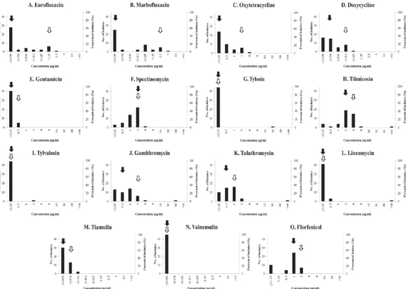
The distribution of the MIC values of fluoroquinolones (enrofloxacin and marbofloxacin) showed one main peak coinciding with MIC50value at the lowest antibiotic concentration (�0.039μg/ml), while the other values represented equipartition with the highest MIC values (2.5μg/ml and 5μg/ml, respectively) (Fig 1A and 1B). One strain (MycSu17) exceeded the unofficial breakpoint [11], with the MIC value of 2.5μg/ml of enrofloxacin. All of the exam- ined tetracyclines had low MIC values with MIC50and MIC90values of�0.25μg/ml and 2μg/
ml of oxytetracycline; and 0.078μg/ml and 0.312μg/ml of doxycycline (Fig 1C and 1D). The
lowest examined concentration of gentamicin (�0.25μg/ml) was effective against most of the studied strains (Fig 1E). MIC50and MIC90values of spectinomycin were 2μg/ml, with MIC 4μg/ml being the highest detected value (Fig 1F). Five macrolides were tested (Fig 1G–1K), out of which tilmicosin showed a Gaussian distribution with 2μg/ml and 4μg/ml MIC50and MIC90values, respectively. One main peak at the lowest antibiotic concentration (�0.25μg/
ml) was observed in the MIC values of tylosin and tylvalosin against the examined strains.
MIC50values of gamithromycin and tulathromycin were 0.5μg/ml, while MIC90values were 2μg/ml and 1μg/ml, respectively. Both MIC50and MIC90values of lincomycin coincided with the lowest examined antibiotic concentration (�0.25μg/ml) (Fig 1L). For all macrolides and for lincomycin high MIC values (>64μg/ml of tilmicosin and tulathromycin; 64μg/ml of gamithromycin; 32μg/ml of tylosin; 2μg/ml of tylvalosin; and>64μg/ml of lincomycin) were detected against an outlier strain (MycSu18). Both studied pleuromutilins had low MIC values (Fig 1M and 1N). The MIC50and MIC90values of valnemulin were�0.039μg/ml, while that of tiamulin�0.039μg/ml and 0.078μg/ml. MIC50and MIC90values of florfenicol were 1μg/
ml and 2μg/ml (Fig 1O).
Single-nucleotide polymorphisms correlating with decreased antibiotic susceptibility
High MIC values of fluoroquinolones, macrolides and lincosamides, exceeding the unofficial breakpoints [11] were found in some cases (e.g. MycSu17-18). Both synonymous and non-
Table 1. MIC values against the type strain and summary of MIC ranges, MIC50and MIC90values (μg/ml) against theM.hyopneumoniaestrains involved in this study.
NCTC 10110 initial
NCTC 10110 final
Range initial
Range final
MIC50
initial
MIC50
final
MIC90
initial
MIC90
final Fluoroquinolones
Enrofloxacin �0.039 0.078 �0.039–2.5 �0.039–5 �0.039 0.312 1.25 2.5
Marbofloxacin �0.039 0.156 �0.039–5 �0.039–10 �0.039 1.25 2.5 5
Tetracyclines
Oxytetracycline �0.25 4 �0.25–4 0.5–32 �0.25 4 2 16
Doxycycline �0.039 0.625 �0.039–0.625 0.078–2.5 0.078 0.625 0.312 2.5
Aminoglycoside
Gentamicin �0.25 1 �0.25–0.5 0.5–2 �0.25 1 0.5 2
Aminocyclitol
Spectinomycin 1 4 �0.25–4 1–8 2 4 2 4
Macrolides
Tylosin �0.25 0.5 �0.25–32 �0.25–64 0.25 0.5 �0.25 0.5
Tilmicosin 2 8 �0.25-�64 2->64 2 8 4 16
Tylvalosin �0.25 �0.25 �0.25–2 �0.25–8 �0.25 �0.25 �0.25 �0.25
Gamithromycin 1 4 �0.25–64 1->64 0.5 4 2 8
Tulathromycin 1 4 �0.25-�64 0.5->64 0.5 2 1 4
Lincosamide
Lincomycin �0.25 1 �0.25-�64 �0.25->64 �0.25 0.5 �0.25 1
Pleuromutilins
Tiamulin �0.039 0.156 �0.039–0.156 0.078–0.312 �0.039 0.156 0.078 0.156
Valnemulin �0.039 �0.039 �0.039 �0.039 �0.039 �0.039 �0.039 �0.039
Phenicol
Florfenicol 1 2 �0.125–2 1–4 1 2 2 4
https://doi.org/10.1371/journal.pone.0209030.t001
synonymous substitutions were observed in genes associated with susceptibility to fluoroquin- olones (gyrA,gyrB,parCandparE); however, only SNPs resulting in amino acid alterations were further examined in the present study. None of the amino acid changes in the genesgyrB andparEshowed correlation with the defined MIC values. On the other hand, amino acid changes in thegyrAgene (Gly81Ala, Ala83Val and Glu87Gly) and in theparCgene (Ser80Phe, Ser80Tyr or Asp84Asn) correlated with decreased susceptibility of fluoroquinolones (S3 Table). Single alterations in theparCgene seem to have no crucial effect on fluoroquinolone susceptibility when initial MIC values are examined. On the other hand, at least 12-fold con- centration difference is observed in the final MIC values against strains, which contain a single alteration in theparCgene. As opposed to the observed slight increase of MIC values of fluoro- quinolones in association with the single substitution event in geneparC, double substitutions in genesparCandgyrAcorrelated with final MIC values higher than 2μg/ml in all cases, with one exception (MycSu44). It is noteworthy, that the double substitutions in strain MycSu44 consisted of Ala83Val in genegyrAand Asp84Asn inparC, while the rest of the strains showed various amino acid substitution types in genegyrAbut only the change of serine at amino acid
Fig 1. Distribution of the minimal inhibitory concentrations of each tested antibiotic against the studiedM.hyopneumoniaeisolates. MIC50and MIC90values are marked with black and white arrows, respectively.
https://doi.org/10.1371/journal.pone.0209030.g001
position 80 in geneparC. The one outlier strain (MycSu17) against which 2.5–5μg/ml initial MIC values of fluoroquinolones were detected contained the double substitution combination Ser80Phe (inparCgene) with Ala83Val (ingyrAgene). Correlation was described between increased MIC values of macrolides and lincosamides against Mycoplasma species/M.hyop- neumoniaeand SNPs in the 23S rRNA sequence [14,19]. A nucleotide substitution at the position A2059G was found in the outlier strain (MycSu18) showing extremely decreased sus- ceptibility to macrolides and lincosamides (S3 Table). The observed SNPs in the strains origi- nating from the same herds were consistent with one exception: the strains originating from Mezőtu´r (MycSu7; 8 and 41), which also clustered into completely different sequence types according to earlier genotyping analysis [31] showed distinct susceptibility profiles and genetic alterations correlating with antibiotic susceptibility.
Discussion
Antibiotic susceptibility testing of porcine mycoplasmas is not performed routinely, because it is fastidious, time-consuming and requires special techniques and media [17]. Furthermore, the lack of official standards makes the interpretation of the results difficult. The Clinical and Laboratory Standards Institute (CLSI) has provided official breakpoints for certain antibiotics but only for human pathogen mycoplasmas [35] and the procedures and media vary according to each of the examined species [36].
Fluoroquinolones are potentially active antimicrobial agents againstM.hyopneumoniae through inhibition of the bacterial DNA gyrase and topoisomerase IV enzymes [19,22]. In the present study, a broad range of MIC values was recorded with low MIC50value of enrofloxa- cin, similarly to previous results in other European publications in the last 20 years [11,13,34].
One of the examined strains (MycSu17) was inhibited by higher enrofloxacin concentration, the MIC value against this strain exceeded the unofficial breakpoint determined by Hannan et al. [11]. Similar observations have already been recorded with high MIC values in Thailand (�2μg/ml) and in Belgium (>1μg/ml) [13,33], which forewarns the importance of suscepti- bility testing before choosing antibiotics for treatment. Although MIC50value of marbofloxa- cin against the studied strains was mostly in accordance with recent data, MIC90value against the Hungarian isolates was higher than those against Belgian, Spanish and British strains (0.5–
1μg/ml) [34].
No amino acid substitutions, correlating with increased MIC values, were observed in the genesgyrBandparE, corroborating earlier publications [18]. Although single amino acid substitutions in theparCgene (Ser80Phe, Ser80Tyr or Asp84Asn) showed correlation with increased MIC values of fluoroquinolones in earlier publications [15,18,19], the degree of increase seems to be negligible according to the initial MIC values detected in the present study. However, definite increase of MIC values was detected when double substitutions in parCandgyrAgenes were described in the examined strains. The observed effect of the double substitutions is in accordance with previous findings of Vicca et al. [18]. Various combinations of amino acid changes were detected in the examined strains containing double substitutions in the genesgyrAandparC, defining a unique combination in the outlier strain (MycSu17).
Moreover, new amino acid alterations (Glu87Gly and Gly81Ala) have been described ingyrA gene ofM.hyopenumoniaein the present study, which had been observed only inM.bovisand M.gallisepticumbefore [23,24]. Factors influencing the degree of the decrease of susceptibility to fluoroquinolones, such as the type of amino acid changes or mechanisms are yet to be dis- covered. Although initial MIC values are advised to be taken into account in the interpretation of the results of antibiotic susceptibility tests [37], correlations between the amino acid substi- tutions and increased final MIC values were more defined in the current examinations and
better supported previous observations, which highlights the usefulness of determining final MIC values also.
The increasing susceptibility against fluoroquinolones is a notable problem, because these agents are important antibiotics for human therapy [38]. To maximize efficacy and reduce mutant selection in case of fluoroquinolones, the ratio of maximum serum concentration to the MIC (Cmax/MIC ratio) of equal or higher than 10 was proposed [39]. Marbofloxacin administered at 4 or 8 mg/kg intramuscularly resulted in 6.3 and 3.38μg/ml Cmaxin pigs [40]
respectively, resulting in maximum activity against strains with MICs of 0.625 and 0.3125μg/
ml or lower in case of the two dosages, respectively.
Tetracyclines are frequently used to controlM.hyopneumoniaeinfections, and they act by binding to the decoding centre of the small ribosomal subunit of the bacterium [4,41]. Most of the previous publications from Europe defined similar MIC50and MIC90values of oxytetracy- cline [11,13,34]; but higher MIC50and MIC90values of doxycycline were described against strains originating from Spain (1μg/ml both) and Thailand (3.12μg/ml and 6.25μg/ml) than against the Hungarian isolates. According to other publications supported also by our results, tetracyclines are still active againstM.hyopneumoniaedespite of their long-standing usage in human and veterinary medicine [32,33].
The aminoglycoside gentamicin seems to be an effective antimicrobial agent against M.hyopneumoniae, as low MIC50and MIC90values were observed in the present study, simi- larly to earlier data [13,32]. Although MIC range of the aminocyclitol spectinomycin was broad similarly to the findings of a previous Spanish study, the MIC50and MIC90values were higher in the present study compared to Spanish and Belgian MIC values [13,32].
Macrolides are among the most frequently used antibiotics in the swine industry to treat M.hyopneumoniaeinfections [4]. Both 16-membered (tylosin, tilmicosin and tylvalosin) and 15-membered (tulathromycin and gamithromycin) macrolides were effective against the stud- ied strains. However, the MIC value of tulathromycin against the type strain was three orders of magnitude higher, than in the literature [34]. The reason of the discrepancy might be a dif- ferent passage number of the type strain, or the different medium/antibiotic solution used dur- ing the test. However, the MIC value of tulathromycin against the type strain did not exceed 16μg/ml (a possible unofficial breakpoint according to other porcine respiratory pathogens [42]) in either case. In the current study, a slight increase of MIC50and MIC90values of macro- lides was described compared to the literature [34], and extremely high MIC values against an outlier strain (MycSu18) was noted. According to the literature, nucleotide substitutions at the bases 2057–2059 of the 23S rRNA sequence play an important role in acquired resistance to macrolides [14,19,43]. Analysis of the 23S rRNA sequence of the strain MycSu18 revealed a nucleotide substitution A2059G (E.colinumbering), which was also described in macrolide and lincosamide resistantM.bovisstrains before [24]. According to the habituation study of Hannanet al. [12] and the high MIC values presented in this study, emergence of macrolide- resistance could be a considerable problem, which was confirmed by earlier reported results from Belgium [13], Thailand [33] and Spain [32].
Lincomycin is also active againstM.hyopneumoniae, but extremely high MIC values appear every now and then [13,32,33], like the outlier strain (MycSu18) in the present study. The rea- son of the decreased susceptibility can be the cross-resistance with macrolides, as reported in an earlier publication, which described decreasing susceptibility against tylosin and lincomy- cin in strains originating from a lincomycin-treated herd [13]. The simultaneously appearing change in susceptibility may lead back to the same mode of action of macrolides and lincosa- mides, inhibiting bacterial protein synthesis on the 50S ribosomal subunit [44].
Pleuromutilins are important antibiotics to controlM.hyopneumoniaeinfections through inhibiting bacterial protein synthesis [45]. According to our results and other publications,
tiamulin seems to be one of the most effective antimicrobial agents againstM.hyopneumoniae with lowin vitroinhibitory concentrations [11–13,32–34]. Valnemulin is the most effective antibiotic against all of the studied strains, which supported the earlier published observations [12,32,34].
The chloramphenicol derivative florfenicol is an inhibitor of bacterial protein synthesis, used exclusively for veterinary purposes [46]. The moderate distribution of the MIC range and the relatively low MIC50and MIC90values of florfenicol, were similar to earlier observations from different parts of Europe and Thailand [13,33,34], and they may indicate that this antibi- otic is an effective agent againstM.hyopneumoniae.
In vitroMIC values do not necessarily correlate with the effectiveness of the antimicrobials in vivoand interpretation of the MIC distributions is difficult asMycoplasmaspecies with vet- erinary relevance do not have official clinical breakpoints [34]. Furthermore, strains with dif- ferent antibiotic susceptibility can coexist within a herd [33]. PK/PD (pharmacokinetic- pharmacodynamic) analysis is an important tool to maximizein vivoantimicrobial activity [47,48]. Most of our results were in accordance with other results of the European region, this involves, that all the tested agents are most probably still suitable to control enzootic pneumo- nia. Nonetheless the results of this study may help veterinarians to choose the proper antimi- crobial agent againstM.hyopneumoniae. Although the isolation ofM.hyopneumoniaestrains is a time-consuming and fastidious process, the regularly accomplished antibiotic susceptibil- ity testing of the swine herds should enable appropriate antibiotic usage during treatment. Fur- thermore, the development of PCR-based susceptibility tests based on SNPs correlating with changes in the MIC values, could improve diagnostics and treatment, similarly to antibiotic susceptibility testing inM.bovis[24].
Conclusion
This study provided current and relevant information about the antibiotic susceptibility profiles ofM.hyopneumoniaestrains circulating in Hungary and surrounding countries.
Low MIC values of all the tested antibiotics were described against most of the studiedM.
hyopneumoniaestrains, and the lowest MIC values were found in case of gentamicin, tylosin, tylvalosin, lincomycin, tiamulin and valnemulin. In certain cases, high MIC values of fluoro- quinolones (MycSu17) or macrolides and lincomycin (MycSu18) were observed. Single or double amino acid substitutions in the genesgyrA(Gly81Ala, Ala83Val, Glu87Gly),parC (Ser80Phe, Ser80Tyr, Asp84Asn) and a SNP in the 23S rRNA sequence (A2059G) were also detected correlating with decreased antibiotic susceptibilities. Macrolides and fluoroquino- lones are frequently used empirically as a first choice for the management of mycoplasmoses in livestock in Europe. The regular testing of the sensitivity profile ofM.hyopneumoniae, the determination of herd specific MICs would promote the use of less critical antibacterials (e.g.:
florfenicol, tetracyclines, pleuromutilins), and might contribute to the preservation of the criti- cally important antibiotics (macrolides and fluoroquinolones) both for veterinary and human medicine.
Supporting information
S1 Table. Background data ofM.hyopneumoniaestrains and initial minimum inhibitory concentration (MIC) values (μg/ml) of 15 antimicrobials against the strains used in the study. Isolation data (Sample ID, Herd of origin and Date of isolation) and MIC values of enrofloxacin (EFX), marbofloxacin (MFX), oxytetracycline (OTC), doxycycline (DX), genta- micin (GTC), spectinomycin (SPC), tylosin (TYL), tilmicosin (TIL), tylvalosin (TVN), gami- thromycin (GTM), tulathromycin (TTM), tiamulin (TIA), valnemulin (VAL), lincomycin
(LCM) and florfenicol (FFC) are presented. Abbreviations for herd of origin are: H-Hungary, CZ-Czech Republic, SK-Slovakia.
(DOCX)
S2 Table. Background data ofM.hyopneumoniaestrains and final minimum inhibitory concentration (MIC) values (μg/ml) of 15 antimicrobials against the strains used in the study. Isolation data (Sample ID, Herd of origin and Date of isolation) and MIC values of enrofloxacin (EFX), marbofloxacin (MFX), oxytetracycline (OTC), doxycycline (DX), genta- micin (GTC), spectinomycin (SPC), tylosin (TYL), tilmicosin (TIL), tylvalosin (TVN), gami- thromycin (GTM), tulathromycin (TTM), tiamulin (TIA), valnemulin (VAL), lincomycin (LCM) and florfenicol (FFC) are presented. Abbreviations for herd of origin are: H-Hungary, CZ-Czech Republic, SK-Slovakia.
(DOCX)
S3 Table. Initial and final minimum inhibitory concentration (MIC) ranges (μg/ml) of fluoroquinolones, macrolides and lincomycin against the examinedM.hyopneumoniae isolates with the amino acid substitutions in thegyrAandparCgenes and nucleotide sub- stitutions in the 23S rRNA sequence.
(DOCX)
Author Contributions Data curation: Miklo´s Gyuranecz.
Formal analysis: Kinga Ma´ria Sulyok.
Investigation: Orsolya Felde, Veronika Hrivna´k, Krisztia´n Kiss, Imre Biksi.
Methodology: Zsuzsa Kreizinger, Kinga Ma´ria Sulyok, Miklo´s Gyuranecz.
Project administration: Veronika Hrivna´k.
Resources: Miklo´s Gyuranecz.
Supervision: Miklo´s Gyuranecz.
Writing – original draft: Orsolya Felde, Zsuzsa Kreizinger.
Writing – review & editing: Kinga Ma´ria Sulyok, A´ kos Jerzsele, Miklo´s Gyuranecz.
References
1. Artiushin S, Minion FC. Arbitrarily primed PCR analysis of Mycoplasma hyopneumoniae field isolates demonstrates genetic heterogeneity. Int J Syst Evol Microbiol. 1996; 46:324–328.
2. Kobisch M, Friis NF. Swine mycoplasmoses. Rev Sci Tech. 1996; 15:1569–1605. PMID:9190026 3. Maes D, Verdonck M, Deluyker H, de Kruif A. Enzootic pneumonia in pigs. Vet Q. 1996; 18:104–109.
https://doi.org/10.1080/01652176.1996.9694628PMID:8903144
4. Maes D, Segales J, Meyns T, Sibila M, Pieters M, Haesebrouck F. Control of Mycoplasma hyopneumo- niae infections in pigs. Vet Microbiol. 2008; 126:297–309.https://doi.org/10.1016/j.vetmic.2007.09.008 PMID:17964089
5. Maes D, Sibila M, Kuhnert P, Segale´ s J, Haesebrouck F, Pieters M. Update on Mycoplasma hyopneu- moniae infections in pigs: Knowledge gaps for improved disease control. Transbound Emerg Dis. 2017;
https://doi.org/10.1111/tbed.12677PMID:28834294
6. Wyns H, Meyer E, Plessers E, Watteyn A, De Baere S, De Backer P, et al. Pharmacokinetics of gami- thromycin after intravenous and subcutaneous administration in pigs. Res Vet Sci. 2014; 96:160–163.
https://doi.org/10.1016/j.rvsc.2013.11.012PMID:24331716
7. Pallare´s FJ, Lasa C, Roozen M, Ramis G. Use of tylvalosin in the control of porcine enzootic pneumo- nia. Vet Rec Open. 2015;
8. El Garch F, de Jong A, Simjee S, Moyaert H, Klein U, Ludwig C, et al. Monitoring of antimicrobial sus- ceptibility of respiratory tract pathogens isolated from diseased cattle and pigs across Europe, 2009–
2012: VetPath results. Vet Microbiol. 2016; 194:11–22.https://doi.org/10.1016/j.vetmic.2016.04.009 PMID:27102206
9. Thacker EL, Thacker BJ, Boettcher TB, Jayappa H. Comparison of antibody production, lymphocyte stimulation, and protection induced by four commercial Mycoplasma hyopneumoniae bacterins. Swine Health Prod. 1998; 6:107–112.
10. McCormack WM. Susceptibility of mycoplasmas to antimicrobial agents: Clinical implications. Clin Infect Dis. 1993; 17:200–201.
11. Hannan PCT, Windsor GD, De Jong A, Schmeer N, Stegemann M. Comparative susceptibilities of vari- ous animal-pathogenic mycoplasmas to fluoroquinolones. Antimicrob Agents Chemother. 1997;
41:2037–2040. PMID:9303412
12. Hannan PCT, Windsor HM, Ripley PH. In vitro susceptibilities of recent field isolates of Mycoplasma hyopneumoniae and Mycoplasma hyosynoviae to valnemulin (Econor), tiamulin and enrofloxacin and the in vitro development of resistance to certain antimicrobial agents in Mycoplasma hyopneumoniae.
Res Vet Sci. 1997; 63:157–160. PMID:9429250
13. Vicca J, Stakenborg T, Maes D, Butaye P, Peeters J, de Kruif A, et al. In vitro susceptibilities of Myco- plasma hyopneumoniae field isolates. Antimicrob Agents Chemother. 2004; 48:4470–4472.https://doi.
org/10.1128/AAC.48.11.4470-4472.2004PMID:15504886
14. Stakenborg T, Vicca J, Butaye P, Maes D, Minion FC, Peeters J, et al. Characterization of in vivo acquired resistance of Mycoplasma hyopneumoniae to macrolides and lincosamides. Microb Drug Resist. 2005; 11:290–294.https://doi.org/10.1089/mdr.2005.11.290PMID:16201934
15. Le Carrou J, Laurentie M, Kobisch M, Gautier-Bouchardon AV. Persistence of Mycoplasma hyopneu- moniae in experimentally infected pigs after marbofloxacin treatment and detection of mutations in the parC gene. Antimicrob Agents Chemother. 2006; 50:1959–1966.https://doi.org/10.1128/AAC.01527- 05PMID:16723552
16. Yamamoto K, Koshimizu K, Ogata M. In vitro susceptibility of Mycoplasma hyopneumoniae to antibiot- ics. Japanese J Vet Sci. 1986; 48:1–5.
17. Hannan PCT. Guidelines and recommendations for antimicrobial minimum inhibitory concentration (MIC) testing against veterinary mycoplasma species. Vet Res. 2000; 31:373–395.https://doi.org/10.
1051/vetres:2000100PMID:10958240
18. Vicca J, Maes D, Stakenborg T, Butaye P, Minion F, Peeters J, et al. Resistance mechanism against flu- oroquinolones in Mycoplasma hyopneumoniae field isolates. Microb Drug Resist. 2007; 13:166–170.
https://doi.org/10.1089/mdr.2007.716PMID:17949302
19. Gautier-Bouchardon AV. Antimicrobial resistance in Mycoplasma spp. Microbiol Spectr. 2018;https://
doi.org/10.1128/microbiolspec.ARBA-0030-2018PMID:30003864
20. Felde O, Kiss K, Biksi I, Jerzsele A´ , Gyuranecz M. A serte´sek Mycoplasma hyopneumoniae okozta tu¨dőgyullada´ sa. (Pneumonia of pigs caused by Mycoplasma hyopneumoniae) Magy A´ llatorvosok Lapja. 2018; 140:337–348.
21. European Centre for Disease Prevention and Control, European Food Safety Authority, and European Medicines Agency, ECDC/EFSA/EMA first joint report on the integrated analysis of the consumption of antimicrobial agents and occurrence of antimicrobial resistance in bacteria from humans and food-pro- ducing animals. EFSA J. 2015;
22. Hooper DC. Mechanisms of action and resistance of older and newer fluoroquinolones. Clin Infect Dis.
2000; 31(Supplement_2):S24–28.
23. Reinhardt AK, Kempf I, Kobisch M, Gautier-Bouchardon AV. Fluoroquinolone resistance in Myco- plasma gallisepticum: DNA gyrase as primary target of enrofloxacin and impact of mutations in topo- isomerases on resistance level. J Antimicrob Chemother. 2002; 50:589–592.https://doi.org/10.1093/
jac/dkf158PMID:12356806
24. Sulyok KM, Kreizinger Z, Wehmann E, Lysnyansky I, Ba´nyai K, Marton S, et al. Mutations associated with decreased susceptibility to seven antimicrobial families in field and laboratory-derived Mycoplasma bovis strains. Antimicrob Agents Chemother. 2017;https://doi.org/10.1128/AAC.01983-16PMID:
27895010
25. Vester B, Douthwaite S. Macrolide Resistance conferred by base substitutions in 23S rRNA Antimicrob Agents Chemother. 2001; 45:1–12.https://doi.org/10.1128/AAC.45.1.1-12.2001PMID:11120937 26. Friis NF. Some recommendations concerning primary isolation of Mycoplasma suipneumoniae and
Mycoplasma flocculare a survey. Nord Vet Med. 1975; 27:337–339. PMID:1098011
27. Etheridge JR, Cottew GS, Lloyd LC. Isolation of Mycoplasma hyopneumoniae from lesions in experi- mentally infected pigs. Aust Vet J. 1979; 55:356–359. PMID:533486
28. Mattsson JG, Bergstro¨ m K, Wallgren P, Johansson KE. Detection of Mycoplasma hyopneumoniae in nose swabs from pigs by in vitro amplification of the 16S rRNA gene. J Clin Microbiol. 1995; 33:893–
897. PMID:7540629
29. Lauermann LH, Chilina AR, Closser JA, Johansen D. Avian Mycoplasma identification using polymer- ase chain reaction amplicon and restriction fragment length polymorphism analysis. Avian Dis. 1995;
39:804–811. PMID:8719214
30. Kearse M, Moir R, Wilson A, Stones-Havas S, Cheung M, Sturrock S, et al. Geneious Basic : An inte- grated and extendable desktop software platform for the organization and analysis of sequence data.
Bioinformatics. 2012; 28:1647–1649.https://doi.org/10.1093/bioinformatics/bts199PMID:22543367 31. Felde O, Kreizinger Z, Sulyok KM, Marton S, Ba´nyai K, Korbuly K, et al. Genotyping Mycoplasma hyop-
neumoniae isolates based on multi-locus sequence typing, multiple-locus variable-number tandem repeat analysis and analysing gene p146. Vet Microbiol. 2018; 222:85–90.https://doi.org/10.1016/j.
vetmic.2018.07.004PMID:30080678
32. Tavı´o MM, Poveda C, Assunc¸ão P, Ramı´rez AS, Poveda JB. In vitro activity of tylvalosin against Span- ish field strains of Mycoplasma hyopneumoniae. Vet Rec. 2014;https://doi.org/10.1136/vr.102458 PMID:25185108
33. Thongkamkoon P, Narongsak W, Kobayashi H, Pathanasophon P, Kishima M, Yamamoto K. In vitro susceptibility of Mycoplasma hyopneumoniae field isolates and occurrence of fluoroquinolone, macro- lides and lincomycin resistance. J Vet Med Sci. 2013; 75:1067–1670. PMID:23503167
34. Klein U, de Jong A, Moyaert H, El Garch F, Leon R, Richard-Mazet A, et al. Antimicrobial susceptibility monitoring of Mycoplasma hyopneumoniae and Mycoplasma bovis isolated in Europe. Vet Microbiol.
2017; 204:188–193.https://doi.org/10.1016/j.vetmic.2017.04.012PMID:28532800
35. Wayne P. Clinical and Laboratory Standards Institute (CLSI). Methods for antimicrobial susceptibility testing for human mycoplasmas; Approved guideline. CLSI document M43-A. 2011.
36. Waites KB, Duffy LB, Be´be´ar CM, Matlow A, Talkington DF, Kenny GE, et al. Standardized methods and quality control limits for agar and broth microdilution susceptibility testing of Mycoplasma pneumo- niae, Mycoplasma hominis, and Ureaplasma urealyticum. J Clin Microbiol. 2012; 50:3542–3547.https://
doi.org/10.1128/JCM.01439-12PMID:22915608
37. Kreizinger Z, Gro´ zner D, Sulyok KM, Nilsson K, Hrivna´k V, Benčina D, et al. Antibiotic susceptibility pro- files of Mycoplasma synoviae strains originating from Central and Eastern Europe. BMC Vet Res. 2017;
https://doi.org/10.1186/s12917-017-1266-2PMID:29149886
38. Collignon P, Powers JH, Chiller TM, Aidara-Kane A, Aarestrup FM. World Health Organization ranking of antimicrobials according to their importance in human medicine: A critical step for developing risk management strategies for the use of antimicrobials in food production animals. Clin Infect Dis. 2009;
49:132–141.https://doi.org/10.1086/599374PMID:19489713
39. Rodvold KA, Neuhauser M. Pharmacokinetics and pharmacodynamics of fluoroquinolones. In: Pharma- cotherapy: the journal of human pharmacology and drug therapy. 2001. 233S–252S.
40. Schneider M, Paulin A, Dron F, Woehrle´ F. Pharmacokinetics of marbofloxacin in pigs after intravenous and intramuscular administration of a single dose of 8 mg/kg: dose proportionality, influence of the age of the animals and urinary elimination. Vet Pharmacol Ther. 2014; 37:523–530.https://doi.org/10.1111/
jvp.12125PMID:24666477
41. Nguyen F, Starosta AL, Arenz S, Sohmen D, Do¨ nho¨fer A, Wilson DN. Tetracycline antibiotics and resis- tance mechanisms. Biol Chem. 2014; 395:559–575.https://doi.org/10.1515/hsz-2013-0292PMID:
24497223
42. Godinho KS. Susceptibility testing of tulathromycin: Interpretative breakpoints and susceptibility of field isolates. Vet Microbiol. 2008; 129:426–432.https://doi.org/10.1016/j.vetmic.2007.11.033PMID:
18187275
43. Hansen JL, Ippolito JA, Ban N, Nissen P, Moore PB, Steitz TA. The structures of four macrolide antibiot- ics bound to the large ribosomal subunit. Mol Cell. 2002; 10:117–128. PMID:12150912
44. Weisblum B. Insights into erythromycin action from studies of its activity as inducer of resistance. Anti- microb Agents Chemother. 1995; 39:797–805. PMID:7785974
45. Poulsen SM, Karlsson M, Johansson LB, Vester B. The pleuromutilin drugs tiamulin and valnemulin bind to the RNA at the peptidyl transferase centre on the ribosome. Mol Microbiol. 2001; 41:1091–1099.
PMID:11555289
46. Priebe S, Schwarz S. In vitro activities of florfenicol against bovine and porcine respiratory tract patho- gens. Antimicrob Agents Chemother. 2003; 47:2703–2705.https://doi.org/10.1128/AAC.47.8.2703- 2705.2003PMID:12878547
47. Ahmad I, Huang L, Hao H, Sanders P, Yuan Z. Application of PK / PD modeling in veterinary field : Dose optimization and drug resistance prediction. Biomed Res Int. 2016;https://doi.org/10.1155/2016/
5465678PMID:26989688
48. Somogyi Z, Karancsi Z, Jerzsele A´ . Farmakokinetika/farmakodina´mia (PK/PD) megko¨zelı´te´s az a´llat- gyo´gya´szatban: Irodalmi o¨sszefoglalo´. (Pharmacokinetics/pharmacodynamics approach in the veteri- nary medicine: Literature review) Magy A´ llatorvosok Lapja. 2018; 14:37–46.