https://doi.org/10.1007/s11120-020-00784-1 ORIGINAL ARTICLE
Chloramphenicol enhances Photosystem II photodamage in intact cells of the cyanobacterium Synechocystis PCC 6803
Sandeesha Kodru1,2 · Ateeq ur Rehman1 · Imre Vass1
Received: 18 May 2020 / Accepted: 9 September 2020 / Published online: 26 September 2020
© The Author(s) 2020
Abstract
The effect of chloramphenicol, an often used protein synthesis inhibitor, in photosynthetic systems was studied on the rate of Photosystem II (PSII) photodamage in the cyanobacterium Synechocystis PCC 6803. Light-induced loss of PSII activity was compared in the presence of chloramphenicol and another protein synthesis inhibitor, lincomycin, by measuring the rate of oxygen evolution in Synechocystis 6803 cells. Our data show that the rate of PSII photodamage was significantly enhanced by chloramphenicol, at the usually applied 200 μg mL−1 concentration, relative to that obtained in the presence of lincomycin. Chloramphenicol-induced enhancement of photodamage has been observed earlier in isolated PSII membrane particles, and has been assigned to the damaging effect of chloramphenicol-mediated superoxide production (Rehman et al.
2016, Front Plant Sci 7:479). This effect points to the involvement of superoxide as damaging agent in the presence of chlo- ramphenicol also in Synechocystis cells. The chloramphenicol-induced enhancement of photodamage was observed not only in wild-type Synechocystis 6803, which contains both Photosystem I (PSI) and PSII, but also in a PSI-less mutant which contains only PSII. Importantly, the rate of PSII photodamage was also enhanced by the absence of PSI when compared to that in the wild-type strain under all conditions studied here, i.e., without addition and in the presence of protein synthesis inhibitors. We conclude that chloramphenicol enhances photodamage mostly by its interaction with PSII, leading probably to superoxide production. The presence of PSI is also an important regulatory factor of PSII photodamage most likely via decreasing excitation pressure on PSII.
Keywords Chloramphenicol · Photoinhibition · Photosystem I · Photosystem II · Superoxide · Synechocystis PCC 6803 Abbreviations
Chl Chlorophyll
DMBQ 2,6-Dimethoxy-1,4-benzoquinone PSI Photosystem I
PSII Photosystem II
Introduction
Photosynthesis is a process in which green plants, algae, and cyanobacteria utilize energy from sunlight to pro- duce carbohydrates from carbon dioxide and water. This process is the ultimate source of energy for all plants to drive their metabolic processes. Too much light reaching the photosynthetic apparatus can cause photodamage and finally can lead to the death of a cell, a phenomenon called photoinhibition (Arntzen et al. 1984; Aro et al. 1993; Vass and Aro 2008; Zavafer et al. 2017). A major impact on the photosynthetic machinery under high light conditions is the impairment of electron transport in the Photosystem II (PSII) complex, as well as damage of its D1 reaction center subunit (Ohad et al. 1984; Prasil et al. 1992; Aro et al. 1993). Important mediators of photodamage in plant cells are the various reactive oxygen species (ROS), such as singlet excited oxygen, free radicals (superoxide and hydroxyl ions), superoxide and peroxides, which are pro- duced mainly in the chloroplasts and mitochondria (Apel
Electronic supplementary material The online version of this article (https ://doi.org/10.1007/s1112 0-020-00784 -1) contains supplementary material, which is available to authorized users.
* Imre Vass vass.imre@brc.hu
1 Institute of Plant Biology, Biological Research Centre, Temesvari krt. 62, Szeged 6726, Hungary
2 Doctoral School of Biology, University of Szeged, Szeged, Hungary
and Hirt 2004; Krieger-Liszkay 2005; Krieger-Liszkay et al. 2008; Pospísil 2012; Vass 2012). The activity of the photodamaged PSII complex can be restored via the so-called PSII repair cycle, in which de novo synthesis of the D1 subunits plays a key role (Aro et al. 1993; Baena- Gonzalez and Aro 2002; Komenda et al. 2007; Nixon et al.
2010; Vass 2012; Järvi et al. 2015; Li et al. 2018).
Light stress to PSII becomes a problem for photosynthetic activity when the rate of photodamage exceeds the rate of repair processes. Therefore, it is important to monitor sepa- rately the rates of photodamage and of the protein synthesis- dependent repair. Decoupling of photodamage and repair can be achieved by protein synthesis inhibitors, such as lincomy- cin or chloramphenicol, which block the initiation of protein synthesis and of peptide bond formation, respectively (Con- treras and Vazquez 1977). These protein synthesis inhibitors can be applied both in chloroplasts (Mulo et al. 2003; Chow et al. 2005; Tikkanen et al. 2014) and cyanobacterial cells (Constant et al. 1997; Nishiyama et al. 2001, 2005; Taka- hashi and Murata 2005; Takahashi et al. 2009; Sicora et al.
2003). While there are no reports concerning the participa- tion of lincomycin in photosynthetic electron transport, chlo- ramphenicol has been reported to accept electrons from the acceptor side of Photosystem I (PSI) and to transfer them to molecular oxygen leading to superoxide production (Okada et al. 1991). Although O2·− has lower reactivity than other ROS, it can induce damage to proteins and membrane com- ponents due to its ability to produce highly oxidizing radi- cals (Pospisil et al. 2019). This side effect of chlorampheni- col has been considered as a source of a potential artifact by several research groups, who used lincomycin instead of chloramphenicol in photoinhibition studies (Constant et al.
1997; Tyystjärvi and Aro 1996; Tyystjarvi et al. 2002; Chow et al. 2005; Miyata et al. 2012; Campbell and Tyystjärvi 2012; Tikkanen et al. 2014). However, other groups kept using chloramphenicol in measurements of PSII photodam- age, and often obtained results which were contradictory to those studies in which lincomycin was used (Nishiyama et al. 2001, 2005; Takahashi and Murata 2005; Takahashi et al. 2009). It is also of note that while chloramphenicol was used at 30–65 or 100 μg mL−1 in some early investigations, it was applied at 200 μg mL−1 or higher concentrations in the majority of photosynthesis related studies.
Recently, we have shown in isolated thylakoid mem- brane systems that chloramphenicol can accept electrons not only from PSI but also from the acceptor side of PSII, and deliver them to molecular oxygen leading to superoxide production (Rehman et al. 2016). In addition, the presence of 200 μg mL−1 chloramphenicol enhances photodamage of PSII electron transport in the isolated systems, an effect that is reversible by superoxide dismutase (Rehman et al. 2016), pointing to the damaging role of superoxide in the process of photoinhibition.
In the present work, we have extended our earlier stud- ies from isolated thylakoid membrane preparations to intact cell cultures of the cyanobacterium Synechocystis PCC 6803, as well as to its PSI-less mutant. Our data show that the presence of 200 μg mL−1 chlorampheni- col enhances photodamage of PSII in intact Synechocys- tis cells even in the absence of PSI. As a consequence, chloramphenicol, when applied at 200 μg mL−1 or higher concentrations, can lead to artifacts in studies aiming at the determination of the true rate of PSII photodamage in intact systems.
Materials and methods
Cell culturesWild-type Synechocystis sp. PCC 6803 (which will be referred to as WT Synechocystis) cells were propagated in BG-11 growth medium in a rotary shaker at 30 °C under a 3% CO2-enriched atmosphere. The intensity of white light during growth was 40 µmol photons m−2 s−1. Cells in the exponential growth phase (A580 of 0.8–1) were used. The PSI-less strain of Synechocystis, was produced by Wim Ver- maas similarly to that described earlier (Shen et al. 1993), but using spectinomycin instead of chloramphenicol as selective antibiotic. PSI-less cells were grown at low light intensity (of 5 µmol photons m−2 s−1) in the presence of 5 mM glucose and 25 μg mL−1 spectinomycin. As a control for the PSI-less strain, WT cells were also cultured under the same conditions, i.e., 5 µmol photons m−2 s−1 light intensity in the presence of 5 mM glucose when indicated.
Light treatment
Cells were harvested by centrifugation at 8000×g for 10 min and resuspended in 100 mL fresh BG-11 medium at 5 μg Chl mL−1 concentration. Before starting high light treatment, cells were incubated for 1 h under growth light (40 μmol photons m−2 s−1) followed by measuring the control value of oxygen evolution, which was used as zero time point for the high light treatment. In experiments with protein synthesis inhibitors, cells were incubated at growth light for 40 min in the absence of inhibitor followed by an additional 20 min incubation in the presence of the inhibitor before measur- ing the control value of oxygen evolution rate. For photoin- hibitory treatment, cells were illuminated with 500 μmol photons m−2 s−1 white light without additions, in the pres- ence of the protein synthesis inhibitor lincomycin (300, or 400 μg mL−1) or chloramphenicol (200 µg mL−1). The tem- perature during illumination was maintained at 30 °C.
Oxygen evolution rate measurements
Steady-state O2 evolution rates were measured by using a Hansatech DW2 O2 electrode at 30 °C under illumination with 2300 μmol m−2 s−1 light intensity in the presence of 0.5 mM DMBQ as an artificial electron acceptor.
Statistical analysis
Statistical analysis was performed by one way ANOVA using the Turkey test for means comparison. The calcula- tions were done by the Origin 2018 graphics software.
Results
Chloramphenicol enhances PSII photodamage in comparison to lincomycin in a concentration dependent way in WT Synechocystis
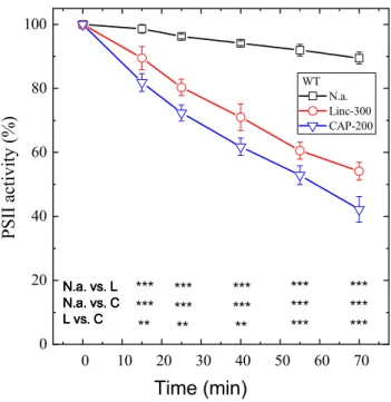
In order to quantify the effect of 200 μg mL−1 chlorampheni- col on the rate of PSII photodamage, WT Synechocystis cells were exposed to 500 μmol photons m−2 s−1 illumination without addition, and in the presence of either lincomycin or chloramphenicol as protein synthesis inhibitor. High light alone induced only a small extent of PSII activity loss, as quantified by the rate of oxygen evolution in the presence of 0.5 mM DMBQ as an artificial acceptor. After 70 min illumination, PSII activity declined to 90% of its initial value (Fig. 1, squares). In the presence of lincomycin, PSII inac- tivation was enhanced and the residual activity declined to 55% of its initial value after 70 min (Fig. 1, circles). When 200 μg mL−1 chloramphenicol was used as a protein syn- thesis inhibitor instead of lincomycin, the extent of PSII deactivation was further enhanced, and the residual activity after 70 min illumination was only ca. 40% of its initial value (Fig. 1, down triangles). The lower part of Fig. 1 shows that the differences in PSII activity values were statistically sig- nificant at each time point for the no addition vs. lincomycin, no addition vs. chloramphenicol, and lincomycin vs. chlo- ramphenicol treatments at p < 0.01 (or 0.001) significance level (see also Supplementary Table 1).
In order to make sure that the enhanced loss of PSII activity in the presence of chloramphenicol was a real effect and not caused by partial inhibition of protein synthesis in the presence of the applied 300 μg mL−1 concentration of lincomycin, the photoinhibitory experiments were also per- formed in the presence of 400 μg mL−1 lincomycin. These data (Supplementary Fig. 1) confirmed that lincomycin at 300 μg mL−1 concentration was sufficient for full inhibi- tion of protein synthesis. Therefore, the enhancement of
photodamage in the presence of 200 μg mL−1 chlorampheni- col is caused by an effect, which is unrelated to protein syn- thesis inhibition.
In the majority of photosynthetic applications chloram- phenicol is applied in 200 μg mL−1 or higher concentra- tions. However, in some studies 100 μg mL−1 or even as low as 30–50 μg mL−1 was also used (Supplementary Table 1).
Therefore, we aimed to check if these lower concentrations also enhance PSII photodamage. Our data show that PSII activity loss in the presence of 100 μg mL−1 chlorampheni- col was practically identical with that observed in the pres- ence of 300 μg mL−1 lincomycin (Supplementary Fig. 1). In contrast, 50 μg mL−1 chloramphenicol resulted in a slower loss of PSII activity than lincomycin. In addition, some recovery was also observed under low light conditions fol- lowing the photoinhibitory treatment, which indicates that 50 μg mL−1 chloramphenicol is not sufficient for complete blocking of PSII repair under our experimental conditions.
The rates of PSII activity loss were calculated by fitting the activity curves with a single exponential decay func- tion (Aie−kt, where Ai is the initial activity, k is the rate of
Fig. 1 Differential effects of lincomycin and chloramphenicol on photoinhibitory activity loss in WT Synechocystis cells. WT Synecho- cystis cultures were exposed to 500 μmole photons m−2 s−1 intensity illumination. The cultures were left untreated (squares), treated with 300 μg mL−1 lincomycin (circles), or 200 μg mL−1 chloramphenicol (down triangles). PSII activity was assessed by measuring the rate of oxygen evolution in the presence of 0.5 mM DMBQ as an artificial acceptor. Data are shown as a percentage of the initial PSII activity, which was obtained from the growth-light-adapted cultures before the onset of high light illumination. The asterisk signs indicate the time points where the PSII activity values are significantly (*p < 0.05,
**p < 0.01, ***p < 0.001) different between the untreated (n.a.), lin- comycin (L) or chloramphenicol (C)-treated samples
activity loss, and t is time). The calculated rates are sum- marized in Table 1, which show that the k = 0.0016 min−1 rate of photodamage, which was observed in the absence protein synthesis inhibitors was increased to 0.0090 min−1 in the presence of 300 μg mL−1 lincomycin. Practically the same photodamage rate (0.0083 min−1) was obtained in the presence of 100 μg mL−1 chloramphenicol. However, the damage rate was increased by ca. 50% (to 0.0120 min−1) when 200 μg mL−1 chloramphenicol was applied showing a concentration dependence of chloramphenicol-induced photodamage.
Chloramphenicol enhances PSII photodamage in comparison to lincomycin in PSI‑less
Synechocystis
In order to clarify if the photodamage-enhancing effect of chloramphenicol is related to its interaction with PSI or PSII, the light treatment experiments were also performed in a Synechocystis mutant, which lacks PSI. In contrast to
the WT strain, high light exposure alone induced a sub- stantial loss of PSII activity in the PSI-less mutant, which resulted in a decline to ca. 77% of the initial activity after a 70-min illumination (Fig. 2, squares). The addition of lincomycin enhanced the activity loss to ca. 35% of the initial activity after a 70-min illumination (Fig. 2, cir- cles). In the presence of 200 μg mL−1 chloramphenicol the extent of PSII deactivation was further enhanced, lead- ing to a residual activity of ca. 20% of the initial activity after 70 min (Fig. 2, down triangles). The lower part in Fig. 2 shows that the differences in PSII activity values were statistically significant in each time point for the no addition vs. lincomycin, no addition vs. chloramphenicol, and lincomycin vs. chloramphenicol treatments at p < 0.05 (0.01, or 0.001) significance level (see also Supplementary Table 2).
The calculated rates of PSII photodamage confirm that 200 μg mL−1 chloramphenicol enhances photodamage also in the PSI-less mutant (k = 0.0217 min−1) relative to that
Table 1 Rates of PSII photodamage
Synechocystis cultures were exposed to 500 μmole m−2 s−1 light and their PSII activity was followed as a function of illumination time by measuring the rate of oxygen evolution in the presence of DMBQ as artificial acceptor as shown in Fig. 1. PSII photodamage rates were calculated by fitting the PSII activity curves with a single exponential decay function (Aie−kt, where Ai is the PSII initial activity, k is the photodamage rate, and t is time) using the built in option of the Ori- gin 2018 Pro software. The Stdev column shows the standard error of rate constants as calculated by the fitting software. The experiments were performed in photoautotrophically grown WT cells, without addition, and in the presence of protein synthesis inhibitors (300 and 400 μg mL−1 lincomycin, 200 μg mL−1 chloramphenicol, abbrevi- ated as CAP). The photoinhibitory treatments were also performed in a PSI-less Synechocystis mutant line, which cannot grow photoau- totrophically, and therefore was cultured photomixotrophically in the presence of 5 mM glucose (PSI-less-gl). As a control for the PSI-less mutant, the WT cells were also grown photomixotrophically (WT-gl).
The PSI-less-gl and WT-gl cultures were exposed to the same high light and inhibitor treatments as the photoautotrophically grown WT
Sample Rate of photodamage (min−1)
Mean Stdev
WT 0.0016 1.42E−04
WT+300 linc 0.0088 3.13E−04
WT+400 linc 0.0090 9.77E−04
WT+100 CAP 0.0083 1.24E−03
WT+200 CAP 0.0120 6.59E−04
WT-gluc 0.0015 1.36E−04
WT-gluc+300 linc 0.0085 3.65E−04
WT-gluc+200 CAP 0.0119 8.23E−04
PSI-less 0.0035 5.41E−04
PSI-less+300 linc 0.0152 5.51E−04
PSI-less+200 CAP 0.0217 1.23E−03
Fig. 2 Differential effects of lincomycin and chloramphenicol on photoinhibitory activity loss in PSI-less Synechocystis cells. PSI-less Synechocystis cultures were exposed to 500 μmole photons m−2 s−1 intensity illumination. The cultures were left untreated (squares), treated with 300 μg mL−1 lincomycin (circles), or 200 μg mL−1 chlo- ramphenicol (down triangles). PSII activity was assessed by measur- ing the rate of oxygen evolution in the presence of 0.5 mM DMBQ as an artificial acceptor. Data are shown as a percentage of the ini- tial PSII activity, which was obtained from the growth-light-adapted cultures before the onset of high light illumination. The asterisk signs indicate the time points where the PSII activity values are sig- nificantly (*p < 0.05, **p < 0.01, ***p < 0.001) different between the untreated (n.a.), lincomycin (L) or chloramphenicol (C)-treated sam- ples
observed in the presence of lincomycin (k = 0.0152 min−1) (Table 1).
The lack of PSI enhances PSII photodamage
Data in Figs. 1, 2 and Table 1 show that the light-induced loss of PSII activity is apparently enhanced in the PSI-less mutant when compared to the WT. However, we have to note that the growth conditions for the WT and the PSI-less strains were different (photoautotrophic growth at 40 μmol photons m−2 s−1 for the WT, and photomixotrophic growth in the presence of 5 mM glucose at 4 μmol photons m−2 s−1 for the PSI-less mutant). In order to clarify if the enhanced photodamage of PSII activity is due to the lack of PSI, or occurs as a consequence of different growth conditions, the light treatments were also performed in WT cells that were grown under the same conditions as the PSI-less mutant.
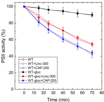
Growing the WT cells in the presence of glucose and low light did not affect their light sensitivity when compared to those grown photoautotrophically at standard light intensity (Fig. 3, see also Table 1). However, pairwise comparison of
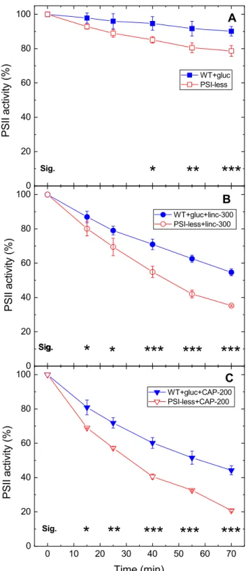
the activity loss curves in cells which were cultured under the same conditions (photomixotrophy and low light) reveal that PSII photodamage is significantly larger in the PSI- less strain than in the WT. This effect was observed in all three treatment conditions, i.e., without addition (Fig. 4a), in the presence of lincomycin (Fig. 4b) or chloramphenicol (Fig. 4c). The calculated rates of PSII photodamage are also ca. twofold higher in the PSI-less mutant than in the WT grown under photomixotrophic (or photoautotrophic) condi- tions (Table 1).
Discussion
Chloramphenicol enhances PSII photodamage Our present data confirm that the rate of photodamage of PSII in intact Synechocystis cells is higher in the presence of the usually applied 200 μg mL−1 chloramphenicol than in the presence of lincomycin (Figs. 1, 2, 3). The data presented in Supplementary Fig. 1 exclude the possibility that this phe- nomenon would be caused by a sub-saturating concentration of lincomycin. However, the different action mechanisms of protein synthesis inhibition by lincomycin and chloram- phenicol should also be considered for the explanation of the results. It is known that high light exposure results in elevated association of psbA mRNA with polysomes (Tyyst- jarvi et al. 2001) and also that lincomycin does not interact with ribosomes with bound nascent peptides (Contreras and Vazquez 1977) and therefore will not act on pre-formed polysomes. However, chloramphenicol is effective against peptide bond formation in ribosomes and polysomes (Con- treras and Vazquez 1977). As a consequence, D1 copies which are pre-formed before the addition of lincomycin can be incorporated into the thylakoid membrane and can sup- port PSII repair to some extent in spite of the presence of the protein synthesis inhibitor, whereas in the presence of chloramphenicol such an effect cannot occur. An interest- ing example of this phenomenon was the observation that after transferring high-light-exposed Synechocystis cells to low light conditions, the oxygen-evolving activity increased above the control level which persisted before the high light treatment, and this effect was not inhibited by lincomycin, only by chloramphenicol (Bentley et al. 2008). Based on the above-described difference in the action mechanisms of the two protein synthesis inhibitors, the smaller extent of PSII activity loss in the presence of lincomycin could in principle arise from a partial blocking of PSII repair due to the incorporation of pre-formed D1 copies, which were produced before the start of high light illumination, into the thylakoid membranes. However, under our experimental protocol cells were incubated in the presence of lincomycin (and chloramphenicol) for 20 min under standard growth
Fig. 3 Effect of photoheterotrophic growth on photoinhibitory activ- ity loss in WT Synechocystis cells. WT Synechocystis cultures were grown either in glucose-free BG-11 medium at 40 μmole photons m−2 s−1 light intensity (open symbols), or in the presence of 5 mM glucose at 5 µmole photons m−2 s−1 light intensity (closed symbols).
The cultures were left untreated (squares), treated with 300 μg mL−1 lincomycin (circles) or 200 μg mL−1 chloramphenicol (down trian- gles) and were exposed to 500 μmole photons m−2 s−1 intensity illu- mination. PSII activity was assessed by measuring the rate of oxygen evolution in the presence of 0.5 mM DMBQ as artificial acceptor.
Data are shown as percentage of the initial PSII activity, which was obtained from the growth-light-adapted cultures before the onset of high light illumination
light intensity (40 µmol photons m−2 s−1) before the onset of the high light treatment, which should be sufficient to empty the pre-D1 pool by the time strong illumination started. In addition, the effect of pre-D1 accumulation at growth light (40 µmol photons m−2 s−1) before lincomycin additions should be negligible for the repair of PSII under high light (500 µmol photons m−2 s−1) exposure. Therefore, we can safely conclude that lincomycin fully blocked PSII repair and the greater loss of PSII activity in the presence of 200 μg mL−1 chloramphenicol is related to a side effect of this protein synthesis inhibitor.
The side effect of chloramphenicol, which is concentra- tion dependent, has been previously demonstrated in isolated PSII membranes (Rehman et al. 2016). Since this effect was reversible by SOD, which eliminated O2·−, it was concluded that chloramphenicol-mediated O2·− production is the cause of chloramphenicol-enhanced photodamage in isolated sys- tems (Rehman et al. 2016). SOD is a large enzyme (32.5 kDa molecular mass), which cannot penetrate through the cell wall of Synechocystis; therefore, SOD addition cannot be used to verify the involvement of O2·− in chloramphenicol- induced damage in intact cells. On the other hand, based on the analogy with the results obtained in isolated PSII mem- brane particles, it is highly likely that O2·− production in the presence of chloramphenicol is the main cause of enhanced photodamage also in the intact cells.
O2·− has lower reactivity than other ROS (Pospisil et al.
2019); however, it can induce damage to proteins and mem- brane components due to its ability to produce highly oxi- dizing radicals. During spontaneous dismutation of O2·− it forms H2O2, which in turn can produce the highly oxidizing hydroxyl radical (HO·). In addition, protonation of the ani- onic form of O2·− leads to the formation of the perhydroxyl radical (HO2·), which is considered to be more reactive than O2·− and damages polyunsaturated fatty acids, amino acids and nucleic acids by H2 abstraction (Aikens and Dix 1991).
O2·− formation has been suggested to occur in PSII during high light exposure in the absence of added electron trans- port mediators (Pospísil 2012; Kale et al. 2017). In addition, O2·−-induced or O2·−-mediated damage of PSII core proteins (D1 and D2) has been observed in isolated PSII preparations by using tandem mass spectrometry, which detects specific oxidative modifications of amino acid residues (Kale et al.
2017). In this case, the O2·− that damages PSII is produced in PSII itself, most likely by reducing oxygen from Phe− or QA− (Pospísil 2012; Kale et al. 2017). However, in the lit- erature there is also a precedent for O2·−-mediated PSII pho- todamage by O2·− which is produced at the acceptor side of PSI in the presence of methyl viologen in isolated thylakoids and intact leaves (Krieger-Liszkay et al. 2011).
Chloramphenicol is known to interact not only with PSI (Okada et al. 1991) but also with PSII (Rehman et al.
2016) by accepting electrons at the acceptor sides of both
Fig. 4 Differential light sensitivity of WT and PSI-less Synechocystis cells.
WT (closed symbols) and PSI-less Synechocystis cultures (open symbols) were grown in the presence of 5 mM glucose at 5 µmole photons m−2 s−1 light intensity. The cultures were left untreated (squares), treated with 300 μg mL−1 lincomycin (circles), or 200 μg mL−1 chloramphenicol (down triangles) and were exposed to 500 μmole photons m−2 s−1 intensity illumination. PSII activ- ity was assessed by measuring the rate of oxygen evolution in the presence of 0.5 mM DMBQ as artificial acceptor. Data are shown as percentage of the ini- tial PSII activity, which was obtained from the growth-light-adapted cultures before the onset of high light illumination. The asterisk signs indicate the time points where the PSII activity values are significantly (*p < 0.05, **p < 0.01,
***p < 0.001) different between the WT and PSI-less samples
photosystems and then reducing oxygen and producing O2·−. Therefore, in principle either PSI, PSII, or both photosys- tems could be the site of superoxide production that damages PSII. Interestingly, the data presented in Figs. 1 and 2, as well as in Table 1 clearly show that the rate of PSII photo- damage is increased practically to the same extent (ca. 37%) both in the WT and the PSI-less strain when chlorampheni- col is used instead of lincomycin as protein synthesis inhibi- tor. This finding indicates that the O2·− produced at PSI has a negligible contribution to the damage of PSII activity under our conditions, therefore the main source of O2·− that dam- ages PSII in the presence of chloramphenicol is PSII itself.
PSII photodamage is enhanced in the absence of PSI An important observation in our work is that light-induced loss of PSII activity in the presence of protein synthesis inhibitors proceeds faster in a PSI-less mutant than in the WT strain containing both PSI and PSII (Fig. 1 vs. Fig. 2, and Fig. 4). The net loss of PSII activity is determined by the balance of the competing photodamage and repair pro- cesses (Aro et al. 1993; Komenda et al. 2012; Järvi et al.
2015). Both sides of the balance can be affected by vari- ous factors. The rate of photodamage is linearly dependent on light intensity (Tyystjärvi and Aro 1996). However, it is also affected by the energetics of electron transfer in PSII.
Namely, the increase of the free energy gap between Phe and QA provides protection against photodamage, whereas the decrease of the Phe and QA free energy gap enhances photodamage (Vass and Cser 2009; Rehman et al. 2013).
This effect is related to the balance of radiative and non- radiative charge recombination pathways modulating the efficiency of singlet oxygen production, which is an impor- tant mediator of PSII photodamage (Cser and Vass 2007;
Rehman et al. 2013). Modification of the environment of QA either by binding of herbicide molecules to the QB site or by site-directed mutagenesis also affects the rate of PSII photo- damage. In both cases, the increase of Em(QA/QA−) provides protection against photodamage, whereas the decrease of Em(QA/QA−) enhances it (Krieger-Liszkay and Rutherford 1998; Fufezan et al. 2007).
The rate of PSII repair can also be influenced by sev- eral environmental factors. Salt, cold, moderate heat, oxi- dative stress and CO2 limitation can all inhibit PSII repair and therefore enhance net photodamage (Murata et al. 2007;
Takahashi and Murata 2008). Production of reactive oxygen species, including singlet oxygen can also specifically inhibit the repair process by suppressing de novo synthesis of the D1 protein (Nishiyama et al. 2006). A large amount of ATP is also required for D1 protein synthesis, which is the key step of PSII repair, therefore the efficiency of cyclic elec- tron flow, which contributes significantly to the pH gradient
that drives ATP synthesis, can also influence the repair rate (Murata and Nishiyama 2018).
In the PSI-less Synechocystis mutant, the PSI electron transport pathways, which can oxidize the PQ pool are almost completely blocked. Therefore, the sustained reduc- tion of the secondary quinone electron acceptors (PQ pool and QB) maintains a highly reduced state of QA, a condi- tion also referred to as high excitation pressure (Maxwell et al. 1995). This condition enhances the production rate of singlet oxygen, which is one of the main mediators of PSII photodamage (Krieger-Liszkay et al. 2008; Vass and Cser 2009; Vass 2011; Fischer et al. 2013). Therefore, our data support the idea that enhancement of PSII photodamage in the absence of PSI is related to the lack of efficient electron transport beyond the PQ pool.
Conclusions
Our data show that chloramphenicol at 200 μg mL−1, which is a frequently applied concentration in most photosynthesis related studies, enhances photodamage of PSII. Therefore, high chloramphenicol amounts should be avoided in studies, which aim at the determination of true rate of PSII photo- damage. 100 μg mL−1 chloramphenicol appears to be a safe choice since it fully inhibits PSII repair without the side effect of enhanced photodamage rate. It is also of note that the side effect of chloramphenicol (and its recommended safe value) could be influenced by cell density of the culture and the temperature of the photoinhibitory treatment, which are expected to decrease and increase the enhancement of photodamage, respectively. Therefore, it is recommended to check the effect of the applied chloramphenicol concentra- tion in comparison with lincomycin when new experimen- tal conditions are designed. The chloramphenicol-induced enhancement of PSII photodamage is probably related to superoxide production. Therefore, superoxide should be considered as an important reactive oxygen species, besides singlet oxygen and hydroxyl radicals, that can damage PSII.
This idea is in agreement with previous results showing superoxide-induced modification of the D1 reaction center protein (Kale et al. 2017) as well as superoxide production in PSII (Pospisil et al. 2019). The observed enhancement of PSII photodamage in the absence of PSI supports the important role of excitation pressure in influencing the rate of photodamage (Maxwell et al. 1995; Kornyeyev et al.
2010; Bersanini et al. 2014).
Acknowledgements We thank Wim Vermaas for providing us the PSI- less Synechocystis 6803 strain. This work was supported by the Hun- garian Ministry for National Economy GINOP-2.3.2-15-2016-00001, OTKA K-116016.
Author contributions SK—Experimental work, correction of the man- uscript. AR—Contribution to supervision of the work, critical reading of the manuscript. IV—Fund generation for the work, manuscript writ- ing, supervision of the work.
Funding Open access funding provided by ELKH Biological Research Center. This work (S.K:, A.R., I.V.) was supported by the Hungarian Ministry for National Economy GINOP-2.3.2-15-2016-00001, OTKA K-116016.
Data availability All data presented are available in the form of figures, and tables in the main text and in the supplementary material.
Compliance with ethical standards
Conflict of interest On behalf of all authors, the corresponding author states that there is no conflict of interest.
Open Access This article is licensed under a Creative Commons Attri- bution 4.0 International License, which permits use, sharing, adapta- tion, distribution and reproduction in any medium or format, as long as you give appropriate credit to the original author(s) and the source, provide a link to the Creative Commons licence, and indicate if changes were made. The images or other third party material in this article are included in the article’s Creative Commons licence, unless indicated otherwise in a credit line to the material. If material is not included in the article’s Creative Commons licence and your intended use is not permitted by statutory regulation or exceeds the permitted use, you will need to obtain permission directly from the copyright holder. To view a copy of this licence, visit http://creat iveco mmons .org/licen ses/by/4.0/.
References
Aikens J, Dix TA (1991) Perhydroxyl radical (HOO.) initiated lipid- peroxidation—the role of fatty-acid hydroperoxides. J Biol Chem 266(23):15091–15098
Apel K, Hirt H (2004) Reactive oxygen species: metabolism, oxidative stress, and signal transduction. Annu Rev Plant Biol 55:373–399 Arntzen CJ, Kyle DJ, Wettern M, Ohad I (1984) Photoinhibition: a
consequence of the accelerated breakdown of the apoprotein of the secondary electron acceptor of Photosystem II. In: Hallick R, Staehelin LA, Thornber JP (eds) Biosynthesis of the photosyn- thetic apparatus: molecular biology development and regulation.
UCLA Symposium Series, New York, pp 313–324
Aro E-M, Virgin I, Andersson B (1993) Photoinhibition of Photosys- tem II. Inactivation, protein damage and turnover. Biochim Bio- phys Acta 1143:113–134
Baena-Gonzalez E, Aro E-M (2002) Biogenesis, assembly and turnover of Photosystem II units. Phyl Trans R Soc Lond B 357:1451–1460 Bentley FK, Luo H, Dilbeck P, Burnap RL, Eaton-Rye J (2008) Effects
of inactivating psbM and psbT on photodamage and assembly of Photosystem II in Synechocystis sp. PCC 6803. Biochemistry 47:11637–11646
Bersanini L, Battchikova N, Jokel M, Rehman AU, Vass I, Allahver- diyeva Y, Aro E-M (2014) Flavodiiron protein Flv2/Flv4-related photoprotective mechanism dissipates excitation pressure of PSII in cooperation with phycobilisomes in cyanobacteria. Plant Phys- iol 164(2):805–818
Campbell DA, Tyystjärvi E (2012) Parameterization of photosys- tem II photoinactivation and repair. Biochim Biophys Acta 4660:258–265
Chow WS, Lee H-Y, He J, Hendrickson L, Hong Y-N, Matsubara S (2005) Photoinactivation of Photosystem II in leaves. Photosynth Res 84:35–41
Constant S, Perewoska I, Alfonso M, Kirilovsky D (1997) Expression of the psbA gene during photoinhibition and recovery in synecho- cystis PCC 6714: inhibition and adamage of transcritptional and translational machinery prevent the restoration of Photosystem II activity. Plant Mol Biol 34:1–13
Contreras A, Vazquez D (1977) Cooperative and antagonistic inter- actions of peptidyl-transfer-RNA and antibiotics with bac- terial-ribosomes. Eur J Biochem 74(3):539–547. https ://doi.
org/10.1111/j.1432-1033.1977.tb114 22.x
Cser K, Vass I (2007) Radiative and non-radiative charge recombina- tion pathways in Photosystem II studied by thermoluminescence and chlorophyll fluorescence in the cyanobacterium Synechocystis 6803. Biochim Biophys Acta 1767:233–243
Fischer BB, Hideg É, Krieger-Liszkay A (2013) Production, detec- tion, and signaling of singlet oxygen in photosynthetic organisms.
Antioxid Redox Signal 18:2145–2162
Fufezan C, Gross CM, Sjödin M, Rutherford AW, Krieger-Liszkay A (2007) Influence of the redox potential of the primary quinone electron acceptor on photoinhibition in Photosystem II. J Biol Chem 282:12492–12502
Järvi S, Suorsa M, Aro E-M (2015) Photosystem II repair in plant chloroplasts—regulation, assisting proteins and shared compo- nents with photosystem II biogenesis. Biochim Biophys Acta 1847(9):900–909
Kale R, Hebert AE, Frankel LK, Sallans L, Bricker TM, Pospísil P (2017) Amino acid oxidation of the D1 and D2 proteins by oxygen radicals during photoinhibition of Photosystem II. Proc Natl Acad Sci USA 114(11):2988–2993
Komenda J, Tichy M, Prasil O, Knoppová J, Kuviková S, de Vries R, Nixon PJ (2007) The exposed N-terminal tail of the D1 subu- nit is required for rapid D1 degradation during Photosystem II repair in Synechocystis sp. PCC 6803. Plant Cell 19:2839–2854 Komenda J, Sobotka R, Nixon PJ (2012) Assembling and maintain- ing the Photosystem II complex in chloroplasts and cyanobac- teria. Curr Opin Plant Biol 15:245–251
Kornyeyev D, Logan BA, Holaday AS (2010) Excitation pressure as a measure of the sensitivity of photosystem II to photoinactiva- tion. Funct Plant Biol 37:943–951
Krieger-Liszkay A (2005) Singlet oxygen production in photosyn- thesis. J Exp Bot 56:337–346
Krieger-Liszkay A, Rutherford AW (1998) Influence of herbicide binding on the redox potential of the quinone acceptor in Pho- tosystem II: relevance to photodamage and phytotoxicity. Bio- chemistry 37(50):17339–17344
Krieger-Liszkay A, Fufezan C, Trebst A (2008) Singlet oxygen pro- duction in photosystem II and related protection mechanism.
Photosynth Res 98:551–564
Krieger-Liszkay A, Kós PB, Hideg É (2011) Superoxide anion radi- cals generated by methylviologen in photosystem I damage pho- tosystem II. Physiol Plant 142:17–25
Li L, Aro EM, Millar AH (2018) Mechanisms of photodamage and protein turnover in photoinhibition. Trends Plant Sci 23(8):667–
676. https ://doi.org/10.1016/j.tplan ts.2018.05.004
Maxwell DP, Falk S, Huner NPA (1995) Photosystem-II excitation pressure and development of resistance to photoinhibtion. 1.
Light-harvesting Complex-II abundance and zeaxanthin content in Chlorella-vulgaris. Plant Physiol 107(3):687–694. https ://doi.
org/10.1104/pp.107.3.687
Miyata K, Noguchi K, Terashima I (2012) Cost and benefit of the repair of photodamaged photosystem II in spinach leaves: roles of acclimation to growth light. Photosynth Res 113:165–180
Mulo P, Pursiheimo S, Hou CX, Tyystjarvi T, Aro E-M (2003) Multi- ple effects of antibiotics on chloroplast and nuclear gene expres- sion. Funct Plant Biol 30:1097–1103
Murata N, Nishiyama Y (2018) ATP is a driving force in the repair of photosystem II during photoinhibition. Plant, Cell Environ 41(2):285–299. https ://doi.org/10.1111/pce.13108
Murata N, Takahashi S, Nishiyama Y, Allakhverdiev SI (2007) Pho- toinhibition of photosystem II under environmental stress. Bio- chim Biophys Acta 1767:414–421
Nishiyama Y, Allakhverdiev SI, Murata N (2005) Inhibition of the repair of Photosystem II by oxidative stress in cyanobacteria.
Photosynth Res 84(1–3):1–7
Nishiyama Y, Allakhverdiev SI, Murata N (2006) A new paradigm for the action of reactive oxygen species in the photoinhibition of Photosystem II. Biochim Biophys Acta 1757:742–749 Nishiyama Y, Yamamoto H, Allakhverdiev SI, Inaba H, Yokota A,
Murata N (2001) Oxidative stress inhibits the repair of photoda- mage to the photosynthetic machinery. EMBO J 20:5587–5594 Nixon PJ, Michoux F, Yu J, Boehm M, Komenda J (2010) Recent
advances in understanding the assembly and repair of photosys- tem II. Ann Bot 106:1–16
Ohad I, Kyle DJ, Arntzen CJ (1984) Membrane protein damage and repair: removal and replacement of inactivated 32-kilodalton poly- peptides in chloroplast membranes. J Cell Biol 99:481–485 Okada K, Satoh K, Katoh S (1991) Chloramphenicol is an inhibitor of
photosynthesis. FEBS Lett 295:155–158
Pospísil P (2012) Molecular mechanisms of production and scavenging of reactive oxygen species by photosystem II. Biochim Biophys Acta 1817:218–231
Pospisil P, Prasad A, Rac M (2019) Mechanism of the formation of electronically excited species by oxidative metabolic processes:
role of reactive oxygen species. Biomolecules. https ://doi.
org/10.3390/biom9 07025 8
Prasil O, Zer H, Godde D, Ohad I (1992) Role of the PSII acceptor site in the lihgt induced degradation of D1. In: Murata N (ed) Research in photosynthesis, vol 4. Kluwer Academic Publishers, Dordrecht, pp 501–504
Rehman AU, Cser K, Sass L, Vass I (2013) Characterization of sin- glet oxygen production and its involvement in photodamage of Photosystem II in the cyanobacterium Synechocystis PCC 6803 by histidine-mediated chemical trapping. Biochim Biophys Acta 1827:689–698
Rehman AU, Kodru S, Vass I (2016) Chloramphenicol mediates super- oxide production in Photosystem II and enhances its photodamage in isolated membrane particles. Front Plant Sci 7:479. https ://doi.
org/10.3389/fpls.2016.00479
Shen G, Boussiba S, Vermaas WFJ (1993) Synechocystis sp. PCC 6803 and phycobilisome function strains lacking photosystem I. Plant Cell 5:1853–1863
Sicora C, Máté Z, Vass I (2003) The interaction of visible and UV-B light during photodamage and repair of Photosystem II. Photo- synth Res 75:127–137
Takahashi S, Murata N (2005) Interruption of the Calvin cycle inhibits the repair of Photosystem II from photodamage. Biochim Biophys Acta 1708:352–361
Takahashi S, Murata N (2008) How do environmental stresses acceler- ate photoinhibition? Trends Plant Sci 13:178–182
Takahashi S, Whitney SM, Badger MR (2009) Different thermal sen- sitivity of the repair of photodamaged photosynthetic machin- ery in cultured Symbiodinium species. Proc Natl Acad Sci USA 106:3237–3242
Tikkanen M, Mekala NR, Aro E-M (2014) Photosystem II photoinhibi- tion-repair cycle protects Photosystem I from irreversible damage.
Biochim Biophys Acta 1837:210–215
Tyystjarvi T, Herranen M, Aro EM (2001) Regulation of translation elongation in cyanobacteria: membrane targeting of the ribo- some nascent-chain complexes controls the synthesis of D1 protein. Mol Microbiol 40(2):476–484. https ://doi.org/10.104 6/j.1365-2958.2001.02402 .x
Tyystjarvi T, Tuominen I, Herranen M, Aro E-M, Tyystjarvi E (2002) Action spectrum of psbA gene transcription is similar to that of photoinhibition in Synechocystis sp. PCC 6803. FEBS Lett 516:167–171
Tyystjärvi E, Aro EM (1996) The rate constant of photoinhibition, measured in lincomycin-treated leaves, is directly proportional to light intensity. Proc Natl Acad Sci USA 93:2213–2218
Vass I (2011) Role of charge recombination processes in photodam- age and photoprotection of the photosystem II complex. Physiol Plant 142:6–16
Vass I (2012) Molecular mechanisms of photodamage in the Photosys- tem II complex. Biochim Biophys Acta 1817:209–217
Vass I, Aro E-M (2008) Photoinhibition of Photosystem II electron transport. In: Renger G (ed) Primary processes of photosynthesis:
basic principles and apparatus. Comprehensice series in photo- chemical and photobiological sciences. Royal Society of Chem- istry Publishing, Cambridge, pp 393–411
Vass I, Cser K (2009) Janus-faced charge recombinations in photosys- tem II photoinhibition. Trends Plant Sci 14:200–205
Zavafer A, Koinuma W, Chow WS, Cheah MH, Mino H (2017) Mecha- nism of photodamage of the oxygen evolving Mn cluster of Pho- tosystem II by excessive light energy. Sci Rep 7:8. https ://doi.
org/10.1038/s4159 8-017-07671 -1
Publisher’s Note Springer Nature remains neutral with regard to jurisdictional claims in published maps and institutional affiliations.


