RESEARCH ARTICLE
Increased sensitivity of heavy metal bioreporters in transporter deficient Synechocystis PCC6803 mutants
Ga´bor Patyi1,2, Barbara Ho´ di1,2, Da´niel SolymosiID1¤, Imre Vass1, Pe´ter B. Ko´ sID1,3*
1 Institute of Plant Biology, Biological Research Centre of the Eo¨tvo¨s Lo´ra´nd Research Network, Szeged, Hungary, 2 Faculty of Science and Informatics, Doctoral School in Biology, University of Szeged, Szeged, Hungary, 3 Department of Biotechnology, Faculty of Science and Informatics, Szeged University, Szeged, Hungary
¤ Current address: Laboratory of Molecular Plant Biology, Department of Biochemistry, University of Turku, Turku, Finland
*kos.peter@brc.hu
Abstract
The detection and identification of heavy metal contaminants are becoming increasingly important as environmental pollution causes an ever-increasing health hazard in the last decades. Bacterial heavy metal reporters, which constitute an environmentally friendly and cheap approach, offer great help in this process. Although their application has great poten- tial in the detection of heavy metal contamination, their sensitivity still needs to be improved.
In this study, we describe a simple molecular biology approach to improve the sensitivity of bacterial heavy metal biosensors. The constructs are luxAB marker genes regulated by the promoters of heavy metal exporter genes. We constructed a mutant strain lacking the clus- ter of genes responsible for heavy metal transport and hence achieved increased intracellu- lar heavy metal content of the Synechocystis PCC6803 cyanobacterium. Taking advantage of this increased intracellular heavy metal concentration the Ni2+; Co2+and Zn2+detection limits of the constructs were three to tenfold decreased compared to the sensitivity of the same constructs in the wild-type cyanobacterium.
Introduction
The continuous accumulation of heavy metals (HMs) is a common environmental phenome- non in aquatic and terrestrial ecosystems through human activities, and this pollution is persis- tent because these compounds cannot be degraded hence they are accumulated by organisms [1,2]. The effluents produced by the processing industry contain a variety of heavy metals (HM), such as nickel, cobalt, zinc, copper, and cadmium. These compounds significantly con- tribute to the increase of toxic HM pollution in the environment. In addition to industrial pol- lution, traffic-related street dust has also become increasingly important in recent years, which can cause an extremely high health risk in big cities [3]. A connection was found between inhalable cobalt and respiratory symptoms and lung dysfunctions in the Swedish hard metal a1111111111
a1111111111 a1111111111 a1111111111 a1111111111
OPEN ACCESS
Citation: Patyi G, Ho´di B, Solymosi D, Vass I, Ko´s PB (2021) Increased sensitivity of heavy metal bioreporters in transporter deficient Synechocystis PCC6803 mutants. PLoS ONE 16(12): e0261135.
https://doi.org/10.1371/journal.pone.0261135 Editor: Md. Asraful Alam, Zhengzhou University, CHINA
Received: September 14, 2021 Accepted: November 28, 2021 Published: December 16, 2021
Copyright:©2021 Patyi et al. This is an open access article distributed under the terms of the Creative Commons Attribution License, which permits unrestricted use, distribution, and reproduction in any medium, provided the original author and source are credited.
Data Availability Statement: All relevant data are within the paper and itsSupporting Information files.
Funding: PBK was partly funded by the project entitled “Drinking water: multidisciplinary assessment of secure supply from the source to the consumers” (project nr. 2018-1.2.1-NKP-2018- 00011) supported by the National Research, Development and Innovation Office. This work was also supported by the Dr. Rollin D. Hotchkiss Foundation. The funders had no role in study
industry [4]. The average inhalable cobalt concentration was 0.0017 mg/m3. In roadside sam- ples, the Co2+level of the soil samples ranged from 1.9 to 3.5 mg/kg [5]. The amount of zinc and nickel contamination is more significant in rivers and lakes. In Europe, reported nickel concentrations in drinking water were generally below 10μg L-1(0.17μM). The levels of zinc in surface water and groundwater normally do not exceed 0.01 and 0.05 mg L-1(0.05–
0.25μM) [6]. According to a study from 2020 in India, the concentration of cobalt was about 0.003μM, nickel 0.22μM, and zinc up to 0.82μM, respectively, in the studied river area [7].
Due to its importance, the detection and removal of toxic heavy metals have increased attention, and various bioremediation materials, such as plants, fungi, and bacteria have been employed [8–10].
Cyanobacteria are present in both aquatic and terrestrial ecosystems. Particularly they can be major primary producers in oligotrophic and freshwater systems. Cyanobacteria are photo- synthetic gram-negative prokaryotes; they are the only known prokaryotes that perform oxy- gen-evolving photosynthesis. Furthermore, cyanobacteria require a variety of metal cations such as Cu2+, Ni2+, Fe2+, and Zn2+to maintain their cellular metabolism and growth [11–13].
Besides requiring high amounts of various metals for living, cyanobacteria are also frequently affected by drastic changes in metal concentrations and are often challenged by heavy metal toxicities. To survive in the polluted environment a sensitive regulation system in metal uptake and removal is required. Cyanobacteria have complex metal resistance system mechanisms including exclusion via active transport by ATPases or chemiosmotic efflux systems. These P- type ATPases transport metal ions from the cytosol into the periplasm [14]. Our knowledge of the cyanobacterial heavy metal regulation system is always increasing as new metabolic path- ways are discovered. However, the systematization and collection of this information is not easy, since the metal tolerance and requirement are different in almost every species. The cells have to acquire the correct element for each cellular function, but different metal ions can compete for the same binding site [15].
The cyanobacteriumSynechocystissp. PCC6803 (hereafter referred to asSynechocystis) has a small well-known genome, and it is easy to manipulate, hence it receives strong attention in basic and applied research. In theSynechocystisgenome, a metal regulated gene cluster was identified, which is involved in the heavy metal resistance via export of zinc, cobalt, and nickel [16]. This cluster is a 12 kb long region and includes 11 open reading frames (ORFs) consisting of six putative transcriptional units as follows: thecoaTandcoaRinvolved in the Co2+toler- ance thenrsBACDoperon involved in the Ni2+and Co2+homeostasis and regulated by the nrsRSoperon region, a hypothetical membrane protein-coding gene and theziaAandziaR involved in the Zn2+tolerance [17].
As we showed earlier [18] the transcriptional repressors are rather sensitive to the corre- sponding heavy metals, hence the above-mentioned transport proteins are upregulated at low heavy metal concentrations, where general stress responses are not triggered. The observation enabled us to construct whole-cell biosensors inSynechocystisby fusing thecoaTandnrsBACD promoters with the reporter genesluxABencoding easily detectable luminescent proteins [19].
These strains can sense the metal ions present in their environment, representing an alterna- tive to traditional analytical chemical methods, with the advantage to detect the bioavailable fraction (rather than total concentration) of an analyte, allowing for a more accurate assess- ment of the biological significance of the pollution. ThesenrsLuxandcoaLuxbiosensors responded to the respective heavy metal ions. After 3 hours of incubation in the case ofcoaLux reporter the detection range was 0,3–6μM Co2+and 1–3μM Zn2+and thenrsLuxstrain was specific in the range of 0,2–6μM Ni2+concentration [19].
In the current study, we aimed to increase the sensitivity of the biosensor strains. To this end, we constructed a knock-outΔnrsRSBACD:ΔcoaRT:ΔziaRAmutant strain, which then
design, data collection and analysis, decision to publish, or preparation of the manuscript.
Competing interests: The authors have declared that no competing interests exist.
lacks the above described heavy-metal responsive gene cluster coding for the export of the given heavy metal ions; hence the ratio of intracellular vs. extracellular heavy metal concentra- tion was expected to be higher than that in the wild type. For easy readability and pronuncia- tion, we designated the knockout mutant as NiCoZia strain, so this designation will be used hereafter. We created thenrsR,coaR, andziaRpromoter-driven heavy metal responsive lumi- nescent biosensors both in wild typeSynechocystisand in this mutant cell line. We found a sig- nificant increase of intracellular heavy metal concentration in the mutant that resulted in up to a tenfold increase in sensitivity of the reporters.
Materials and methods
Strains, growth conditions, and heavy metal salt treatment
Synechocystiscells were grown in photoautotrophic 3% CO2-rich atmospheric condition under 40μmol photon m−2s−1white light intensity and 30˚C in BG-11 [20] liquid medium.
For the preparation of the starter culture, 1.5 mL frozen stock culture was inoculated to 50 mL liquid BG-11 medium containing the appropriate antibiotic. From the fully-grown starter cul- ture, 1 mL was inoculated into 200 mL BG-11 without antibiotics and incubated until logarith- mic phase (OD720nm: 0.8). Heavy metal salt stress treatments were carried out in test tubes with the appropriate (ZnSO4, CoCl2, NiCl2) salt supplemented BG-11 liquid media without glucose. For the segregation ofΔnrsRSBACD:ΔcoaRT:ΔziaRA(NiCoZia) strain 50μg mL-1 spectinomycin (Spe), and for the biosensor constructs 50μg/ml kanamycin (Km) were used.
The BG-11 medium contains 0,137μM Co2+and 0.77μM Zn2+.
Escherichia colistrain DH5αwas used for routine DNA manipulations [21] and construc- tions in Luria broth (LB) medium at 37˚C [22]
Construction of NiCoZia strain
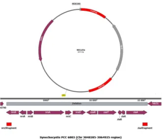
We constructed a mutant where the gene cluster was replaced by a spectinomycin cassette (Sp- R). An insert was built up into pUC19 vector from PCR amplified fragments with flanking restriction sites of the distal ends ofziaR(PstI, XbaI),nrsS(KpnI, EcoR) usingSynechocystis genome DNA and the spectinomycin cassette (XbaI, KpnI) using the vector pDF-trc [23] as the template (Fig 1). The plasmid was amplified inE.coliand transformed toSynechocystisvia natural transformation. The mutant formed by double crossover was grown on a selective BG- 11 plate containing 50μg mL-1spectinomycin (Spe).
Construction of bioreporter strains in WT and NiCoZia
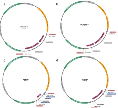
We used the pILA promoter probe vector [24] utilizing theLuxABluminescence reporter sys- tem. The insert of thecoaR; ziaRandnrsS + nrsR(in short:nrsRS) promoter region was ampli- fied by PCR using chromosomal DNA of WTSynechocystisPCC6803 as a template and the appropriate primer pair with a KpnI flanking at the 5’ end of both primers (Table 1) The pro- moter fragments were inserted into the unique KpnI site of pILA. Transformants ofE.coli with the right orientation were selected via colony PCR using test primer pairs. The newly cre- ated pILAcoaR, pILAziaR and pILAnrsRS-1 and pILAnrsRS-2 plasmids (Fig 2) were amplified and isolated fromE.coli. Wild type and NiCoZiaSynechocystisstrains were transformed and the clones were selected with Km selection as described above.
Bioluminescence assay
Heavy metal salt treatments were carried out in 96 well black (Opti-Plate) cell culture plates with low autofluorescence (Perkin-Elmer) in 25˚C and 40μE light intensity. Each well
contained 200μL of cell culture (OD720: 0,8–1) and 100μL of salt solution in different concen- trations. The plates were covered with punctured transparent foil [19]. After the 3-hour long treatment added 6μL of 50 mM decanal dissolved in 50% Methanol to 300μL of the samples placed per well. Luminescence of the luciferase reaction is induced by the addition of the decanal to the 300μL cultures in 1 mM final concentration [24]. After the addition of the sub- strate, the samples were preincubated in dark for 2 minutes before the light emission
Fig 1. Schematic representation of the cloning strategy used for construction of the NiCoZia strain. The upper part of the figure shows the plasmid map of the pUC19 with the nrs-SpeR-zia fragment. The lower part of the figure shows the construction of the heavy metal cluster and the homologous regions.
https://doi.org/10.1371/journal.pone.0261135.g001
Table 1. Oligonucleotides used in this work.
Gene symbol Oligonucleotide sequence
nrsS-Frg-Fw 5’TTGAATTCGTAACTGGATGGTGAATAAACTTCCCTT
nrsS-Frg-Rev 5’TTGGTACCTACGGATTTATTGCTACTAAGTCGCTTA
ziaA-Frg-Fw 5’TTTCTAGAGAGACCTAAAACGCATGGGAGTTGAAAA
ziaA-Frg-Rev 5’TTCTGCAGCTTAGCAATCCGAGTAGCATTCAAAATC
SpecR-XbaI 5’TTTCTAGACCGGAGACGGTCACAGCTTGTCTGTAAG
SpecR-KpnI 5’TTGGTACCATGTATGCTCTTCTGCTCCTGCCGGCCGA
CoaR-pILA-Fw 5’AAAAAGGTACCTTCACCATCCTTTCCCTATC
CoaR-pILA-Rev 5’AAAAAGGTACCACCTTCTCAGCCTAAACC
CoaR-Test-Fw 5’CAGGGCTTTCAGTTGTCT
CoaR-Test-Rev 5’GGTGATATGGGGAATGGG
NrsRS-pILA-Fw 5’AAAAAGGTACCGACGGCGTAAAGTTGATAAA
NrsRS-pILA-Rev 5’AAAAAGGTACCTCCCCCGCTAAGATCAGA
NrsRS -Test-Fw 5’TATTAGCAAGACTGCGGG
NrsRS-Test-Rev 5’TGTTGTTGTTGTTGGTAGG
ZiaR-pILA-Fw 5’AAAAAGGTACCCATCGTCCATCTCCTTAATC
ZiaA-pILA-Rev 5’AAAAAGGTACCCCGACTTGCATTTGCTGA
ZiaR-Test-Fw 5’TCCTAACGCCAACCTCTA
ZiaR-Test-Rev 5’CCCGATACAAATTCATCACA
pILA-Test-Fw 5’ACAACCAAATTTTCCCCAAG
pILA-Test-Rev 5’TCGATAGTGGCTCCAAGT
https://doi.org/10.1371/journal.pone.0261135.t001
monitoring. Assays were performed with four parallel samples at room temperature. Lumines- cence was determined by a Top Count NXT luminometer (Packard Instruments), with the expression of counts per second (CPS) and normalized to the OD720values of the cells.
Measurement of growth inhibition
The differences in growth intensity caused by excess metal ions in WT and NiCoZia cyanobac- terial cultures were quantified by measuring the optical density at 680 nm and 720 nm in 30μE light intensity and 30˚C temperature for a period of 2 to 4 days. We set the starterSynechocystis culture’s optical density to OD720= 0.1 and added heavy metal salt in 8 different concentra- tions (0μM; 0.05μM; 0.1μM; 0.25μM; 0.5μM; 1μM; 2.5μM; 5μM) in 50 mL final volume.
For the measurement, we used a Photo Multi Cultivator MC-1000 (Photon Systems Instru- ment) with automatic OD measurement every hour. Two inhibition parameters were deter- mined for each metal ion, minimal inhibitory concentration (ICmin) and maximal inhibitory concentration (ICmax). The ICminrefers to the lowest tested concentration leading to growth inhibition and ICmaxrefers to the highest tested concentration where no further growth was observed.
Determination of the intracellular heavy metal content
Synechocystiscultures were supplemented with 5μM of the respective salts. Cells were col- lected after 3-hour incubation by centrifugation (6000 rpm 10 min), washed once with BG-11 solution, and freeze-dried. From 50 mL cultures 40±5 mg wet weight cell pellet could be har- vested, from which 5±1 mg dry material could be obtained. The heavy metal content of the samples was determined as described earlier [18].
Fig 2. Schematic figure of the promoter probe vectors. Schematic figure of the promoter probe vector pILA with the inserts ofcoaR(d),ziaR(c), and with the two differently orientednrsRS(a; b). We used the appropriate primers (Table 1) for the fragments’ amplification from the WTSynechocystisgenome and applied KpnI digestion.LuxAand LuxBgenes code for the Luciferase reporter proteins, which are required for the detection. TheaphIIandblarefer to genes conferring resistance to ampicillin and kanamycin, respectively.
https://doi.org/10.1371/journal.pone.0261135.g002
Results
Growth inhibition in the NiCoZia (ΔnrsRSBACD :ΔcoaRT :ΔziaAR) and the wild type strain
First, we examined the effect of the removal of the heavy metal-specific cluster from the genome ofSynechocystison the growth of the cultures. To this end we treated the WT and NiCoZia cells for 98-hours with CoCl2; ZnSO4and NiCl2in 8 different concentrations (0μM;
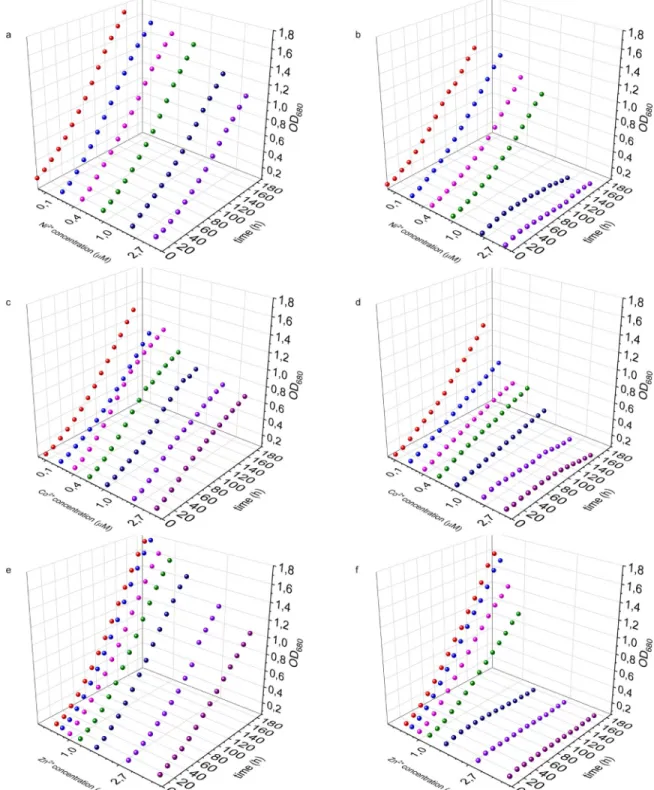
0.05μM; 0.1μM; 0.25μM; 0.5μM; 1μM; 2.5μM; 5μM) and simultaneously measured the cells optical density (S1 File). The WT shows a faster growth rate compared to the mutant, even under control conditions without heavy metal supplementation (Fig 3).
In the investigated concentration range, increasing cobalt concentrations resulted in increasingly pronounced growth inhibition in NiCoZia strain, reaching complete growth arrest at 2.5μM and 5μM. In contrast, the growth inhibition was also concentration-depen- dent, but much less pronounced in the WT strain (Fig 3C and 3D). Similarly, zinc concentra- tions of 0.24μM and up caused concentration-dependent severe growth inhibition in NiCoZia strain while this inhibition was mild and could be only observed from 1μM Zn2+in WT (Fig 3E and 3F). Lower Ni2+concentrations did not cause severe growth inhibition in NiCoZia strain but also complete arrest happened at 2.5μM and 5μM, at which concentrations only mild growth inhibition could be observed in the WT strain (Fig 3A and 3B).
Intracellular heavy metal content
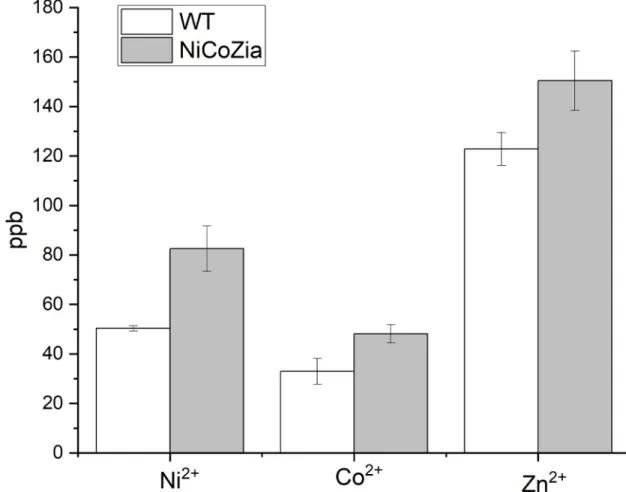
Inferring from the growth curves, we concluded that in the absence of cation efflux systems encoded by thecoa,zia, andnrsgenes is likely that the intracellular heavy metal concentrations are increased as compared to the wild type. To clarify this, the intracellular heavy metal con- tent was measured using ICP-MS in both the WT and the NiCoZia strains after 3 h long cobalt, nickel, and zinc treatments. BG-11 culture media were supplemented with 5μM heavy metals, and then their intracellular heavy metal concentrations were compared with the untreated samples. The data (S2 File) clearly showed that the NiCoZia strain accumulates more heavy metals within cells than its wild-type counterpart does. This phenomenon was obvious with all three studied heavy metals, the largest accumulation difference observed for nickel (Fig 4), where an approximately 40% increase in intracellular nickel concentration was observed in the nickel-treated NiCoZia strain. The mutation led to an increase in about 20% Zn2+and about 20% Co2+content compared to WT. It is in agreement with the expectations concerning that knocked out region coded for a heavy metal exporter system.
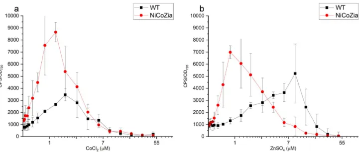
pILAcoaR bioreporter’s luminescent response to Co
2+and Zn
2+in WT and NiCoZia strains
We aimed to lower the detection limits of the bioreporters, in other words, to obtain a pheno- type that would be observed at higher extracellular concentrations in the wild type cyanobacte- rium. Hence, the luminescence of the bioluminescent pILAcoaRSynechocystisbioreporter strains driven by theCoaRpromoter generated from both WT and NiCoZia strains were assessed at different cobalt or zinc concentrations to determine and compare their sensitivity and specificity for the given heavy metal. As expected, during the heavy metal treatments, the bioreporter strains with NiCoZia genomic background showed greater sensitivity. The maxi- mum luminescence was found at 3μM cobalt concentrations in WT, in agreement with previ- ous studies, where the detection range was 1–3μM [19], while the maximum luminescence response was observed at 1μM cobalt concentration in NiCoZia. Moreover, while we found an about threefold increase in Co2+sensitivity (Fig 5A) the improvement in Zn2+sensitivity
was about tenfold. The maximal bioluminescence response was found around 7 and 10μM zinc concentration in WT (like earlier, Peca et al. 2008), and this value changed to 1μM in NiCoZia (Fig 5B).
Fig 3. Growth inhibition. The growth curves of the WT (a, c, e) and NiCoZia (b, d, f)Synechocystiscell lines in different Ni2+(a,b); Co2
+(c,d) and Zn2+(e,f) concentration.
https://doi.org/10.1371/journal.pone.0261135.g003
pILAnrsR bioreporter’s luminescent response to Ni
2+in WT and NiCoZia strains and the effect of the orientation of the bidirectional nrs promoter on Ni
2+sensitivity
The WT strain with pILAnrsRS promoter controlled construct showed luminescence from 0.2μM to 50μM nickel concentration. In the NiCoZia bioreporter strain with the same con- struct, this range shifted by about tenfold lower concentrations compared to WT, from 0.05μM NiCl2to approximately 5μM NiCl2(Fig 6). The maximum induction changed to 0.2μM NiCl2(in the mutant) from 4μM NiCl2(observed in the WT).
Considering that these operons have bidirectional promoters, we wanted to see which direction of the promoter is to be used for the lux genes, which orientation provides better util- ity to our purpose. For this study, we chose the nrs promoter. We cloned the insert containing the promoter and the nrsRS gene pair in both orientations: in the first orientation (nrsRS-1) the promoter is immediately in front of theluxAgene, similarly to as it is oriented in theSyne- chocystisgenome upstream to thenrsBgene. In the second, opposite orientation (nrsRS-2) LuxAgene is downstream ofnrsRandnrsSat the other end of the insert (Fig 2A and 2B).
Comparing the two constructs in WT and NiCoZia strains, it is clear that the original nrsRS-1 orientation (Fig 6A) exerts a higher luminescence induction during nickel treatment than the nrsRS-2 orientation (Fig 6B). However, the nrsRS-2 orientation is also capable of significant
Fig 4. Content of the intracellular Co2+; Ni2+and Zn2+in WT and NiCoZia mutantSynechocystiscells. After 3-hour treatment with 5μM heavy metal supplemented culture medium at 30μE light intensity at 25˚C temperature, the HM content was determined by ICP-MS measurement and presented in ppb (parts per billion).
https://doi.org/10.1371/journal.pone.0261135.g004
luminescence induction. The greater induction efficiency of nrsRS-1 orientation is beneficial in our case, nevertheless, in applications requiring lower induction levels, the opposite orienta- tion may be more advantageous.
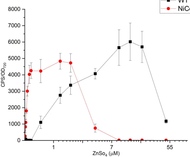
pILAziaR bioreporter’s luminescent response to Zn
2+in WT and NiCoZia strains
Bioreporter strains with theziaRpromoter were treated with 0.015–50μM ZnSO4supple- mented BG-11 for 3 hours, and the difference in luminescence induction (Fig 7) was similar to what we have obtained withcoaRandnrsRSpromoter-driven reporter genes. pILAziaR WT
Fig 5. The bioluminescent response for CoCl2(a) and ZnSO4(b) of pILACoaR_WT and pILACoaR_NiCoZia. Cells were incubated for three hours in BG-11 medium supplemented with different concentrations of heavy metal salts before the bioluminescence was measurement. Each point represents the mean of four parallels (S3 File).
https://doi.org/10.1371/journal.pone.0261135.g005
Fig 6. The bioluminescent responses of the pILAnrsRS constructs in the two nrsRS promoter orientations in WT and NiCoZia strains. Cells were incubated for three hours in BG-11 medium supplemented with different concentrations of (from 30 nM to 48μM) heavy metal salt. The bioluminescence was measured as described before. Each point represents the mean of three parallels (S4andS5Files).
https://doi.org/10.1371/journal.pone.0261135.g006
showed a luminescent signal in the concentration range of 0.5–50μM ZnSO4with a peak at 14μM ZnSO4treatment. This construct is more sensitive in the NiCoZia strain than in the WT background, namely, its induction can be observed from 20 nM to about 5μM ZnSO4
concentration range with a peak around 1.5μM ZnSO4treatment. Thus, the construct in NiCoZia strain proved to be about 10-fold more sensitive than in WT and can be used to detect up to 0.03μM ZnSO4concentration. This value is much lower than the previous mini- mum detection limit of 0.5μM ZnSO4measured in WT.
Discussion
Numerous whole-cell reporter organisms have been developed inSynechocystisdetermining a variety of metal cations in polluted soils or wastewater samples. However, in many cases, the lack of sufficient sensitivity [25] is an obstacle to their proper application (Table 2). The cellu- lar responses to individual metal ions are dependent on their intracellular concentration, which is determined by the combined function of efflux and influx systems and ion channels.
By eliminating the genes responsible for the efflux of three heavy metals we created a mutant
Fig 7. The bioluminescent response of the pILAziaR WT and pILAziaR NiCoZia for ZnSO4. Cells were incubated for three hours in BG-11 medium supplemented with different concentrations of (from 48μM to 30 nM) heavy metal salts. The bioluminescence was measured as described before. Each point represents the mean of three parallels (S6 File).
https://doi.org/10.1371/journal.pone.0261135.g007
with increased intracellular heavy metal concentration as compared to the wild type. It shows an inhibited growth compared to the wild-type strain even below the previously established [17] ICminconcentrations. The ICminvalue for cobalt is 2μM in the WT strain, whereas it is 0.1μM CoCl2in NiCoZia mutant, which means a 20-fold increased sensitivity in growth for cobalt (Fig 3D). The NiCoZia mutant was also more sensitive than the WT to zinc, showing an ICminof 0.5μM ZnSO4(Fig 3F) and 1μM NiCl2concentration (Fig 3B).
The increased sensitivity was the effect of the increased intracellular heavy metal concen- trations resulted by the lack of the corresponding transporters, as confirmed by ICP-MS determination of the intracellular content (Fig 4). This feature could be utilized for improv- ing the sensitivity of the specific bioreporter constructs that use heavy metal-sensitive pro- moter-driven expression of luminescent proteins. The luminescence measurements (Fig 5) showed that the pILAcoaR NiCoZia bioreporter strain exhibited a threefold higher sensitiv- ity to cobalt and a 10-fold higher sensitivity for zinc than the bioreporter in the WT back- ground. The pILAziaR NiCoZia bioreporter strain showed almost 10-fold higher sensitivity for Zn2+than the WT variant (Fig 7). The pILAnrsRS NiCoZia construct also showed increased sensitivity.
Considering that we used bidirectional promoter systems, we also investigated the effect of the orientation of the cloned DNA segment. Both nrsRS-1 and nrsRS-2 oriented constructs showed concentration-dependent fluorescence induction. The nrsRS-1 orientation where the original transporters inSynechocystiswere replaced by the reporter genes and are coded diver- gently from the regulator gene results in a higher level of induction and seems to be more effective than the opposite orientation (Fig 6).
The current investigation revealed that by eliminating efflux transporter genes from the cyanobacterial genome and choosing the right orientation of the bidirectional promoter sys- tem a remarkable, up to tenfold, increase in sensitivity can be obtained in constructing biore- porter strains.
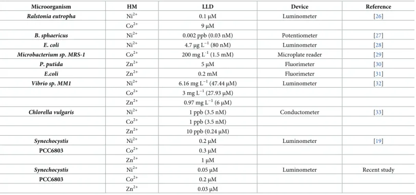
Table 2. Genetically engineered microorganisms as Ni2+, Co2+,and Zn2+biosensors and their lowest limit of detection (LLD).
Microorganism HM LLD Device Reference
Ralstonia eutropha Ni2+ 0.1μM Luminometer [26]
Co2+ 9μM
B.sphaericus Ni2+ 0.002 ppb (0.03 nM) Potentiometer [27]
E.coli Ni2+ 4.7μg L−1(80 nM) Luminometer [28]
Microbacterium sp.MRS-1 Co2+ 200 mg L-1(1.5 mM) Microplate reader [29]
P.putida Zn2+ 5μM Fluorimeter [30]
E.coli Zn2+ 0.2 mM Fluorimeter [31]
Vibrio sp.MM1 Ni2+ 6.16 mg L−1(47.44μM) Luminometer [32]
Co2+ 3 mg L−1(27.93μM)
Zn2+ 0.97 mg L−1(6μM)
Chlorella vulgaris Ni2+ 1 ppb (3.5 nM) Conductometer [33]
Co2+ 1 ppb (3.5 nM)
Zn2+ 10 ppb (0.24μM)
Synechocystis Ni2+ 0.2μM Luminometer [19]
PCC6803 Co2+ 0.3μM
Zn2+ 1μM
Synechocystis Ni2+ 0.05μM Luminometer Recent study
PCC6803 Co2+ 0.2μM
Zn2+ 0.03μM
https://doi.org/10.1371/journal.pone.0261135.t002
It is also noteworthy that a roughly 20% increase in the internal Zn2+concentration resulted in a tenfold decrease in the detection limit of the corresponding bioreporter strain. This find- ing shows that with limited manipulation of the genome significant improvements can be achieved, which may make the application of biosensors even more competitive and valuable method in environmental monitoring.
Biosensors for Ni2+, Co2+or Zn2+detection published in the last few years are listed in Table 2. Although their sensitivity may be remarkable, these bioanalytical tools are yet to be improved. Our current study helps to further enhance the utility of whole cell-based biosensors by facilitating the detection of lower contaminant levels in environmental samples than before.
Supporting information
S1 File. The optical density of the cultures.
(XLSX)
S2 File. The intracellular heavy metal concentrations in the treated and untreated cultures of WT and mutant strains.
(XLSX)
S3 File. Luminescence of the strains with CoaR construct upon cobalt and zinc treatment.
(XLSX)
S4 File. Luminescence of the strains with NrsRS-1 construct upon nickel treatment.
(XLSX)
S5 File. Luminescence of the strains with NrsRS-2 construct upon nickel treatment.
(XLSX)
S6 File. Luminescence of the strains with ZiaR construct upon zinc treatment.
(XLSX)
Author Contributions Conceptualization: Pe´ter B. Ko´s.
Data curation: Barbara Ho´di, Pe´ter B. Ko´s.
Formal analysis: Ga´bor Patyi, Barbara Ho´di, Da´niel Solymosi, Pe´ter B. Ko´s.
Funding acquisition: Imre Vass, Pe´ter B. Ko´s.
Investigation: Ga´bor Patyi, Da´niel Solymosi, Pe´ter B. Ko´s.
Methodology: Pe´ter B. Ko´s.
Project administration: Pe´ter B. Ko´s.
Resources: Pe´ter B. Ko´s.
Supervision: Pe´ter B. Ko´s.
Validation: Ga´bor Patyi, Imre Vass, Pe´ter B. Ko´s.
Visualization: Ga´bor Patyi.
Writing – original draft: Ga´bor Patyi.
Writing – review & editing: Ga´bor Patyi, Barbara Ho´di, Imre Vass, Pe´ter B. Ko´s.
References
1. Nahlik AM, Blocksom KA, Herlihy AT, Kentula ME, Magee TK, Paulsen SG. Use of national-scale data to examine human-mediated additions of heavy metals to wetland soils of the US. Environ Monit Assess. 2019;191.https://doi.org/10.1007/s10661-019-7311-9PMID:30810872
2. Real MIH, Azam HM, Majed N. Consumption of heavy metal contaminated foods and associated risks in Bangladesh. Environ Monit Assess. 2017;189.https://doi.org/10.1007/s10661-017-5902-xPMID:
28353206
3. Dytłow S, Go´rka-Kostrubiec B. Concentration of heavy metals in street dust: an implication of using dif- ferent geochemical background data in estimating the level of heavy metal pollution. Environ Geochem Health. 2021; 43: 521–535.https://doi.org/10.1007/s10653-020-00726-9PMID:33037955
4. Andersson L, Hedbrant A, Bryngelsson I-L, Persson A, Johansson A, Ericsson A, et al. Respiratory Health and Inflammatory Markers—Exposure to Cobalt in the Swedish Hard Metal Industry. J Occup Environ Med. 2020;62.https://doi.org/10.1097/JOM.0000000000001952PMID:33009343
5. Khan ZI, Arshad N, Ahmad K, Nadeem M, Ashfaq A, Wajid K, et al. Toxicological potential of cobalt in forage for ruminants grown in polluted soil: a health risk assessment from trace metal pollution for live- stock. Environ Sci Pollut Res. 2019; 15381–15389.https://doi.org/10.1007/s11356-019-04959-9PMID:
30937740
6. WHO WHO. Guidelines for drinking-water quality. incorporating the first addendum. Geneva, Switzer- land: WHO; 2017. ISBN 978-92-4-154995-0; 2017.
7. Rampley CPN, Whitehead PG, Softley L, Hossain MA, Jin L, David J, et al. River toxicity assessment using molecular biosensors: Heavy metal contamination in the Turag-Balu-Buriganga river systems, Dhaka, Bangladesh. Sci Total Environ. 2020; 703: 134760.https://doi.org/10.1016/j.scitotenv.2019.
134760PMID:31744697
8. Ahmed DAEA, Gheda SF, Ismail GA. Efficacy of two seaweeds dry mass in bioremediation of heavy metal polluted soil and growth of radish (Raphanus sativus L.) plant. Environ Sci Pollut Res. 2021; 28:
12831–12846.https://doi.org/10.1007/s11356-020-11289-8PMID:33089464
9. Kumar A, Chaturvedi AK, Yadav K, Arunkumar KP, Malyan SK, Raja P, et al. Fungal Phytoremediation of Heavy Metal-Contaminated Resources: Current Scenario and Future Prospects. In: Yadav AN, Singh S, Mishra S, Gupta A, editors. Recent Advancement in White Biotechnology Through Fungi: Vol- ume 3: Perspective for Sustainable Environments. Cham: Springer International Publishing; 2019. pp.
437–461.https://doi.org/10.1007/978-3-030-25506-0_18
10. Abdel-Razek MA, Abozeid AM, Eltholth MM, Abouelenien FA, El-Midany SA, Moustafa NY, et al. Bioreme- diation of a pesticide and selected heavy metals in wastewater from various sources using a consortium of microalgae and cyanobacteria. Slov Vet Res. 2019; 56: 61–73.https://doi.org/10.26873/SVR-744-2019 11. Baptista MS, Vasconcelos MT. Cyanobacteria metal interactions: Requirements, toxicity, and ecologi-
cal implications. Crit Rev Microbiol. 2006; 32: 127–137.https://doi.org/10.1080/10408410600822934 PMID:16893750
12. Nies DH. Efflux-mediated heavy metal resistance in prokaryotes. FEMS Microbiol Rev. 2003; 27: 313–
339.https://doi.org/10.1016/S0168-6445(03)00048-2PMID:12829273
13. Cavet Jennifer S.; Borrelly GPM. Zn, Cu and Co in cyanobacteria: selective control of metal availability.
FEMS Microbiol Rev. 2003; 27: 165–181.https://doi.org/10.1016/S0168-6445(03)00050-0PMID:
12829266
14. Patterson C. J., Pernil R., Foster A. W., & Robinson NJ. Cyanobacterial models that address cross-talk in metal homeostasis. Metals in Cells. 2016.
15. Pennella MA, Shokes JE, Cosper NJ, Scott RA, Giedroc DP. Structural elements of metal selectivity in metal sensor proteins. Proc Natl Acad Sci U S A. 2003; 100: 3713–3718.https://doi.org/10.1073/pnas.
0636943100PMID:12651949
16. Garcı´a-Domı´nguez M, Lopez-Maury L, Florencio FJ, Reyes JC. A gene cluster involved in metal homeostasis in the cyanobacterium Synechocystis sp. strain PCC 6803. J Bacteriol. 2000; 182: 1507–
1514.https://doi.org/10.1128/JB.182.6.1507-1514.2000PMID:10692354
17. Peca L, Kos PB, Vass I. Characterization of the activity of heavy metal-responsive promoters in the cya- nobacterium Synechocystis PCC 6803. Acta Biol Hung. 2007; 58 Suppl: 11–22.https://doi.org/10.1556/
ABiol.58.2007.Suppl.2PMID:18297791
18. Blasi B, Peca L, Vass I, Ko´s PB. Characterization of stress responses of heavy metal and metalloid inducible promoters in synechocystis PCC6803. J Microbiol Biotechnol. 2012; 22: 166–169.https://doi.
org/10.4014/jmb.1106.06050PMID:22370344
19. Peca L, Ko´s PB, Ma´te´ Z, Farsang A, Vass I. Construction of bioluminescent cyanobacterial reporter strains for detection of nickel, cobalt and zinc. FEMS Microbiol Lett. 2008; 289: 258–264.https://doi.org/
10.1111/j.1574-6968.2008.01393.xPMID:19016871
20. Rippka R, Deruelles J, Waterbury JB. Generic assignments, strain histories and properties of pure cul- tures of cyanobacteria. J Gen Microbiol. 1979; 111: 1–61.https://doi.org/10.1099/00221287-111-1-1 21. Kirtania P, Ho´di B, Mallick I, Vass IZ, Fehe´r T, Vass I, et al. A single plasmid based CRISPR interfer- ence in Synechocystis 6803 –A proof of concept. PLoS One. 2019; 14: e0225375.https://doi.org/10.
1371/journal.pone.0225375PMID:31770415
22. J. Sambrook, D.W. Russell. Molecular cloning: a laboratory manual, 3rd ed., Cold Spring Harbor Labo- ratory Press, Cold Spring Harbor, N.Y. Zool. Res. 2001.https://doi.org/10.3724/SP.J.1141.2012.01075 23. Guerrero F, Carbonell V, Cossu M, Correddu D, Jones PR. Ethylene Synthesis and Regulated Expres-
sion of Recombinant Protein in Synechocystis sp. PCC 6803. PLoS One. 2012;7.https://doi.org/10.
1371/journal.pone.0050470PMID:23185630
24. Kunert A, Hagemann M, Erdmann N. Construction of promoter probe vectors for Synechocystis sp.
PCC 6803 using the light-emitting reporter systems Gfp and LuxAB. J Microbiol Methods. 2000; 41:
185–194.https://doi.org/10.1016/s0167-7012(00)00162-7PMID:10958963
25. Peca L, Nagy CI, O¨ rdo¨g A, Vass I, Ko´s PB. Deveopment of a bioluminescent cyanobacterial reporter strain for detection of Arsenite, Arsenate and Antimonite. Environ Eng Manag J. 2017; 16: 62599711.
Available:http://omicron.ch.tuiasi.ro/EEMJ/accepted.htm
26. Tibazarwa C, Corbisier P, Mench M, Bossus A, Solda P, Mergeay M, et al. A microbial biosensor to pre- dict bioavailable nickel in soil and its transfer to plants. Environ Pollut. 2001; 113: 19–26.https://doi.org/
10.1016/s0269-7491(00)00177-9PMID:11351758
27. Verma N, Singh M. A Bacillus sphaericus based biosensor for monitoring nickel ions in industrial efflu- ents and foods. J Autom Methods Manag Chem. 2006; 2006: 1–4.https://doi.org/10.1155/JAMMC/
2006/83427PMID:17671626
28. Cayron J, Prudent E, Escoffier C, Gueguen E, Mandrand-Berthelot MA, Pignol D, et al. Pushing the lim- its of nickel detection to nanomolar range using a set of engineered bioluminescent Escherichia coli.
Environ Sci Pollut Res. 2017; 24: 4–14.https://doi.org/10.1007/s11356-015-5580-6PMID:26498802 29. Sundararaju S, Arumugam M, Bhuyar P. Microbacterium sp. MRS-1, a potential bacterium for cobalt
reduction and synthesis of less/non-toxic cobalt oxide nanoparticles (Co3O4). Beni-Suef Univ J Basic Appl Sci. 2020;9.https://doi.org/10.1186/s43088-020-00070-y
30. Liu P, Huang Q, Chen W. Construction and application of a zinc-specific biosensor for assessing the immobilization and bioavailability of zinc in different soils. Environ Pollut. 2012; 164: 66–72.https://doi.
org/10.1016/j.envpol.2012.01.023PMID:22336732
31. Ravikumar S, Ganesh I, Yoo IK, Hong SH. Construction of a bacterial biosensor for zinc and copper and its application to the development of multifunctional heavy metal adsorption bacteria. Process Bio- chem. 2012; 47: 758–765.https://doi.org/10.1016/j.procbio.2012.02.007
32. Mohseni M, Abbaszadeh J, Maghool SS, Chaichi MJ. Heavy metals detection using biosensor cells of a novel marine luminescent bacterium Vibrio sp. MM1 isolated from the Caspian Sea. Ecotoxicol Environ Saf. 2018; 148: 555–560.https://doi.org/10.1016/j.ecoenv.2017.11.002PMID:29127817
33. Berezhetskyy AL, Durrieu C, Nguyen-Ngoc H, Chovelon JM, Dzyadevych S V., Tran-Minh C. Conducto- metric biosensor based on whole-cell microalgae for assessment of heavy metals in wastewater. Biopo- lym Cell. 2007; 23: 511–518.https://doi.org/10.7124/bc.000786