tears and aqueous humour reveals potential biomarkers for trabeculectomy complications
Eva Cs}osz1,2 , Eszter Deak1,3, Noemi Toth1,3, Carlo Enrico Traverso4, Adrienne Csutak3 and Jozsef T}ozser1,2
1 Biomarker Research Group, Department of Biochemistry and Molecular Biology, Faculty of Medicine, University of Debrecen, Hungary 2 Proteomics Core Facility, Department of Biochemistry and Molecular Biology, Faculty of Medicine, University of Debrecen, Hungary 3 Department of Ophthalmology, Faculty of Medicine, University of Debrecen, Hungary
4 Clinica Oculistica, DiNOGMI, University of Genoa and IRCCS Ospedale Policlinico San Martino, Genova, Italy
Keywords
aqueous humour; biomarker; cytokine profile; glaucoma; tear; trabeculectomy Correspondence
E. Cs}osz, Biomarker Research Group, Department of Biochemistry and Molecular Biology, Faculty of Medicine, University of Debrecen, Debrecen, Hungary
Tel: +36 52 416432 E-mail: cseva@med.unideb.hu Adrienne Csutak and Jozsef T }ozser contributed equally to this work (Received 11 February 2019, revised 11 March 2019, accepted 5 April 2019) doi:10.1002/2211-5463.12637
Glaucoma is a multifactorial neurodegenerative disease that causes impaired vision and, in advanced cases, blindness. The increasing preva- lence of glaucoma due to an ageing population has necessitated the identifi- cation of suitable biomarkers for the early detection of the disease.
Aqueous humour (AH) has been proposed as a source of biomarkers, but it can only be collected using a minor, yet invasive surgical intervention.
Tears, however, are constantly available and can be collected any time via noninvasive methods. In order to examine the utility of tear as a surrogate for aqueous humour in biomarker development, we compared the levels of 27 cytokines and chemokines in paired samples of tear and aqueous humour using a Luminex multiplex immunobead-based technique. Signifi- cantly higher levels of cytokines in tear compared to aqueous humour were detected suggesting that tear and aqueous humour are not identical in terms of inflammation response. Furthermore, the levels of IFN-c, GM- CSF and IL-5 in tear were significantly lower in patients who developed complications after one year, but no statistically significant changes in cytokine levels were observed in aqueous humour. These three molecules may have potential as predictive biomarkers for the appearance of late flap-related complications of trabeculectomy.
Glaucoma is a multifactorial neurodegenerative disease affecting the optic nerve, leading to impaired vision and, in advanced cases, blindness [1]. There are different types of glaucoma, primarily categorized by the irido- corneal angle. Both the open-angle glaucoma and the angle-closure glaucoma can be divided into primary and
secondary forms, and can also be classified as acute or chronic [2]. In glaucoma, neuropathy of the optic nerve and the loss of retinal ganglion cells can be observed, resulting in atrophy of the optic nerve and impairment of visual functions, leading to blindness [3,4]. Taking into consideration the increasing prevalence of
Abbreviations
AH, aqueous humour; bFGF, fibroblast growth factor; CV, coefficient of variation; ELISA, enzyme-linked immunosorbent assay; G-CSF, granulocyte colony-stimulating factor; GM-CSF, granulocyte-macrophage colony-stimulating factor; IFN, interferon; IL, interleukin; IOP, intraocular pressure; IP-10, interferon gamma-induced protein 10; LC-MS, liquid chromatography coupled mass spectrometry; MCP-1, monocyte chemoattractant protein 1; MIP, macrophage inflammatory proteins; MMC, mitomycin C; PACG, primary angle-closure glaucoma;
PDGF, platelet-derived growth factor; POAG, primary open-angle glaucoma; RANTES, regulated on activation, normal T cell expressed and secreted; TNF, tumour necrosis factor; VEGF, vascular endothelial growth factor.
glaucoma due to an ageing demographics of modern society, it is crucial to find suitable biomarkers for the early detection of the disease [5]. Several studies were carried out to investigate proteins related to the patho- physiological changes observed in glaucoma, most of them focusing on the analysis of the retina, optic nerve, trabecular meshwork and aqueous humour (AH). There were also studies conducted on blood samples, while only few used tear fluid [6–10].
Using various proteomic methods such as LC-MS- based analyses and immunological methods, hundreds of proteins characteristic of glaucoma were identified.
Most of these specimens were obtained from the retina and the trabecular meshwork by highly invasive tech- niques [9–14]. Biological fluids are often used instead of tissue samples for biomarker studies [15]. As such, AH is frequently analysed in the examination of glaucoma as a surrogate for ocular biological fluids [9]. AH is the product of the ciliary body and is drained from the eye through the trabecular meshwork and the uveoscleral pathway. Proteomic analyses of AH samples from patients with glaucoma revealed important proteins involved in metabolism, inflammatory response and antioxidant defence [9,16]. Extensive studies analysing the amount of inflammatory cytokines revealed higher levels of pro-inflammatory cytokines in the AH originat- ing from patients with glaucoma, as compared to controls [17–21].
Despite the usefulness of AH as a source of biomarkers of trabecular meshwork and retinal gan- glion cell damage, the invasive collection of this bio- logical fluid prevents its application for the purpose of screening or follow-ups. On the other hand, tear fluid provides a better alternative, given its availability and ease of collection along with a composition of glau- coma-related proteins traced directly to the AH. Tears are a valuable biological fluid, widely studied in eye diseases for the purpose of identifying potential biomarkers for eye-related and systemic diseases [15].
In glaucoma, only few studies used tear fluid as a bio- logical source [22], and with proteomic analyses, 27 proteins were identified as potential glaucoma biomarkers, mainly for primary open-angle glaucoma (POAG) [12]. The pro- and anti-inflammatory cytoki- nes were also studied in tear fluid, showing a shifted balance towards the pro-inflammatory state in glau- coma upon topical medication [23].
Multiplex immunobead-based assays are often used to examine the level of multiple cytokine and chemo- kine molecules in a single sample. This is very benefi- cial when analysing certain bodily fluids that can be collected only in small amounts, as in the case of tear and AH, respectively [24,25]. In this pilot study, we
compared the concentrations of 27 cytokine and che- mokine molecules in the tear and AH samples origi- nating from the same patient with glaucoma, using a commercially available multiplex immunobead kit. Our aim was to determine whether the readily available tear fluid can replace the use of AH in studying patients with glaucoma following trabeculectomy.
Results and Discussion
Multiplex immunobead assays provide a versatile tool for the analysis of bodily fluids with low volume. The application of antibodies provides high specificity, while the Luminex beads offer the possibility of multi- plexing, allowing for the analysis of multiple analytes in one sample [26]. This technique was used in the form of a validated, commercially available kit for the measurement of the concentration of 27 cytokine and chemokine molecules in tear and AH samples originat- ing from patients with glaucoma, who underwent tra- beculectomy in order to lower their intraocular pressure. All the 27 analytes were detected in both sample types; however, in cases where the amount of the studied molecule was under the limit of detection (Table S1), the number 0 was assigned for the concen- tration of the analyte. The most dilute standards from the calibration curve (S8) had high CV% in case of most of the analytes, indicating that the kit cannot reliably measure the concentration of the individual proteins in very dilute samples (Table S1).
The analysis of tear
All of the examined cytokines, except for IL2 and IL10, were present in at least 90% of the tear samples.
IL2 could be detected only in 45% of the samples, while IL10 was present in 85% of the samples (Table S2). Considering that we did not include healthy control because our aim was to compare the two types of samples originating from the same patient, first we compared our results to data available in the literature.
Comparing our results to those obtained by Marti- nez-de-la-Casa et al. [27] regarding the concentration of the same 27 cytokines and chemokines on control and glaucoma subjects using the same sample collec- tion procedure, we can conclude that the levels of eotaxin, G-CSF, IL-12, IL-13, IL-15, IL-1b, IL-4, IL-7 and MIP1awere in the same range and were very sim- ilar in the two studies. A similar trend in the levels of bFGF, GM-CSF, IFN-c, IL-10, IL-17, IL-5, MCP1, PDGF-BB, TNFa and VEGF was observed as com- pared to controls in the two studies; however, in our
case, the levels of these cytokines were even higher.
The levels of IL-1Ra, IL-2, IL-6, IL-8, IP-10, MIP1b and RANTES were lower in our study as compared to the control tears from healthy subjects published by Martinez-de-la-Casa et al. (Table S3) [27]. However, when we compared the cytokine levels measured in our samples to the results reported by Mrugacz et.al.
examining physiological tear with ELISA method [28], the levels of IL-6, TNFaand IL-17 were much higher in our samples. A similar phenomenon was observed when we compared our data to those obtained by Gupta et al. [29] examining eye-drop naive patients with newly diagnosed POAG compared to controls using a different sample collection procedure and a different method than ours.
It should be noted that it is indeed difficult to com- pare results obtained by different studies, as the appli- cation of different analytical strategies hinders the proper comparison of results between studies. For tear analysis, Gupta et al. and Mrugacz et.al. have used ELISA methods to examine the cytokine levels. Their results are comparable to each other but display mag- nitude lower values compared to our results and the results obtained by Martinez de la Casaet al.[27–29].
The higher level of some cytokines observed in our study compared to the values from the literature might indicate a more pronounced pro-inflammatory condi- tion, which might be due to the fact that the patients recruited into our study had a more advanced phase of glaucoma, finally succumbing to trabeculectomy.
Another factor that can influence tear composition is the method of collection. Depending on the proce- dure, significantly higher levels of pro-inflammatory cytokines were observed in basal tears as compared to reflex tears [30]. In our study, basal tear was collected by the same person throughout the study, and the presence of ocular surface disease or autoimmune dis- ease was regarded as exclusion criteria. It is important to note that neither the tear collection nor the analysis method applied by Gupta et al. was the same as the ones used in this study and by Martinez-de-la-Casa et. al., and the patients with glaucoma recruited in these two later studies were eye-drop users [27,29].
According to the currently used treatment protocols, eye-drops are widely used in the therapy of glaucoma.
Evidence from the literature shows that the adminis- tration of eye-drops can distort the level of cytokines in tears, leading to higher pro-inflammatory cytokine concentrations, in agreement with the findings from our study [23,31].
Gender and the type of glaucoma were not associ- ated with a statistically significant difference on the cytokine levels, tear production rate and tear protein
concentration in any of the studied groups. The group of patients using three or more different eye-drops was compared to the group using less than three eye-drops, but we could not find any statistically significant dif- ference (Table S4). Based on these data, we think that the administration of different drugs does not modu- late the level of the analysed cytokines, tear protein concentration or tear production rate, at least in case of patients examined in this study.
The IOP of the patients and the presence of compli- cations following trabeculectomy were monitored, including infection, blebitis, endophthalmitis, reduced visual acuity, flap-related complications, choroidal ablation, and bleb failure leading to blocked AH filtra- tion to the subconjunctival space.
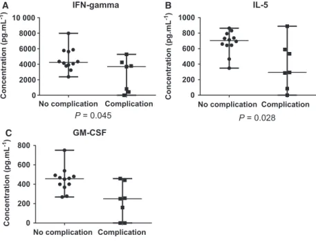
We collected ophthalmological data for 19 out of 20 recruited patients, and we examined the effect of the presence of complications on the level of tear cytoki- nes, tear total protein concentration and tear produc- tion rate. In case of three proteins, statistically significant differences could be observed. The concen- tration of IFN-c was higher in the group of patients without complication, and the same tendency was observed in case of GM-CSF and IL-5, as well (Fig. 1).
IFN-cis secreted by T helper cells, cytotoxic T cells and natural killer cells and has antiviral, immunoregu- latory and antitumour properties [32]. The increase of IFN-c may inhibit collagen synthesis and lead to delayed epidermal wound healing [33,34]. According to another study, the application of mesenchymal stem cells following chemical injury of the cornea reduced IFN-c levels and thus reduces the corneal inflamma- tion and neovascularization resulting in wound healing with less epithelial defects [35].
IL-5 is mainly produced by Th2-type helper T cells and was shown to have an important role in the acti- vation, proliferation and survival of eosinophils and the differentiation of B cells [36]. It was observed in mice overexpressing IL-5 that the wound healing is delayed and re-epithelization is impaired [37].
GM-CSF is a pleiotropic molecule activating granu- locyte and macrophage cell lineages, with an important role in wound healing through the induction of ker- atinocyte proliferation and promoting migration of epithelial cells [38]. Recombinant GM-CSF can be applied to help the wound healing process [38]. It was shown that GM-CSF plays an important role in the early, so-called acute inflammatory phase of ocular wound healing [39]. With the administration of recom- binant human GM-CSF, it was demonstrated that GM-CSF has no effect on cell proliferation but on cell migration [40].
According to our data, ocular complications are associated with reduced levels of each observed cyto- kine. However, according to the scientific literature, faster wound healing, which might be one reason for bleb failure, would require increased levels of these factors. It should be noted that none of the studies involving IL-5, IFN-c and GM-CSF were carried out on tear samples. The phenomena leading to bleb fail- ure are not known in detail, and most probably, multi- ple, more complex mechanisms take part in the regulation of the process.
Despite the fact that our results observed in human tear contradict the results observed by others in skin or in rodent ocular wound healing models, IL-5, IFN- cand GM-CSF can serve as a good starting point for further biomarker studies and verifications.
The analysis of aqueous humour
Aqueous humour was next analysed in the same man- ner as tear. More than half of cytokines detected were present in more than 90% of the samples. However, IL-1b, IL-7, IL-8, IL-9, IL-10, G-CSF and IFN-cwere present in less than 50% of the samples. IL-2, IL-4, IL-5, IL-13, bFGF and MIP1a were present in 70– 85% of the samples.
Next, we compared our data to data acquired from the analysis of human AH removed during cataract surgery by Sharmaet al., POAG and cataract samples by Duvesh et al. and control and PEX samples by Garweget al.(Table S3) [24,41,42]. We found that the
level of bFGF, G-CSF, IL-1Ra, IL-6 and RANTES was higher in our samples; the level of IL8, MIP1a, MIP1b and eotaxin was higher in the study carried out by Sharma et. al.; and the MCP1 levels were in the same range in both studies [24]. Based on other studies, where similar examination methods were used to those applied by our group, the levels of eotaxin, GM-CSF, IL-1b, IL-6, IL-8, IL-9, IL-10, IP-10, MCP1, MIP1a and VEGF were in the same range, and the level of IL-12, IL-4 and MIP1b was lower in our study. At the same time, the levels of bFGF, G- CSF, IL-1ra, IL-2, IL-5, IL-7, IL-13, IL-15, IL-17 and TNFawere higher in our study compared to the stud- ies carried out by Duvesh et al. and Garweg et al.
(Table S3) [41,42].
The effect of complication, gender and type of glau- coma on the cytokine levels was examined but no sta- tistically significant difference could be found between any of the studied groups (data not shown).
Comparative analysis
Tear, as a noninvasively collectable bodily fluid, is constantly available and an important resource for fol- low-up studies. Different studies have examined the composition of tears and of aqueous humour in glau- coma and other eye diseases, but no previous compar- ative study examining the cytokine levels in tears and aqueous humour was carried out. After critical exami- nation of the data obtained for tear and AH, we have compared the level of cytokines between these two
No complication Complication 0
200 400 600 800
GM-CSF
P = 0.007
Concentration (pg.mL-1) Concentration (pg.mL-1)
Concentration (pg.mL-1)
No complication Complication 0
2000 4000 6000 8000 10 000
IFN-gamma
P = 0.045
No complication Complication 0
200 400 600 800 1000
IL-5
P = 0.028 A
C
B
Fig. 1.Concentrations of cytokines with statistically significant differences between the groups with complications and no complication. The concentration (pgmL 1) of IFN-gamma (A), IL-5 (B) and GM-CSF (C) in the two groups is plotted. Below each plot, thePvalues obtained by the utilization of nonparametric Mann–Whitney U-test are indicated.
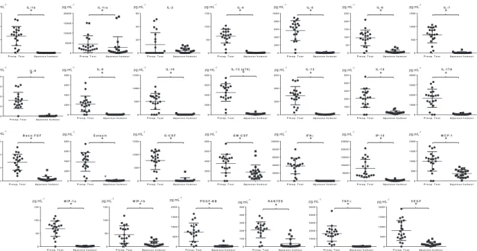
sample types. All studied cytokines and chemokine molecules, except IL-2, showed statistically signifi- cantly higher levels in tears compared to AH (Fig.2, Table1).
Based on our data, we can conclude that tear and aqueous humour are not identical from the inflamma- tory point of view and tear cannot substitute for aque- ous humour for diagnostic purposes. Most probably, the levels of cytokines in the two sample types are con- trolled by different mechanisms. The higher cytokine levels in tear compared to aqueous humour might reflect not only the glaucoma-related, but also the eye- drop- or other factors-induced pro-inflammatory con- ditions, as well. A limitation of the study is the rela- tively low number of the participants recruited. Larger studies are needed to validate our findings and also to be able to do a proper patient stratification based on the drugs used.
Materials and methods
Subjects and sample collection
All participants recruited into this study were patients of the Department of Ophthalmology, Faculty of Medicine, University of Debrecen. The participants provided written informed consent for tear and aqueous humour collection in
accordance with the Declaration of Helsinki. The study was approved by the Ethical Committee of the University of Debrecen (approval number: 4234-2014). All patients under- went trabeculectomy surgery to reduce intraocular pressure.
Exclusion criteria were the presence of autoimmune and/or ocular surface diseases and previous history of ocular sur- gery within 6 months. For the comparison of the tear and aqueous humour, 20 patients (11 male and 9 female; mean age: 58.8 14.8) with glaucoma [eight patients with pri- mary angle-closure glaucoma (PACG) and 12 patients with POAG] were recruited and the tears and aqueous humour were collected. Basal tear was collected only once for 2 min from patients except G002 and G093 right before the tra- beculectomy. In case of patients G002 and G093, sample collection was not possible on the day of trabeculectomy; in their case, a previously collected tear sample was used.
These cases are marked with *in Table S2. The nontrau- matic tear collection was carried out before the trabeculec- tomy, using sterile glass capillary tubes (VWR Ltd, Hungary) from the lateral inferior meniscus without local anaesthesia or stimulation [43]. Tear samples were collected for 2 min, and then promptly transferred to 0.2-mL PCR tubes and centrifuged on 4°C (264g) for 10 min, and the supernatants were then deep-frozen and stored at 70°C until analysis. The AH samples were collected during tra- beculectomy surgery through a limbal paracentesis by the same operator using sterile glass capillary. Care was taken to prevent blood and intraocular tissue contamination. The
pg.mL-1
pg.mL-1 pg.mL-1 pg.mL-1 pg.mL-1 pg.mL-1 pg.mL-1 pg.mL-1
pg.mL-1 pg.mL-1
pg.mL-1 pg.mL-1
pg.mL-1 pg.mL-1
pg.mL-1
pg.mL-1 pg.mL-1 pg.mL-1 pg.mL-1 pg.mL-1 pg.mL-1 pg.mL-1
pg.mL-1 IL -1 b
P r e o p . T e a r Aq u e o u s h u m o u r 0
5 0 1 0 0 1 5 0
IL -1 r a
P r e o p . T e a r Aq u e o u s h u m o u r 0
5 0 0 0 1 0 0 0 0 1 5 0 0 0 2 0 0 0 0
IL -2
P r e o p . T e a r Aq u e o u s h u m o u r 0
2 0 4 0 6 0
IL -4
P r e o p . T e a r Aq u e o u s h u m o u r 0
5 0 1 0 0 1 5 0
IL -5
P r e o p . T e a r Aq u e o u s h u m o u r 0
2 0 0 4 0 0 6 0 0 8 0 0 1 0 0 0
IL -6
P r e o p . T e a r Aq u e o u s h u m o u r 0
5 0 1 0 0 1 5 0 2 0 0 2 5 0
IL -7
P r e o p . T e a r Aq u e o u s h u m o u r 0
5 0 0 1 0 0 0 1 5 0 0
IL -8
P r e o p . T e a r Aq u e o u s h u m o u r 0
2 0 0 4 0 0 6 0 0 8 0 0
IL -9
P r e o p . T e a r Aq u e o u s h u m o u r 0
2 0 0 4 0 0 6 0 0 8 0 0
IL -1 0
P r e o p . T e a r Aq u e o u s h u m o u r 0
5 0 0 1 0 0 0 1 5 0 0
IL -1 2 (p 7 0 )
P r e o p . T e a r Aq u e o u s h u m o u r 0
2 0 0 4 0 0 6 0 0 8 0 0
IL -1 3
P r e o p . T e a r Aq u e o u s h u m o u r 0
2 0 0 4 0 0 6 0 0
IL -1 5
P r e o p . T e a r Aq u e o u s h u m o u r 0
1 0 0 2 0 0 3 0 0 4 0 0 5 0 0
IL -1 7 A
P r e o p . T e a r Aq u e o u s h u m o u r 0
1 0 0 0 2 0 0 0 3 0 0 0 4 0 0 0
B a s ic F G F
P r e o p . T e a r Aq u e o u s h u m o u r 0
5 0 0 1 0 0 0 1 5 0 0
E o ta x in
P r e o p . T e a r Aq u e o u s h u m o u r 0
2 0 0 4 0 0 6 0 0 8 0 0
G -C S F
P r e o p . T e a r Aq u e o u s h u m o u r 0
5 0 0 1 0 0 0 1 5 0 0
G M -C S F
P r e o p . T e a r Aq u e o u s h u m o u r 0
2 0 0 4 0 0 6 0 0 8 0 0
IF Nγ
P r e o p . T e a r Aq u e o u s h u m o u r 0
2 0 0 0 4 0 0 0 6 0 0 0 8 0 0 0 1 0 0 0 0
IP -1 0
P r e o p . T e a r Aq u e o u s h u m o u r 0
5 0 0 0 1 0 0 0 0 1 5 0 0 0 2 0 0 0 0 2 5 0 0 0
M C P - 1
P r e o p . T e a r Aq u e o u s h u m o u r 0
5 0 0 1 0 0 0 1 5 0 0 2 0 0 0
M IP -1 a
P r e o p . T e a r Aq u e o u s h u m o u r 0
5 0 1 0 0 1 5 0
M IP -1 b
P r e o p . T e a r Aq u e o u s h u m o u r 0
5 0 1 0 0 1 5 0
P D G F -B B
P r e o p . T e a r Aq u e o u s h u m o u r 0
5 0 0 1 0 0 0 1 5 0 0 2 0 0 0
R A N T E S
P r e o p . T e a r Aq u e o u s h u m o u r 0
1 0 0 2 0 0 3 0 0 4 0 0 5 0 0
T N Fα
P r e o p . T e a r Aq u e o u s h u m o u r 0
1 0 0 0 2 0 0 0 3 0 0 0 4 0 0 0 5 0 0 0
V E G F
P r e o p . T e a r Aq u e o u s h u m o u r 0
5 0 0 1 0 0 0 1 5 0 0 2 0 0 0
*
* * * *
*
*
*
* * *
*
*
*
* *
*
*
* *
*
* *
*
* * *
pg.mL-1 pg.mL-1 pg.mL-1 pg.mL-1
Fig. 2.Concentrations of 27 cytokines and chemokines in preoperative tear and aqueous humour samples from patients with glaucoma.
The ‘x’ axis shows the sample type, while ‘y’ axis shows the concentration of the analyte in pgmL 1. For statistical analysis, paired nonparametric Wilcoxon test was used, and*indicates statistically significant (P<0.05) difference.
samples were expelled from the capillaries to 0.2-mL PCR tubes and processed in an identical way as tears. Protein concentration of tear and aqueous humour samples was determined using the Bradford method [44].
Multiplex analysis of cytokines in tear and aqueous humour samples
The concentration of the interleukins IL-1b, IL-1Ra, IL-2, IL-4, IL-5, IL-6, IL-7, IL-8, IL-9, IL-10, IL-12p70, IL-13, IL-15, IL-17, eotaxin, basic fibroblast growth factor (bFGF), granulocyte colony-stimulating factor (G-CSF), granulocyte-macrophage colony-stimulating factor (GM- CSF), interferon (IFN)-c, interferon gamma-induced pro- tein 10 (IP-10), monocyte chemoattractant protein 1
(MCP1), macrophage inflammatory proteins MIP1a and MIP1b, platelet-derived growth factor (PDGF-BB), regu- lated on activation, normal T cell expressed and secreted (RANTES), tumour necrosis factora(TNFa) and vascular endothelial growth factor (VEGF) was analysed using a multiplex immunobead system based on xMAP technology (Luminex, Austin, TX, USA). The 27-plex Bio-Plex kit (Bio-Rad Laboratories, Hercules, CA, USA) was utilized strictly adhering to the manufacturer’s instructions. The tear and aqueous humour samples were diluted. Based on previous experiments, 1 : 25 dilution was applied for tears and 1 : 2 dilution for AH samples, where the amount of the AH was sufficient. In cases where the amount of the AH collected was not enough, the dilutions indicated in Table S1 were used. Two technical replicates were used in each case; however, in case of G041 and G107 the tear sample amount was enough only for one replicate (samples marked with # in Table S2).
Fluorescence intensity data were recorded and analysed using the BIO-PLEX MANAGER software (version 4.1.1.; Bio- Rad Laboratories). Concentrations of the 27 cytokines and chemokines were calculated based on the recorded calibra- tion curve and the dilution factor used. For curve fitting, logistic regression was applied.
Statistical analysis
Statistical analysis was performed using SPSS 25.0 (IBM, Armonk, NY, USA) for Windows. Nonparametric Mann– Whitney U-test was used to compare the protein concentra- tions among groups. For the comparison of tear and aque- ous humour samples originating from the same patient, paired nonparametric Wilcoxon test was used. All data with p<0.05 are defined as significantly different.
Conclusion
In our study, we compared the levels of 27 cytokines in tear and AH originating from patients with glau- coma who underwent trabeculectomy. Our findings are largely in agreement with the data in the scientific liter- ature; however, in the case of some cytokines differ- ences were detected. According to our data, tear and AH are not identical from the inflammatory point of view; significantly higher levels of cytokines were found in tear compared to AH.
The concentrations of three tear proteins, IFN-c, GM-CSF and IL-5, were associated with the presence of complications. However, more patients have to be recruited to be able to identify/verify these proteins as potential risk factors for the occurrence of flap-related complications following trabeculectomy.
Our data highlight the promising potential of this continuously available, easy-to-collect body fluid for
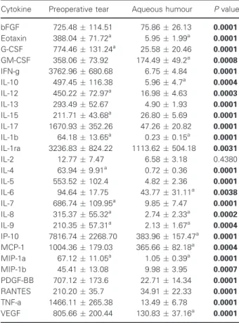
Table 1. Concentrations of cytokines and chemokines in preoperative tear and aqueous humour samples by multiplex bead immunoassay. Data are expressed as mean (pgmL 1)SEM. The concentrations were calculated based on the recorded 8 point calibration curves and applied dilutions shown in Table S1.
Statistical significance between groups was determined using paired nonparametric Wilcoxon test. ThePvalues in bold indicate statistically significant (P<0.05) values. SEM, standard error of the means.
Cytokine Preoperative tear Aqueous humour Pvalue
bFGF 725.48114.51 75.8626.13 0.0001
Eotaxin 388.0471.72a 5.951.99a 0.0001 G-CSF 774.46131.24a 25.5820.46 0.0001 GM-CSF 358.0673.92 174.4949.2a 0.0008
IFN-g 3762.96680.68 6.754.84 0.0001
IL-10 497.45116.38 5.964.7a 0.0004
IL-12 450.2272.97a 16.984.63 0.0003
IL-13 293.4952.67 4.901.93 0.0001
IL-15 211.7143.68a 26.805.69 0.0001
IL-17 1670.93352.26 47.2620.82 0.0001
IL-1b 64.1813.65a 0.230.15a 0.0001
IL-1ra 3236.83824.22 1113.62504.18 0.0031
IL-2 12.777.47 6.583.18 0.4380
IL-4 63.949.91a 0.720.36 0.0001
IL-5 553.52102.4 4.822.36 0.0001
IL-6 94.6417.75 43.7731.11a 0.0038
IL-7 686.74109.95a 9.857.47 0.0001
IL-8 315.3755.32a 2.742.33a 0.0002
IL-9 210.3557.31a 2.131.67a 0.0004
IP-10 7816.742268.70 383.96157.47a 0.0001 MCP-1 1004.36179.03 365.6682.18a 0.0004 MIP-1a 67.1211.05a 1.050.39a 0.0001
MIP-1b 45.4113.08 9.983.95 0.0007
PDGF-BB 707.12173.6 22.7114.34 0.0001
RANTES 210.2035.7 34.9122.33 0.0001
TNF-a 1466.11265.38 13.496.78 0.0001
VEGF 805.66200.44 130.8337.16a 0.0001
aIndicate values which are in the same range with data obtained by other studies.
dynamic testing, making it worth exploring as an alter- native to samples collectable by invasive methods.
Acknowledgements
This research was funded by Janos Bolyai Research Scholarship of the Hungarian Academy of Sciences, UNKP-18-4-DE-436, and by PD116817 and PD121075 National Research, Development and Innovation Office (NKFI) grants. The technical help of Kamilla Solyom is greatly acknowledged. We thank Dr. Mohamed Mahdi and Dr. Ivan Uray for the English proofreading.
Conflict of interest
The authors declare no conflict of interest.
Author contributions
EC and AC conceptualized the study. EC and NT curated the data. EC and JT acquired funding. EC, ED and NT contributed to methodology. EC, JT and AC provided resources. EC visualized the data. EC wrote the original draft of the manuscript. CET, JT and AC reviewed and edited the manuscript.
References
1 Kingman S (2004) Glaucoma is second leading cause of blindness globally.Bull World Health Organ82, 887– 888.
2 Andersson Geimer S (2013) Glaucoma diagnostics.Acta Ophthalmol91, 390–391.
3 Krizaj D, Ryskamp DA, Tian N, Tezel G, Mitchell CH, Slepak VZet al.(2014) From mechanosensitivity to inflammatory responses: new players in the pathology of glaucoma.Curr Eye Res39, 105–119.
4 Chang EE & Goldberg JL (2012) Glaucoma 2.0:
Neuroprotection, neuroregeneration,
neuroenhancement.Ophthalmology119, 979–986.
5 Tham Y-C, Li X, Wong TY, Quigley HA, Aung T and Cheng C-Y (2014) Global Prevalence of glaucoma and projections of glaucoma burden through 2040: a systematic review and meta-analysis.Ophthalmology 121, 2081–2090.
6 Bollinger KE, Crabb JS, Yuan X, Putliwala T, Clark AF and Crabb JW (2011) Quantitative proteomics:
TGFb₂signaling in trabecular meshwork cells.Invest Ophthalmol Vis Sci52, 8287–8294.
7 Crabb JW, Yuan X, Dvoriantchikova G, Ivanov D, Crabb JS and Shestopalov VI (2010) Preliminary quantitative proteomic characterization of
glaucomatous rat retinal ganglion cells.Exp Eye Res91, 107–110.
8 Bollinger KE, Crabb JS, Yuan X, Putliwala T, Clark AF and Crabb JW (2012) Proteomic similarities in steroid responsiveness in normal and glaucomatous trabecular meshwork cells.Mol Vis18, 2001–2011.
9 Funke S, Perumal N, Bell K, Pfeiffer N and Grus FH (2017) The potential impact of recent insights into proteomic changes associated with glaucoma.Expert Rev Proteomics14, 311–334.
10 Tezel G (2013) A proteomics view of the molecular mechanisms and biomarkers of glaucomatous neurodegeneration.Prog Retin Eye Res35, 18–43.
11 Hagan S, Martin E, Enrıquez-de-Salamanca A, Aass C, Norheim I, Eriksen Eet al.(2016) Tear fluid
biomarkers in ocular and systemic disease: potential use for predictive, preventive and personalised medicine.
EPMA J7, 15.
12 Pieragostino D, Agnifili L, Fasanella V, D’Aguanno S, Mastropasqua R, Di Ilio Cet al.(2013) Shotgun proteomics reveals specific modulated protein patterns in tears of patients with primary open angle glaucoma na€ıve to therapy.Mol BioSyst9, 1108–1116.
13 Yang X, Hondur G, Li M, Cai J, Klein JB, Kuehn MH et al.(2015) Proteomics analysis of molecular risk factors in the ocular hypertensive human retina.
Investig Ophthalmol Vis Sci56, 5816–5830.
14 H€ohn R, Mirshahi A, Nickels S, Schulz A, Wild PS, Blettner Met al.(2017) Cardiovascular medication and intraocular pressure: results from the Gutenberg Health Study.Br J Ophthalmol101, 1633–1637.
15 CsoszE, Kall o G, Jakob BM, Deak E, Csutak A and Tozser J (2016) Quantitative body fluid proteomics in medicine–a focus on minimal invasiveness.J Proteomics153, 30–43.
16 Kaeslin MA, Killer HE, Fuhrer CA, Zeleny N, Huber AR and Neutzner A (2016) Changes to the aqueous humor proteome during glaucoma.PLoS One11, 1–15.
17 Takai Y, Tanito M and Ohira A (2012) Multiplex cytokine analysis of aqueous humor in eyes with primary open-angle glaucoma, exfoliation glaucoma, and cataract.Investig Ophthalmol Vis Sci53, 241–247.
18 Khalef N, Labib H, Helmy H, Abd M, Hamid E, Moemen Let al.(2017) Levels of cytokine in the aqueous humor of eyes with primary open angle glaucoma, pseudoexfoliation glaucoma and cataract.
Elect Phys9, 3833–3837.
19 Ohira S, Inoue T, Iwao K, Takahashi E and Tanihara H (2016) Factors influencing aqueous proinflammatory cytokines and growth factors in uveitic glaucoma.PLoS One11, 1–13.
20 Shaolin Det al.(2016) Multiplex cytokine levels of aqueous humor in acute primary angle-closure patients:
fellow eye comparison.BMC Ophthalmol16, 1–8.
21 Gu R, Zhou M, Jiang C, Yu J and Xu G (2016) Elevated concentration of cytokines in aqueous in post- vitrectomy eyes.Clin Exp Ophthalmol44, 128–134.
22 Pieragostino D, D’Alessandro M, di Ioia M, Di Ilio C, Sacchetta P and Del Boccio P (2015) Unraveling the molecular repertoire of tears as a source of biomarkers:
beyond ocular diseases.Proteomics - Clin Appl.9, 169–186.
23 Malvitte L, Montange T, Vejux A, Baudouin C, Bron AM, Creuzot-Garcher Cet al.(2007) Measurement of inflammatory cytokines by multicytokine assay in tears of patients with glaucoma topically treated with chronic drugs.Br J Ophthalmol91, 29–32.
24 Sharma RK, Rogojina AT and Chalam KV (2009) Multiplex immunoassay analysis of biomarkers in clinically accessible quantities of human aqueous humor.Mol Vis15, 60–69.
25 Borkenstein A, Faschinger C, Maier R, Weger M, Theisl A, Demel Uet al.(2013) Measurement of tumor necrosis factor-alpha, interleukin-6, Fas ligand, interleukin-1a, and interleukin-1bin the aqueous humor of patients with open angle glaucoma using multiplex bead analysis.Mol Vis19, 2306–2311.
26 De Jager W, Velthuis H, Prakken BJ, Rijkers GT and Kuis W (2003) Simultaneous detection of 15 human cytokines in a single sample of stimulated peripheral blood mononuclear cells simultaneous detection of 15 human cytokines in a single sample of stimulated peripheral blood mononuclear cells.Clin Diagn Lab Immunol10, 133–139.
27 Martinez-de-la-Casa JM, Perez-Bartolome F, Urcelay E, Santiago JL, Moreno-Monta~nes J, Arriola-Villalobos Pet al.(2017) Tear cytokine profile of glaucoma patients treated with preservative-free or preserved latanoprost.Ocul Surf15, 723–729.
28 Mrugacz M, Ostrowska L, Bryl A, Szulc A, Zelazowska- Rutkowska B and Mrugacz G (2017) Pro-inflammatory cytokines associated with clinical severity of dry eye disease of patients with depression.Adv Med Sci62, 338–344.
29 Gupta D, Wen JC, Huebner JL, Stinnett S, Kraus VB, Tseng HCet al.(2017) Cytokine biomarkers in tear film for primary open-angle glaucoma.Clin Ophthalmol11, 411–416.
30 Zakaria N, Van Grasdorff S, Wouters K, Rozema J, Koppen C, Lion Eet al.(2012) Human tears reveal insights into corneal neovascularization.PLoS One7, 1–18.
31 Funke S, Beck S, Lorenz K, Kotterer M, Wolters D, Perumal Net al.(2016) Analysis of the effects of preservative-free tafluprost on the tear proteome.Am J Transl Res8, 4025–4039.
32 Schroder K, Hertzog PJ, Ravasi T and Hume DA (2004) Interferon-gamma: an overview of signals, mechanisms and functions.J Leukoc Biol75, 163–189.
33 Granstein RD, Flotte TJ and Amento EP (1990) Interferons and collagen production.J Invest Dermatol 95, 75S–80S.
34 Laato M, Heino J, Gerdin B, Kahari VM & Niinikoski J (2001) Interferon-gamma-induced inhibition of wound
healing in vivo and in vitro.Ann Chir Gynaecol90 (Suppl. 215), 19–23.
35 Oh JY, Kim MK, Shin MS, Lee HJ, Ko JH, Wee WR et al.(2008) The anti-inflammatory and anti-angiogenic role of mesenchymal stem cells in corneal wound healing following chemical injury.Stem Cells26, 1047– 1055.
36 Lee S, Lee E, Kim S, Jeong P, Cho Y, Yun SJet al.
(2012) Identification of pro-inflammatory cytokines associated with muscle invasive bladder cancer ; The Roles of IL-5, IL-20, and IL-28A.PLoS One7, 1–18.
37 Leitch VD, Strudwick XL, Matthaei KI, Dent LA and Cowin AJ (2009) IL-5-overexpressing mice exhibit eosinophilia and altered wound healing through mechanisms involving prolonged inflammation.
Immunol Cell Biol87, 131–140.
38 Groves RW and Schmidt-Lucke JA (2000) Recombinant human GM-CSF in the treatment of poorly healing wounds.Adv Skin Wound Care13, 107–112.
39 Seet L-F, Finger SN, Chu SWL, Toh LZ and Wong TT (2013) Novel insight into the inflammatory and cellular responses following experimental glaucoma surgery: a roadmap for inhibiting fibrosis.Curr Mol Med13, 911–928.
40 Rho CR, Park M and Kang S (2015) Effects of granulocyte-macrophage colony- stimulating (GM-CSF) factor on corneal epithelial cells in corneal wound healing model.PLoS One10, 1–11.
41 Duvesh R, Puthuran G, Srinivasan K, Rengaraj V, Krishnadas SR, Rajendrababu Set al.(2017) Multiplex cytokine analysis of aqueous humor from the patients with chronic primary angle closure glaucoma.Curr Eye Res42, 1608–1613.
42 Garweg JG, Zandi S, Pfister IB, Skowronska M and Gerhardt C (2017) Comparison of cytokine profiles in the aqueous humor of eyes with pseudoexfoliation syndrome and glaucoma.PLoS One12, 1–13.
43 Berta A (1983) Collection of tear samples with or without stimulation.Am J Ophthalmol96, 115–116.
44 Bradford MM (1976) A rapid and sensitive method for the quantitation of microgram quantities of protein utilizing the principle of protein-dye binding.Anal Biochem72, 248–254.
Supporting information
Additional supporting information may be found online in the Supporting Information section at the end of the article.
Table S1.Raw data obtained by the multiplex analysis of cytokines and chemokines in preoperative tear and aqueous humour samples from patients with glau- coma.
Table S2. Clinical features and sample characteristics of patients with glaucoma. In case of each patient recruited the type of glaucoma, age, gender, number of preoperatively used drugs, the tear flow rate, collected tear volume and tear protein concentration, the col- lected AH volume and AH protein concentration and the presence of postoperative complications are pre- sented along with the concentrations of the 27 cytoki- nes and chemokines.
Table S3. Comparison of the concentrations of cytoki- nes and chemokines measured in tears and aqueous humour to data published in the scientific literature.
Table S4. The effect of gender, type of glaucoma and the number of eye drops administrated on the concen- tration of cytokines and chemokines. The number of cases in each group, the mean rank, the sum of ranks and the p values given by the nonparametric Manny- Whitney U-test are shown in case of each analyte.