Copper-Coated Cellulose-Based Water Filters for Virus Retention
Gergő P. Szekeres,
†,‡,#,∇Zolta n Ne ́ ́meth,
†Krisztina Schrantz,
†,§Krisztián Németh,
‡Mateusz Schabikowski,
†,∥Jacqueline Traber,
⊥Wouter Pronk,
⊥Klára Hernádi,*
,‡and Thomas Graule
††Laboratory for High Performance Ceramics, Empa, Swiss Federal Laboratories for Materials Science and Technology, Überlandstrasse 129, Dübendorf CH-8600, Switzerland
‡Department of Applied and Environmental Chemistry, University of Szeged, Rerrich ter 1, Szeged H-6720, Hungarý
§Department of Inorganic and Analytical Chemistry, University of Szeged, Dóm ter 7, Szeged H-6720, Hungarý
∥Department of Magnetic Materials and Nanostructures, Institute of Nuclear Physics Polish Academy of Sciences, Krakow PL-31342, Poland
⊥Department of Process Engineering, Eawag, Swiss Federal Institute of Aquatic Science and Technology, Überlandstrasse 133, Dübendorf CH-8600, Switzerland
ABSTRACT: Despite recent efforts in modernization of water treatment facilities, the problem of access to healthy drinking water for hundreds of millions of people has still not been solved. A waterfilter based on Cu-coated nanofibrillated cellulose with controlled porosity was prepared by the“paper- making” method. We have optimized the proper mass and ratio of functionalized and pure nanofibrillated cellulose for the preparation of the filter. MS2 bacteriophages were used to model human pathogenic virions. We tested ourfilter material in batch experiments and thefixedfilters inflow experiments.
The fabricated Cu-coated nanofibrillated cellulosefilters were
characterized by scanning electron microscopy, X-ray diffraction, specific surface area measurement (Brunauer−Emmett−Teller), dynamic light scattering, and inductively coupled plasma mass spectroscopy. Our measurements proved that the fixation of cellulose nanofibers plays a significant role in the degree of virus retention and it greatly enhances the efficiency of thefiltration.
By using these functionalized waterfilters, we were able to achieve a virus retention of at least 5 magnitudes (5Log) at three different pH values: 5.0, 7.5, and 9.
1. INTRODUCTION
According to World Health Organization (WHO),1 the drinking water of at least 1.8 billion people is contaminated with feces, which leads to about 502 000 gastrointestinal infections, resulting in death from dehydration each year. For countries struggling with providing the access to healthy drinking water, the criteria for adequate water treatment are difficult to fulfill. In such regions, point-of-use (POU) water treatment can be a possible solution.2,3
Membrane processes have proven to be the vanguard of water purification methods, as the conventional techniques that have been used for decades (i.e., coagulation, flocculation, sedimentation, etc.) are not efficient enough in regard to virus removal.4,5 These membrane processes can rely on many different techniques on the scale from micro- to nano- filtration.6−9 Therefore, nanomaterials, i.e., nanofibers10 are thus found in the emerging focus of membrane research for their unique properties. However, size-exclusion processes are not completely suitable for the removal of viral contaminations because of their small size, typically in the range of 20−100 nm.
Recent promising attempts on virus retention rely on electrostatically enhanced adsorption forces2,11 because most waterborne virions possess a net negative surface charge.12,13
Nanofibrillated cellulose (NFC), which is built by the aggregation of elementary cellulose fibrils, is a nanomaterial with a high length-to-width aspect ratio and remarkable mechanical properties, such as toughness, flexibility, and high relative surface area. Cellulose nanofibers have an average diameter of 20−60 nm, as proposed by Lavoine et al.14NFC has the ability to form fibrillar networks due to its strong affinity to make interfibrillar hydrogen bonds. By compressing the NFC content of a suspension followed by the elimination of the continuous medium, a strong cellulose film, called nanopaper, can be produced.15 The porosity of such samples is rather low16 due to the capillary action that leads to the compaction of thefibers.17
There are recent studies investigating the use of cellulose in virus filtration. Metreveli et al.18 prepared a filter of highly crystalline nanocellulose fibers, and from their experimental data they calculated a log retention value (LRV) probability of
≥5.3 for virions with a diameter of≥30 nm, and an LRV≥6.3 for virions with≥80 nm diameter. Another study, by Huang et
Received: October 6, 2017 Accepted: December 27, 2017 Published: January 16, 2018
Article Cite This:ACS Omega2018, 3, 446−454
copying and redistribution of the article or any adaptations for non-commercial purposes.
al.,19 described the application of cellulose acetate nanofibers (ordered by electrospinning) for virus filtration experiments, and they concluded a rejection value of 98.68 ± 0.71% for Hepatitis C virus (d= 55−65 nm). Gustafsson and Mihranyan prepared virus filters of controlled pore sizes by using heat pressing of highly crystalline, nonfunctionalized Cladophora cellulose-based wet mass at different temperatures.20They used surrogate Fluorosphere nanoparticles of 23 ± 3 nm in their filtration experiments, which resulted in an average LRV of 3.8.
Even though these results are very promising, we believed that the virusfiltration properties of cellulose-based membranes can be enhanced to an even higher level of performance.
Therefore, in this study, NFC was used as afilter matrix, being a potential low-cost and biodegradable alternative for the less developed countries in need of reformed water treatment. The filter material was functionalized with in situ synthesized Cu
nano- and microparticles, as Cu reportedly shows antiviral properties.21To understand the key factors in virusfiltration, four different kinds of samples have been investigated: pure NFC suspension as control, Cu-functionalized NFC suspension to see the effects of functionalization, Cu-functionalized NFC filter (three prototypes with three different Cu contents), which gives information about the impact of defined membrane structure, and furthermore, to see whether the amount of materials used in the membrane fabrication plays any role, the same filter but with doubled amount of NFC and Cu was prepared and investigated. All virusfiltration experiments were performed with MS2 bacteriophages, as their size (approx- imately 27 nm) and behavior are comparable with those of human pathogenic viruses2 and because currently there is no published research about virusfiltration performed on cellulose- basedfilters that work with virions smaller than 30 nm.18,19Our Figure 1.Photos and SEM micrographs of pure NFC paper (a, c) and Cu-NFC paper (b, d, e).
ACS Omega
virusfiltration experiments rely on membrane processes as well, and although thesefilters are not heavy-duty membranes, they still have the potential of great membrane development (Figure 1).
2. RESULTS AND DISCUSSION
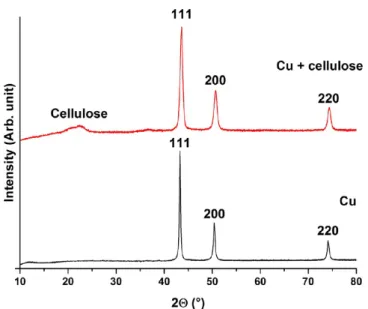
2.1. Investigations of the Cu-NFC Film.The results of X- ray diffraction (XRD) studies are presented inFigure 2. These
studies revealed the 111, 200, and 220 reflections of metallic Cu,25and the reflection with the full width at half-maximum of 2θ= 22°can be assigned to NFC.26The results indicate that the surface of NFC was functionalized with metallic Cu. Even though Fehling’s reaction is known to result in Cu2O, our studies relied on the phenomenon discussed in 1915 that applying excess of reductant can result in metallic Cu.27
Representative scanning electron microscopy (SEM) micro- graphs are shown inFigure 1c−e. Spherical structures 1−5μm in diameter (in orange after applying manual false coloring for better visibility) indicate the metallic Cu on the surface of the fibers. SEM studies at higher magnifications (Figure 1e) showed that many particles are partially embedded into the three-dimensional (3D) structure of thefilters. This facilitates the longer residence time of Cu particles inside the filter by restricting their escape into the drinking water and therefore the overdosage of Cu by the customers. The longer Cu residence time also plays an important role in preserving the activity, therefore promoting the longevity of the as-prepared filters. SEM micrographs also indicated that the microsized Cu spheres are presumably aggregates of nanosized Cu particles;
therefore, we estimated the particle sizes by using the Scherrer equation28on the basis of the X-ray diffractographs. The most intensive reflection (111) was evaluated for the estimations, and they suggest that the average size of the Cu particles is approximately 15 nm. This, combined with the SEM studies, proves that the Cu microspheres are indeed hierarchical structures of Cu nanoparticles organized into spheres. To ensure the reproducibility of the functionalization, the synthesis was repeated without the presence of NFC as well (Figure 1). It is worth noting that the reflections of this sample are narrower than those in the presence of cellulose. We estimated that without the presence of cellulose, the synthesized average Cu
particle diameter is 25 nm, which means approx. 67% of increment compared to that in the previously discussed case.
This particle enlargement shall only be explained by the presence or lack of NFC. The fibers tend to form aggregates (Figure 1e), and the aggregation is facilitated by the increased ion concentration in the reaction mixture.29This aggregation results in temporarily well-defined micro- or nanocompart- ments wherein the in situ Cu nanoparticle synthesis occurs.
Thefibers stand as obstacles, which delay the equilibration of the reactant concentrations by steric hindrance. On the other hand, when no cellulose is present in the reaction mixture, such a steric hindrance does not occur and the synthesis of the nanoparticles only ceases when one of the reactants is completely consumed.
Figure 1d shows that as a result of continuously stirring the reaction mixture, the distribution of the synthesized hierarchical Cu structures is homogeneous on the surface of cellulose;
significant grouping of the microstructures is not visible. These hierarchical structures with dimensions of several micrometers can significantly disrupt the homogeneity of the NFCfilm by their intercalation between NFC aggregates. This phenomenon must be taken into account, as it does not only enhance the permeability of the membrane but it can seriously influence its stability as well.
N2 adsorption Brunauer−Emmett−Teller (BET) experi- ments were performed to evaluate the specific surface area, which was determined to be 6.0 and 2.3 m2/g for pure and Cu- NFC, respectively. This decreasing tendency is the result of the strongly promoted aggregation of NFC by the higher ion concentration in the reaction mixture,29 as an increased nanoparticle aggregation leads to the decrement of specific surface area.
The WHO suggests that the safe concentration of copper in drinking water is <2 mg/L.30Samples for inductively coupled plasma mass spectrometry (ICP-MS) measurements have been prepared to determine the total dissolved Cu content by filtering deionized water of neutral pH through the membrane under the previously detailed experimental conditions. The results showed that the sum of the concentration of63Cu and
65Cu was 336.4 μg/L; thus, the dissolution of Cu during filtration does not overstep in extent the international guidelines.
By analyzing theζpotential of NFC, Cu-NFC, and MS2, we can conclude whether the efficiency of virus retention can be improved by the functionalization. Fall et al. thoroughly discussed the surface charge and theζpotential of pure NFC at different ionic strengths. At low ionic strengths, where all of our experiments were performed, NFC exhibits a strong negativeζpotential (below −40 mV).31If we compare these results to theζpotential of MS2, measured by Syngouna et al.32 (between −10 and −30 mV), we can conclude that the repellence of MS2 from the surface is significant; therefore, we cannot expect a high virus retention from pure NFC. In contrast to this, Cu nanoparticles have been shown to have less negative ζ potential.33 Our dynamic light scattering (DLS) results showed that theζpotentials of our Cu-NFCfilters are
−18, −24, and −25 mV for pH values of 5.0, 7.5, and 9.0, respectively. This is in agreement with the previously mentioned literary data; as Michen et al. discussed, positive patches (or as in our case, Cu patches of less negative ζ potential) on surfaces with very negativeζpotential can result in a generally less negative bruttoζpotential.11Thesefindings Figure 2.XRD analysis of copper (black) and Cu-NFCfilm (red).
ACS Omega
mean that the electric properties of the surface of our Cu-NFC filter are more favorable for the retention of MS2.
2.2. Virus Filtration Experiments. 2.1.1. Batch Experi- ments. The batch adsorption experiments revealed that pure NFC retained less than one logarithmic reduction value of the virions (Table 1). 1Log reduction value (LRV) represents that the reduction was successful at the extent of 1 magnitude (LRV
= Log(initial value)−Log(final value)).
The results of batch experiments where Cu-NFC was applied as adsorbent showed 0 PFU in all 12 tested plates. This led to the conclusion that Cu-NFC has a virus adsorption property of at least 2Log under these conditions. To determine the maximal virus retention capacity of our material, a higher concentration (103−105 PFU/mL) was also applied in batch experiments. Fifty milligrams of the Cu-NFC could show a virus retention of up to 3Log (99.9%). These experimental results reinforce our conclusions that Cu-NFC exhibits higher
virus retention efficiency. The very low but detectable LRV of pure NFC can come from either steric effects, such as the trapping of MS2 bacteriophages between aggregates, or from a less negative secondary energy minimum in the interaction energy profile, such as the one proposed by Syngouna et al.32 2.1.2. Flow Experiments. The control experiment was performed without applying our filter to check the possible adsorption on the glass insert of the funnel. The results showed no measurable bacteriophage retention. Some of the prototype filters were damaged in the flow experiments due to the negative differential pressure applied to enhance thefiltration. A virus retention of up to 3Log could be achieved at pH = 7.5 and 9.0 and up to 4Log at pH = 5.0 (Figure 3) by thesefilters.
Thefilters containing double amounts of each material (DA filters) gave more promising results, as they were also significantly more stable and could sustain their coherence against the applied negative differential pressure. The DAfilters showed a virus retention of at least 5 magnitudes at all three pH values, which refers to an efficiency of at least 99.999% (Figure 4).
Samples of the same composition show significantly distinct antiviral properties in batch and flow experiments. The only difference is in the structure and consistency of the adsorbent;
therefore, the enhancement must derive from the scaffold of the membrane. Presumably, water is able to slacken in the 3D structure of thefilters due to their porosity. Thus, the retention time of a certain volume of filtered liquid is longer. In these
“dead-water pockets”, more contact time is available for the inactivation of the viruses. Consequently, this can result in slightly higher local Cu(II) concentration in these microscopic Table 1. Numbers of Plaque Forming Units (PFUs) on MS2-
InfectedEscherichia coliPlates When the Adsorbent Used in the Batch Experiment Was Pure NFCa
sampling time (min)
batch 1 (PFU)
batch 2 (PFU)
batch 3 (PFU)
average (PFU)
5 19 8 18 15
10 19 22 11 17
15 19 21 22 21
20 18 11 37 22
aThe initial PFU concentration was 100 PFU/mL.
Figure 3.Photograph of MS2-infectedE. colicultures after virusfiltration with Cu-NFCfilters.
ACS Omega
volumes, which has proven antiviral effects because of the traces of electrolytically formed hydrogen peroxide21and possibly due to the potential denaturation of capside proteins as well.34The retention of virions can also be supported by battering to the NFC aggregates that provides more time for their inactivation.
As the continuously stirred system does not have an organized structure, the virus retention can only rely on the electric properties of the Cu-NFC surface. We also assume that an even higher virus retention can occur in normal drinking water than in this model system, as phosphate ions (present in the buffer solution) have been discussed to have a negative effect on the antiviral activity of Cu(II).21
3. CONCLUSIONS
In this study, we show that the preparation of an effective virus filter based on a cheap organic material (NFC), coated with a small amount of Cu, was successful, standing as a candidate for a prototype of a further optimized, high-performance POU virus filter. As Cu coating reduced the stability of the filter membranes, half the amount of NFC existed in its pure, nonfunctionalized state in thefilters.
The control studies of batch experiments investigated the impact of stirring on air, the retention by the poly- (tetrafluoroethylene) (PTFE) membrane used for adsorbent elimination, and the adsorption by pure NFC. They showed that except for the pure NFC, which demonstrated a <1Log retention, none of them exhibited any measurable effect on the virus concentration. The retention by pure NFC was caused by its direct interaction with MS2 bacteriophages, yet the nature of this interaction is still not completely clear. In batch
experiments with Cu-NFC, a 3Log virus retention was observed.
Cu-functionalizedfilters of 50 mg of NFC were used in the flow experiments at pH = 5.0, 7.5, and 9.0. Despite their low robustness, these filters exhibited a virus retention of 3 magnitudes at pH = 7.5 and 9.0 and a retention of 4 magnitudes at pH = 5.0. Using DAfilters, the virus retention exceeded 5 magnitudes (LRV > 5) at all three pH values; from these results, we concluded that both the amount of Cu and the amount of NFC play significant roles in the preparation of an efficient filter. These findings are highly complementary to those already present in the scientific literature: not only do they demonstrate the usefulness of cellulose structures in small- virus filtration but with a very high initial virus stock concentration applied in this study, we could represent the removal efficiency based on exact experimental results, not solely probability.
4. EXPERIMENTAL SECTION
All sample preparations and experiments were performed in a sterile environment in the proximity of a propane−butane laboratory torch to avoid contamination and therefore false results. To sterilize them, refractory instruments and sample holders were autoclaved at 121°C (±1°C) for at least 20 min.
Tools that cannot be heat-treated were disinfected with ethanol (technical grade).
4.1. Materials. D-Glucose, bacteriological agar, NaH2PO4· 2H2O, CuSO4·5H2O, K-Na-tartrate, and NaOH were pur- chased from Sigma-Aldrich Chemie GmbH (Switzerland).
CaCl2·2H2O, NaCl and glycerol were purchased from Merck Eurolab AG (Switzerland). Streptomycin, tryptone (Difco Figure 4.Photograph of MS2-infectedE. colicultures after virusfiltration with DA Cu-NFCfilters.
ACS Omega
0123), and NaCl were obtained from AppliChem PanReac (Germany), Becton Dickinson AG (Switzerland), and VWR International (Switzerland), respectively. Elemental Chlorine Free (ECF) fibers from bleached soft-wood-based pulp-fibers (Picea abies and Pinus spp.) were purchased from Stendal (Berlin, Germany). Acetone and ethanol were obtained from Thommen Furler AG (Büren, Switzerland) and Alko Swiss GmbH (Wildegg, Switzerland), respectively. Ultrapure water (later simply referred to as water), purified by a Barnstead NANOPure Diamond device (Thermo Scientific; equipped with an mdi DNSZ capsulefilter with the pore size of 0.1μm), was used for rinsing and as a solvent.
E. coli (Migula 1895) Castellani and Chalmers 1919 (DSM No.: 5695) colonies were used as hosts for MS2 bacteriophage propagation (DSM No.: 13767). Dry E. coli pellets and the MS2 phage suspension were purchased from DSMZ (Braunschweig, Germany).
4.2. Media Preparation and Bacteria Growth. The necessary media for the growth of bacteria, MS2 propagation, and filtration (i.e., CaCl2, antibiotic solution, broth, virus dilution buffer, and hard and soft agar) were prepared as proposed by Pecson et al.22
The bacteriophages were propagated inE. coli, then purified and concentrated in different steps, according to the protocol provided by DSMZ. The as-received bacteria pellets were rehydrated by 0.5 mL of the broth for 30 min, after which they were transferred into 5 mL of broth. TheE. coliprovided by DSMZ is noncolonial; therefore, a stock was grown from a single bacterium to reach a colony as uniform as possible.
Hundred microliters of the suspended pellet was pipetted onto a hard agar plate and streaked, as shown inFigure 5, by aflame- sterilized inoculation loop. The streaking was performed after a single immersion of the inoculation loop into the pellet suspension.
The inoculated plate was incubated at 37°C for 24 h. After the incubation, several white dots appeared on the hard agar plate, each one representing one uniform colony. One of the colonies was separated and transferred into 25 mL of broth.
The suspension was left at 37 °C to grow, until its optical density measured at 600 nm by spectrophotometry (OD600nm) reached the value of 0.2. Afterward, a sterile glycerol solution (containing 60 vol % glycerol and 40 vol % ultrapure water) was added to reach thefinal glycerol concentration of 9 vol %, to improve the survival rate of bacteria at freezing conditions.
This suspension was then divided into 100μL aliquots. The as- prepared bacterial stocks were stored at−80°C for a maximum of 6 months. After a frozen bacterial colony was thawed and kept at 37°C for up to 10 min, the same procedure was applied for the recultivation.
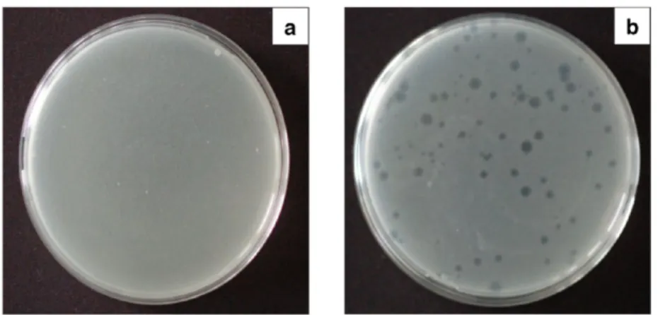
4.3. Determination of MS2 Concentration. As MS2 is inactivated easily, it is required to regularly determine the suspension concentration in PFU/mL (PFU stands for plaque forming units). A dilution series of the original stock was prepared for each determination process (N2, N4, N6, N8, N9, N10, N11, N12, N13, and N14, where Nxrepresents a dilution of 10xtimes of the original stock). Hundred microliters of the diluted virus suspension with 200μL of bacterium suspension (OD600nm= 0.2) was added to 7 mL of the liquid soft agar that was previously kept at 56 °C. After homogenization, the suspension was poured onto a hard agar plate at room temperature and incubated at 37 °C for 24 h. Following the incubation, the amount of active MS2 bacteriophages was determined by the well-known method: counting the trans- parent circles in the opaque white bacterium layer, caused by the phage infection. Each circle represented one PFU of the bacteriophage (Figure 6). The concentration of the original MS2 stock (5×10⁶PFU/mL) was calculated from the number of the infection zones considering the used volumes and concentrations.
4.4. Functionalization of NFC. To prepare the NFC suspension of approximately 1.5 wt %, 2% ECF aqueous suspension was processed by a “Supermass Colloider” (MKZA10-20J CE) equipped with nonporous grinding stones rotating at the nominal velocity of approximately 1500 rpm (at the motor load of 15 kW). The instrument was purchased from Masuko Sangyo Co., Ltd. (Kawaguchi/Saitama, Japan).
Metallic Cu particles were synthesized in situ in the presence of NFC by Fehling’s reaction23by applying an excess reductive agent. NFC suspension (7.500 g, containing approximately 150 mg NFC) was diluted with 30 mL of deionized water; then, 0.456 g of CuSO4·5H2O, 1.380 g of K-Na-tartrate, and 4.200 g of NaOH were added to the suspension. This suspension was stirred at 350 rpm for 30 min by a magnetic stirrer, followed by adding of 0.5 mL of formaldehyde (aqueous solution with the concentration of 37 wt %). The temperature was set to 60°C, and the mixture was stirred for another 60 min. The synthesis of Cu particles became visible as the previously blue mixture turned deep reddish brown. The as-prepared sample was centrifuged at 4000 rpm for 1 min; then, the supernatant was removed and water was added to the sediment. This step was introduced to reach neutral pH that is important to prevent the degradation of cellulose chains in a strongly basic environment.
After the redispersion of the sediment, the previously described washing process was repeated twice and the sediment was resuspended in water, eventually. The as-prepared material is referred to as Cu-NFC in further discussions.
4.5. Preparation of the Nanopaper-Based Filter.Pure cellulose nanopapers were prepared by the “paper-making” method as follows. NFC suspension (6.67 g, containing approximately 100 mg of NFC) was diluted to 30−40 mL by adding water, and it was subsequently filtered through a Durapore poly(vinylidene difluoride) membrane with the uniform pore size of 0.1 μm. The filtration process was enhanced by a Divac 1.2 L vacuum pump. The membrane with thefilter cake was dried on air after eliminating as much water as possible. The resulting white, nontransparent, flexible cellulose nanopaper spontaneously detached from the filter Figure 5.Schematic drawing of the streaked lines on a hard agar plate.
ACS Omega
membrane. In preliminary experiments, these pure cellulose nanopaper membranes (Figure 1a) showed relatively low water permeability because cellulose fibers adsorb water and swell, reducing the size of the pores accessible for the liquid. Studies have shown that the permeability of the nanopaper can be regulated by using different solvents.17To avoid losing the size- exclusion effect during the filtration, a method for controlling the pore size distribution in NFC papers has been developed and proven in a previous study.24
Nanopapers prepared by the above-described method of Cu- modified NFC were conveniently permeable for waterfiltration.
However, they were significantly more fragile than the membranes made of pure NFC. To achieve a sufficient permeability and enhance the mechanical properties of thefinal filter, the 50−50 vol % mixture of modified and unmodified NFC was used for fabrication.
The experimental setup was planned for membranes with 25 nm diameter, containing 50−100 mg of NFC in total. The mixture of modified and unmodified NFC was suspended in water; then, the water was removed by vacuum filtration.
Afterward, the wet filter was washed with either ethanol or acetone with the use of vacuumfiltration. After two repetition cycles, water was considered to be eliminated from the filter cake, which was dried on air for 10−15 min afterward. When ethanol was used as a washing agent, the resulting permeability of the filter material was insufficient, whereas in the case of acetone, the permeability improved. By using this method, a cellulose nanopaper containing 50 mg of cellulose (25 mg of Cu-NFC) was prepared (Figure 1b) with sufficient permeability to performfiltration experiments. Not only the solvent but the amount of Cu-modified NFC also affected the stability and permeability of thefilters. The samefilters were prepared with 12.5 and 37.5 mg of Cu-NFC as well; the former prototype had very low permeability, which would make its use unfavorable, whereas the latter one was too brittle and broke during the filtration. On the basis of these factors, we decided to conduct the further virus filtration experiments with the 50 mg of cellulosefilter, with 25 mg of Cu-NFC.
To determine the potential influence of the thickness of the nanopaper, the quantity of all of the materials used for filter preparation was doubled in subsequent experiments. Thefilters prepared in such a way are referred to as “DA filters” (DA stands for double amount) in further discussions.
4.6. Virus Adsorption Experiments.4.6.1. Batch Experi- ments.Batch experiments were performed in a 250 mL glass beaker with continuous stirring (300 rpm by a magnetic stirrer) at pH = 7.5. Two hundred milliliters of the virus dilution buffer
and 50 mg of the adsorbent (either pure or Cu-NFC) was added to the beaker and stirred for 5 min. Next, 20μL of the original virus stock was pipetted into the mixture (leading to the concentration of 500 PFU/mL) and the beaker was covered with Parafilm M. From this point, samples (1 mL) were collected every 5 min to determine the concentration of active MS2 bacteriophages. Each batch experiment was performed for 20 min. The collected samples werefiltered through Rotilabo PTFE syringefilters of 25 mm diameter, with the uniform pore size of 5.0μm, to remove adsorbents from the suspension; thus, we could prevent further virus adsorption after sample collection. We confirmed experimentally that neither this filtration step (by possible adsorption on PTFE membrane) nor the exposure to air (causing the inactivation of bacteriophages) influences the virus concentration to an extent that is higher than its observable biostatistical fluctuation.
Following thefiltration, 100μL of the purified infected liquid and 200 μL of bacterium suspension were transferred into approximately 7 mL of soft agar, previously kept at 56°C; then, the active virus concentration was determined by the previously described method. Every experiment was performed three times to prove reproducibility.
So as to determine the maximal logarithmic removal rate of thefilter materials, the batch experiments were repeated with the increased starting concentration of MS2 of 1014PFU/mL, which was 107times more concentrated than that of Metreveli et al.18 This allowed the experimental determination of LRV values at the maximal possible precision allowed by our samples of biological nature.
4.6.2. Flow Experiments. In flow experiments, the as- prepared filter was placed into a 30 mL glass funnel with a porous glass insert at the bottom. Vacuum suction was applied to enhance theflux of thefiltered liquid. A control experiment was also performed without afilter to investigate the influence of the equipment on the retention of the bacteriophages, given that the system is porous. Thirty milliliters of the virus dilution buffer was infected with the calculated volume of bacteriophage stock to reach the active phage concentration of 105PFU/mL.
This allowed the investigation of retention of up to 5 magnitudes (5Log). Separate experiments were performed both with membranes containing 25 mg of pure NFC and 25 mg of Cu-NFC and with the DA filters. One milliliter of the filtered liquid was collected after each filtration and used to infectE. colifor virus enumeration.
Theflow experiments were performed at three different pH values: 5.0, 7.5, and 9.0, respectively, to simulate real-life situations.
Figure 6.Uninfected and thus a continuous layer ofE. coli(a) and an infected culture counting 95 PFUs (b).
ACS Omega
4.7. Characterization Methods.The crystalline structure of the membranes was studied by X-ray diffraction (XRD) by a PANalytical X’Pert Pro MPD machine operating with Cu Kα(λ
= 1.5405 Å) radiation. The X-ray scanning was performed over a 2θrange of 10−80°, with a step size of 0.0167°.
Scanning electron microscopy (SEM) studies were per- formed in a FEI Nova NanoSEM 230 that operated in the 10− 15 kV range, after the samples were attached to a conductive carbon tape and sputtered by Leica EM ACE 200 sputter coater with a 5 nm thick platinum layer.
The specific surface area of the samples was determined by single-point BET measurements (Coulter SA3100) carried out at 77 K. The samples were pretreated at 300°C for 15 min in heliumflow (50 cm3/min).
Inductively coupled plasma mass spectrometry (ICP-MS) measurements were performed in an Agilent 7500 ce ICP-MS device to determine the amount of Cu dissolved during filtration.
ζ Potential values were defined by using laser Doppler electrophoresis (DLS, Zetasizer Nano S, Malvern Instruments) and solid surface ζ potentiometry (Anton-Paar SurPASS Electrokinetic Analyzer).
■
AUTHOR INFORMATION Corresponding Author*E-mail: hernadi@chem.u-szeged.hu.
ORCID
Klára Hernádi:0000-0001-9419-689X Present Addresses
∇School of Analytical Sciences Adlershof, Humboldt-Universi- tat zu Berlin, Albert-Einstein-Straße 5-11, Berlin 12489,̈ Germany (G.P.Sz.).
#Department of Chemistry, Humboldt-Universität zu Berlin, Brook-Taylor-Straße 2, Berlin 12489, Germany (G.P.Sz.).
Notes
The authors declare no competingfinancial interest.
■
ACKNOWLEDGMENTSThis work was supported by Switzerland through the Swiss Contribution to the enlarged European Union within the project“Novel nanocompositefilter media for adsorption-based water treatment-NANOSORP”, PSRP: 209/2010. Zoltan Nemeth is thankful for the financial support from the Sciex Programme (project No. 14.119). We would like to thank the Laboratory of Applied Wood Material (EMPA) for providing the prepared NFC gel.
■
(1) WHO and UNICEF Progress on Drinking Water and Sanitation.REFERENCES In Hydrologie Und Wasserbewirtschaftung; WHO, 2014; Vol. 58, pp 244−245.(2) Michen, B. Virus Removal in Ceramic Depth Filters: The Electrostatic Enhanced Adsorption Approach. Ph.D. Thesis, Techni- schen Universität Bergakademie, Freiberg, 2010.
(3) Huang, H.; Young, T. A.; Schwab, K. J.; Jacangelo, J. G.
Mechanisms of virus removal from secondary wastewater effluent by low pressure membrane filtration.J. Membr. Sci.2012,409−410, 1−8.
(4) Viessman, W.; Hammer, M. J.; Perez, E. M.; Chadik, P. A.
Introduction. InWater Supply and Pollution Control, 8th ed.; Addison- Wesley Longman Publishers: Menlo Park, 2008; Chapter 1, pp 1−41.
(5) Strathmann, H. Membrane separation processes: current relevance and future opportunities.AIChE J.2001,47, 1077−1087.
(6) Peltier, S.; Cotte, E.; Gatel, D.; Herremans, L.; Cavard, J.
Nanofiltration: improvements of water quality in a large distribution system.Water Supply2003,3, 193−200.
(7) Ahmad, A. L.; Ooi, B. S.; Mohammad, A. W.; Choudhury, J. P.
Development of a highly hydrophobic nanofiltration membrane for desalination and water treatment.Desalination2004,168, 215−221.
(8) Qin, J.-J.; Oo, M. H.; Kekre, K. A. Nanofiltration for recovering wastewater from a specific dyeing facility.Sep. Purif. Technol.2007,56, 199−203.
(9) Bartels, J.; Souza, M. N.; Schaper, A.; Árki, P.; Kroll, S.; Rezwan, K. Amino-Functionalized Ceramic Capillary Membranes for Con- trolled Virus Retention.Environ. Sci. Technol.2016,50, 1973−1981.
(10) Liu, L.; Bai, H.; Sun, D. D.; et al. Concurrent filtration and solar photocatalytic disinfection/degradation using high-performance Ag/
TiO2 nanofiber membrane.Water Res.2012,46, 1102−1112.
(11) Michen, B.; Fritsch, J.; Aneziris, C.; Graule, T. Improved virus removal in ceramic depth filters modified with MgO. Environ. Sci.
Technol.2013,47, 1526−1533.
(12) Michen, B.; Graule, T. Isoelectric Points of Viruses. J. Appl.
Microbiol.2010,109, 388−397.
(13) Altintas, Z.; Gittens, M.; Pocock, J.; Tothill, I. E. Biosensors for waterborne viruses: Detection and removal.Biochimie2015,115, 144−
157.
(14) Lavoine, N.; Desloges, I.; Dufresne, A.; Bras, J. Microfibrillated cellulose - its barrier properties and applications in cellulosic materials:
a review.Carbohydr. Polym.2012,90, 735−64.
(15) Siró, I.; Plackett, D. Microfibrillated cellulose and new nanocomposite materials: a review.Cellulose2010,17, 459−494.
(16) Henriksson, M.; Berglund, L. A.; Isaksson, P.; Lindstrom, T.;
Nishino, T. Cellulose nanopaper structures of high toughness.
Biomacromolecules2008,9, 1579−1585.
(17) Sehaqui, H.; Zhou, Q.; Ikkala, O.; Berglund, L. A. Strong and Tough Cellulose Nanopaper with High Specific Surface Area and Porosity.Biomacromolecules2011,12, 3638−3644.
(18) Metreveli, G.; Wågberg, L.; Emmoth, E.; Belák, S.; Strømme, M.; Mihranyan, A. A Size-Exclusion Nanocellulose Filter Paper for Virus Removal.Adv. Healthcare Mater.2014,3, 1546−1550.
(19) Huang, W.; Wang, Y.; Chen, C.; Law, J. L. M.; Houghton, M.;
Chen, L. Fabrication of flexible self-standing all-cellulose nanofibrous composite membranes for virus removal.Carbohydr. Polym.2016,143, 9−17.
(20) Gustafsson, S.; Mihranyan, A. Strategies for Tailoring the Pore- Size Distribution of Virus Retention Filter Papers.ACS Appl. Mater.
Interfaces2016,8, 13759−13767.
(21) Thurman, R. B.; Gerba, C. P.; Bitton, G. The molecular mechanisms of copper and silver ion disinfection of bacteria and viruses.Crit. Rev. Environ. Control1989,18, 295−315.
(22) Pecson, B. M.; Martin, L. V.; Kohn, T. Quantitative PCR for Determining the Infectivity of Bacteriophage MS2 upon Inactivation by Heat, UV-B Radiation, and Singlet Oxygen: Advantages and Limitations of an Enzymatic Treatment To Reduce False-Positive Results.Appl. Environ. Microbiol.2009,75, 5544−5554.
(23) Reddy, K. R.; Sin, B. C.; Yoo, C. H.; Park, W. J.; Ryu, K. S.; Lee, J. S.; Sohn, D. W.; Lee, Y. I. A new one-step synthesis method for coating multi-walled carbon nanotubes with cuprous oxide nano- particles.Scr. Mater.2008,58, 1010−1013.
(24) Szekeres, G. P.; Nemeth, Z.; Schrantz, K.; Hernadi, K.; Graule, T. Insights into pore size control in cellulose nanopapers through modeling and experiments.J. Nanosci. Nanotechnol.2018, 3000−3005.
(25) Raffi, M.; Mehrwan, S.; Bhatti, T. M.; Akhter, J. I.; Hameed, A.;
Yawar, W.; ul Hasan, M. M. Investigations into the antibacterial behavior of copper nanoparticles against Escherichia coli. Ann.
Microbiol.2010,60, 75−80.
(26) An, X.; Wen, Y.; Almujil, A.; Cheng, D.; Li, J.; Jia, X.; Zou, J.; Ni, Y. Nano-fibrillated cellulose (NFC) as versatile carriers of TiO2 nanoparticles (TNPs) for photocatalytic hydrogen generation. RSC Adv.2016,6, 89457−89466.
ACS Omega
(27) Cramer, W. On the cause and significance of an abnormal reaction obtained in testing urine for sugar with Fehling’s solution.
Biochem. J.1915,9, 71−77.
(28) Scherrer, P. Bestimmung der Grösse und der inneren Struktur von Kolloidteilchen mittels Röntgenstrahlens. Kolloidchemie Ein Lehrbuch1912, 387−409.
(29) Qi, W.; Xu, H.-N.; Zhang, L. The aggregation behavior of cellulose micro/nanoparticles in aqueous media.RSC Adv. 2015, 5, 8770−8777.
(30) Organization, W. H. Copper in drinking water. http://www.
who.int/water_sanitation_health/dwq/chemicals/copper.pdf.
(31) Fall, A. B.; Lindstrom, S. B.; Sundman, O.; Odberg, L.; Wagberg, L. Colloidal stability of aqueous nanofibrillated cellulose dispersions.
Langmuir2011,27, 11332−11338.
(32) Syngouna, V. I.; Chrysikopoulos, C. V. Interaction between viruses and clays in static and dynamic batch systems.Environ. Sci.
Technol.2010,44, 4539−4544.
(33) Paszkiewicz, M.; Gołąbiewska, A.; Rajski,Ł.; Kowal, E.; Sajdak, A.; Zaleska-Medynska, A. Synthesis and Characterization of Mono- metallic (Ag, Cu) and Bimetallic Ag-Cu Particles for Antibacterial and Antifungal Applications.J. Nanomater.2016,2016, 1−11.
(34) Karlsson, H. L.; Cronholm, P.; Hedberg, Y.; Tornberg, M.; De Battice, L.; Svedhem, S.; Wallinder, I. O. Cell membrane damage and protein interaction induced by copper containing nanoparticles- Importance of the metal release process.Toxicology2013,313, 59−69.
ACS Omega