Chapter 6
Enzymes of the Tryptophan -* Nicotinic Acid Pathway
FRANCESCO M . CHIANCONE Medical Department Lepetit S.pA.
Milan, Italy
I. Introduction 249 II. Enzymatic Activities and Their Variations under Different Experimental
Conditions 252 A. Tryptophan Pyrrolase 252
B. Formylase 256 C. Kynurenine Hydroxylase 257
D. Kynurenine Transaminase 257 E. Kynureninase and 3-Hydroxykynureninase 258
F. 3-Hydroxyanthranilic Oxidase 259 G. Picolinic Carboxylase 260 III. Relationship between Tryptophan —> Nicotinic Acid Pathway and
Lipid Metabolism 260 IV. Significance of the Variations in Enzymatic Activities 261
V. General Considerations 267
VI. Summary 269 VII. Analytical Methods 270
A. Tryptophan Pyrrolase 270
B. Formylase 273 C. Kynurenine Hydroxylase 273
D. Kynurenine Transaminase 275 E. Kynureninase and 3-Hydroxykynureninase 277
F. 3-Hydroxyanthranilic Oxidase 279
G. Picolinic Carboxylase 281
References 282 I . INTRODUCTION
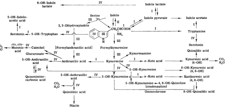
In a recently published symposium on tryptophan metabolism held in Japan (1), a thorough review of the available knowledge and concepts in this field was presented and discussed in depth. In addition to the historical aspects of the subject, the proceedings included sessions on tryptophan pyrrolase, tryptophanase, kynurenine, kynurenic acid, and xanthurenic acid, nicotinic acid, picolinic acid, and anthranilic acid; and a survey of tryptophan metabolism. The evidence for the known meta- bolic pathways in man and other mammals is summarized graphically in Fig. 1.
249
O Μ. CHIANCONE
2
οFIG. 1. Metabolism of tryptophan: I. change of side chain (transaminase, decarboxylase, etc.); II. indole nucleus split off (tryptophanase); III. change of pyrrole nucleus (tryptophan pyrrolase, kynureninase, etc.); IV. change of benzene nucleus (hydroxylase, etc.). From Ichihara (1).
6. ENZYMES OF TRYPTOPHAN —» NICOTINIC ACID PATHWAY 251 In this chapter, because of limitations of space, we shall concern ourselves primarily with the elucidation of various steps in the enzymatic transformation of tryptophan to nicotinic acid (Pathway III, Fig. 1) and a description of the methods by which these metabolic changes
(INDOLES)- TRYPTOPHAN• - (5-OH-DERTVATIVES)
— Tryptophan pyrrolase
FORMYLKYNURENINE
KYNURENIC ACID
Kynurenine transaminase
•
- Formylase
KYNURENINE
Kynureninase
t . ANTHRANILIC ACID
XANTHURENIC ACID
Kynurenine transaminase +
- Kynurenine hydroxylase
3-OH-KYNURENINE
- 3-OH-Kynureninase 3-OH-ANTHRANILIC ACID
3-OH-Anthranilic oxidase-
QUINOLINIC ACID- (INTERMEDIATE)
Picolinic carboxylase
t PICOLINIC ACff)
NICOTINIC ACID (AND DERIVATIVES)
DPN TPN
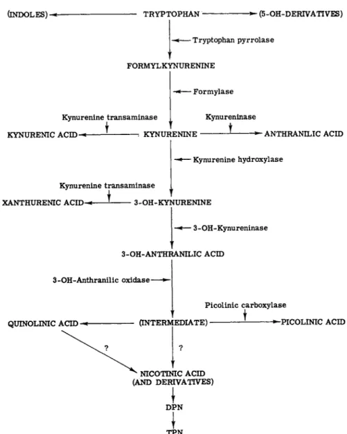
FIG. 2. A simplified scheme of the tryptophan metabolism (via kynurenine). The evidence that 3-OH-kynureninase is distinct from kynureninase is not clear. Recent observations indicate that nicotinic acid is derived from quinolinic acid and not from an intermediate.
may be followed under some physiological and pathological conditions.
Research on the urinary metabolites of the tryptophan -» nicotinic acid pathway has led to numerous investigations in the past decade to determine, principally in the liver, the enzymatic activities in
volved in this metabolic pathway under various physiological and pathological conditions. Directly or indirectly, many of these ob
servations concern problems of nutritional biochemistry. Some of the results are also of interest because, in certain abnormal conditions, e.g., pyridoxine deficiency and experimental diabetes, it is possible to com
pare the enzymatic activity pattern in terms of the tryptophan metab
olites of the urine. Furthermore, knowledge of the data obtained on laboratory animals may serve as a guide for researches to be carried out in humans.
The enzymatic systems which will be considered here are shown in Fig. 2, where the compound on which each of these enzymes operates, as well as the metabolic step which is thereby regulated, and the resultant metabolite are indicated in a simplified form. Discussion of the en
zymes of the tryptophan —» nicotinic acid pathway will be limited to those amenable to measurement by our procedures. The analytical pro
cedures employed permit an evaluation of the enzymatic activity, and not of the enzyme directly—the term "enzymatic activity" being gen
erally used here synonymously with "enzyme."
I I . ENZYMATIC ACTIVITIES AND THEIR VARIATIONS UNDER DIFFERENT EXPERIMENTAL CONDITIONS
A. Tryptophan Pyrrolase (TPy)
Tryptophan pyrrolase (formerly known as tryptophan peroxidase- oxidase—TPO) is the first enzyme operating on the metabolic pathway with which we are concerned. It governs the tryptophan —> formylkyn- urenine conversion (Fig. 2 ) . TPy is an iron porphyrin protein (la) pres
ent in the liver of rats ( 2 ) , mice, guinea pigs, rabbits ( 3 ) , and frogs ( 4 ) , in Drosophila melanogaster ( 5 ) , and in Pseudomonas (la).
It is an adaptive enzyme (la). Administration of tryptophan (sub
strate-induced enzyme formation) or of cortisone (hormone-induced en
zyme formation) causes an increase in tryptophan pyrrolase activity in rat liver. The mechanisms and modes of induction were recently studied by Greengard and Feigelson (6) by means of an immunologic technique.
These direct measurements demonstrated for the first time an increase in the amount of a mammalian enzymatic protein after administration of its substrate. These authors advanced the hypothesis that the in vivo
6. ENZYMES OF TRYPTOPHAN —» NICOTINIC ACID PATHWAY 253 saturation of apotryptophan pyrrolase with heme, mediated by trypto
phan, stimulates synthesis of the apotryptophan pyrrolase molecule.
The variations of TPy activity of the liver with respect to species differences of normal animals, as well as those observed under the in
fluence of tryptophan and cortisone, are shown in Table I (3). In the
T A B L E I
TRYPTOPHAN PYRROLASE IN DIFFERENT ANIMAL SPECIES AND UNDER THE INFLUENCE OF INDUCING AGENTS0
Animal species
Inducing agent6
Animal species — Tryptophan Cortisone
Rat 3.20 48.70 9.30
Mouse 9.76 13.00 16.58
Guinea pig 1.15 2.05 9.93
Rabbit 1.20 — —
β Adapted from Canal and Maffei Faccioli (3).
6 All values in /mioles of kynurenine/gm liver fresh weight /hr.
liver of male Rana pipiens after hibernation, TPy is increased 40- to 50-fold after tryptophan administration, but remains unchanged after cortisone (4). This represents the first example of a lack of hormonal induction.
The maturation of the enzyme also differs in the various animal species. In fetal life, TPy (Table II) is never present in the liver of rats or guinea pigs and rabbits. The same is true even after admin-
T A B L E II
TRYPTOPHAN PYRROLASE IN RELATION TO AGE IN DIFFERENT ANIMAL SPECIES
Animal species
Days from birth Rat Guinea pig Rabbit - 5 Absent Absent" Absent"
1 Absent Present6 Present6
12 Absent — —
15 Present — —
38 Increasing — —
120 Increasing — —
400 Stationary — —
800 Stationary — —
° Even after administration of tryptophan or of adrenocortical extract both to the fetus and to the mother at the end of pregnancy.
6 At levels equal to those of the adult animal.
istration of tryptophan or adrenocortical extract to the fetus or the pregnant mother (7). In the first 24 hours after birth it appears in the liver of guinea pigs and rabbits (9); in rats at the end of the second week of extrauterine life it has already reached levels equal to those of adult animals (8). Substrate and hormone induction also appear in the rabbit parallel to the maturation of "spontaneous" activity, which continues to increase after the first few weeks of life, and for some months.
The onset of TPy activity in the liver of guinea pigs and rabbits during the first 24 hours after birth may be taken as proof that in these animals there are mechanisms operating on the fetus through the placenta or the maternal blood (9). The speed at which this metabolic activity appears in the liver during development is noticeable; the ac
tivity involves adaptive mechanisms which remain to be elucidated.
Although it is unknown why this enzymatic activity is established much later in the rat than in the rabbit or the guinea pig, or how the biogenesis of nicotinic acid takes place in this animal during the first few weeks of extrauterine life, this phenomenon may be associated with the known fact that the rat does not need nicotinic acid or its derivatives for normal growth (9a).
After observing the absence of TPy activity in the liver homogenate and supernatant fluid obtained from day-old rats, our group demon
strated that the microsomes isolated from the same liver will activate tryptophan pyrrolase in adult rat liver homogenate to a higher degree than the microsomes of adult rat liver itself (10, 11). TPy is increased in the liver of female rats around the twentieth day of pregnancy (12), but no differences were observed between adult and old rats (13).
Many different chemical compounds, irrespective of their structure and biological activity, are capable of increasing liver TPy, but only in the intact animal. These data are summarized in our recent review on TPy, which also records the action of physical agents and carcinogenic substances (14). It is interesting to note here the influence of carbo
hydrates and certain amino acids and their derivatives. In addition to tryptophan, there are inducing and noninducing amino acids (Table III). The same holds true for carbohydrates.
Diets having an unbalanced protein, lipid, or carbohydrate content decrease TPy in the liver, as does a complete and balanced diet in which the protein content is exclusively of vegetable origin (Table IV).
Only a diet rich in animal protein, with its greater supply of tryptophan, causes an increase in TPy (14). TPy is increased on a diet deficient in pantothenic acid, while it remains unchanged in rats maintained on diets deficient in biotin and thiamine (14). Ginoulhiac (15) showed that
6. ENZYMES OF TRYPTOPHAN-» NICOTINIC ACID PATHWAY 255
T A B L E I I I
TRYPTOPHAN PYRROLASE: INFLUENCE OF AMINO ACIDS, CARBOHYDRATES, AND OTHER COMPOUNDS ON THE ENZYMATIC ACTIVITY OF R A T LIVER
Compounds
Enzymatic activity
Increased Unchanged
Amino acids
Carbohydrates
Other compounds
Histidine0
Leucine Tyrosine Tryptophan6
Phenylalanine Glucose Fructose Anthranilate Kynurenine Histamine Serotonin
DL-Alanine Lysine Glutamic acid
Sucrose
α Unchanged in adrenalectomized animals.
6 Increased even in adrenalectomized or hypophysectomized animals.
no decrease in TPy occurs in thiamine deficiency, thereby demonstrating that this vitamin does not take part in the conversion of tryptophan to formylkynurenine, as Dalgliesh (16) had thought. Ginoulhiac's findings were confirmed by other workers (17-19).
TPy activity remains unchanged in the liver of fasting rats, but
T A B L E IV
TRYPTOPHAN PYRROLASE: INFLUENCE OF D I E T Enzymatic
Diet activity Comments High animal protein Increased
Low animal protein Decreased With vegetable protein Decreased
High fat Decreased
High carbohydrate Decreased Thiamine deficiency Unchanged Pyridoxine deficiency Unchanged Biotin deficiency Unchanged Pantothenic acid deficiency Increased Water (fasting) Unchanged
Normalizes with restoration of standard diet Normalizes after cortisone i.p.°
Influences the response to S T H6
Influences the response to S T H
Does not affect the response to tryptophan or cortisone derivatives
Lowered after tryptophan load
Does not affect the response to tryptophan Does not affect the response to tryptophan
or A C T H
a Intraperitoneal.
6 Somatotropic hormone.
when the animals are starved it increases (20). In the latter case the increase is found in the supernatant fraction, and the relevant micro
somes exert a lower activating effect than those obtained from non- fasting rats (21). Just before the starved animals were sacrificed, extremely high TPy values were found in the liver, suggesting a sudden breakdown of balance which was manifested by a rapid increase in enzyme activity.
Studies on the relationship of endocrine glands to this enzyme showed a decrease in TPy in rats after adrenalectomy (22) and thy
roidectomy (23), but an increase after pancreatectomy (24). TPy remained unchanged after hypophysectomy (25). All the hormones studies to date [ACTH (adrenocorticotropic hormone), STH (somato
tropic hormone), epinephrine, thyroxine] increase TPy in the liver of intact rats, but not in that of adrenalectomized animals. In these, only insulin, in addition to tryptophan and cortisone, induces an increase of the enzymatic activity. However, in the hypophysectomized or adrenal
ectomized rat, the TPy response is inferior to that observed in the in
tact animal. It is well known that the increase in TPy with cortisone is common to all the glucocorticoids, and that it is possible to utilize the determination of TPy in the liver as a biological activity test for these compounds (26-28).
TPy remained constant in certain experimental toxic hepatopathies, such as those induced by the endotoxin of Salmonella typhi murium, by Amanita phalloides, and by CC14 (29), as well as in the regenerating liver after subtotal hepatectomy in the rat (30). In experimental dia
betes, we noted an increase of TPy in the liver of diabetic rats 6 months after pancreatectomy. In the alloxanized rat, TPy is increased, accord
ing to some investigators (31, 32), and unchanged according to others (33) who, however, found it increased in the phase of acidosis.
B. Formylase
This enzyme is responsible for the formylkynurenine -> kynurenine conversion, and it acts on various formamide-aromatic substrates (Fig.
2). It is far more active on formylkynurenine than on other molecules (34). Formylase has been found in mammalian liver, kidney, spleen, and intestine (34). Its activity in rat liver exceeds that of tryptophan pyrro
lase 600-fold, and, for this reason, the two activities are combined in current determinations.
In fact tryptophan pyrrolase, which should refer to formylkynu- renine, is instead expressed as kynurenine, the metabolite obtained after the enzymatic breakdown of formylkynurenine by formylase. The kynurenine values by which we indicate tryptophan pyrrolase activity
6. ENZYMES OF TRYPTOPHAN - > NICOTINIC ACID PATHWAY 257 thus represent the final result of the action of two enzymes operating in succession: tryptophan pyrrolase and formylase.
C. Kynurenine Hydroxylase
This is one of three enzymes (kynureninase and kynurenine trans- aminase being the other two) concerned with the metabolism of kynu- renine. It governs the kynurenine —»3-hydroxykynurenine conversion.
Kynurenine hydroxylase has been found in the mitochondria of rat and cat kidney and liver (35), and it has been partially purified from the mitochondria of rat liver (36). It acts in the presence of TPNH (reduced triphosphopyridine nucleotide) and 02 (36) or with TPN (triphospho- pyridine nucleotide) and citrate, to form 3-hydroxykynurenine (37).
Our study of its variations in the liver and kidney of hypophysec- tomized or adrenalectomized rats showed an increase in the liver and kidney of hypophysectomized animals, but only in the liver after adrenal- ectomy (38-40).
Kynurenine hydroxylase remains unchanged in the liver of diabetic rats (Table V) 6 months after pancreatectomy (24). This activity decreases by 50 to 70% in the mitochondria of the liver of riboflavin- deficient rats, even after the in vitro addition of riboflavin phosphate or
T A B L E V
LIVER KYNURENINE HYDROXYLASE, KYNURENINE TRANSAMINASE, AND KYNURENINASE UNDER DIFFERENT EXPERIMENTAL CONDITIONS
Kynurenine Kynurenine
Experimental conditions hydroxylase transaminase Kynureninase Riboflavin deficiency Decreased — —
Pyridoxine deficiency
1. Diet Unchanged Decreased Decreased 2. Penicillamine — Decreased" Decreased Experimental diabetes6 Unchanged Unchanged Unchanged
a Unchanged in mitochondria.
6 After pancreatectomy.
of flavin adenine dinucleotide from boiled liver extract (41). In rats subjected to a pyridoxine-deficient diet (Table V) this enzymatic ac- tivity is unchanged in both the liver and the kidney (42). It appeared unaltered also in the liver of diabetic rats, 6 months after pancre- atectomy (24).
D. Kynurenine Transaminase
This enzyme acts both on kynurenine and 3-hydroxykynurenine, giving rise to two different metabolites: kynurenine kynurenic acid
and 3-hydroxykynurenine -> xanthurenic acid. Its coenzyme is pyridoxal phosphate (43), and it has been found in the kidney and liver of rats (44) and cats (37), both in the mitochondria and in the supernatant fluid. Variations of kynurenine transaminase have been studied using chiefly kynurenine as substrate, and this should be borne in mind when interpreting their significance.
This enzyme appears unchanged after hypophysectomy, adrenal
ectomy, or administration of thyroxine, while it is decreased (Table V) in the liver of pyridoxine-deficient rats (42). In liver mitochondria, this decrease is found only in the deficiency due to diet and not in that induced by penicillamine; in the supernatant fluid, it is much less marked in the deficiency due to the antivitamin (45).
Kynurenine transaminase remained unchanged in the liver of diabetic rats 6 months after pancreatectomy (24).
E. Kynureninase and 3-Hydroxykynureninase
These enzymes give rise to anthranilic acid and to 3-hydroxyan- thranilic acid by acting on kynurenine and 3-hydroxykynurenine, re
spectively. The question has not yet been solved whether kynureninase and 3-hydroxykynureninase are distinct enzymes.
Kynureninase, which is the more investigated of the two, has pyridoxal phosphate as coenzyme (47). It is present in the liver of rats, guinea pigs, oxen, pigs (48), cats (35), humans (49), in the kidney of rats and cats (35), in Pseudomonas fluorescens (50), and in Ν euro- spora crassa (51). It has been partially purified from the supernatant fluid of rat liver homogenate (22), from Pseudomonas fluorescens (52), and from Neurospora crassa (51).
The decrease found by Ginoulhiac and Tenconi (53) in the hypoph
ysectomized rat for kynureninase has recently been observed also for 3-hydroxykynureninase by McCoy and Chung (49), who recorded a 75% variation. Tenconi and Ginoulhiac (39, 42) also found that in the liver of adrenalectomized rats, kynureninase was reduced 5 and 15 days after the operation, and that tryptophan administration failed to affect the result of adrenalectomy. Kynureninase is decreased in the rat even after administration of thyroxine (54).
In pyridoxine deficiency (Table V), the higher the degree of de
ficiency, the greater is the decrease observed in the liver, as was shown by Mason and Berg (55), who added different quantities of pyridoxal phosphate to the in vitro reaction system.
In the diabetic rat liver, 6 months after pancreatectomy, both kynureninase and 3-hydroxykynureninase remained unchanged (24).
6. ENZYMES OF TRYPTOPHAN - » NICOTINIC ACID PATHWAY 259 F. 3-Hydroxyanthranilic Oxidase
Boeri and associates (56-58) have shown that the action of this enzyme results in the formation of an intermediate from 3-hydroxyan- thranilic acid. From this intermediate, which has a maximum absorption
at a wavelength of 360 //,m, nicotinic acid, picolinic acid, and quino- linic acid are derived. This enzyme, which has been partially purified from ox liver (56, 58, 59), acts in the presence of Fe+ + and of SH— groups It is also found in the kidney as well as in the liver of the rat and pig (60) and in the blood of the rat (61). It is of interest to note that 3-hydroxyanthranilic oxidase is almost absent in the liver of rats 5 days before birth. At the moment of birth its value is about one-half that in the adult rat liver, and it matures rapidly in the first hours of extrauterine life. It is not influenced by the administration of cortisone, and it is not increased in the liver of pregnant rats (12).
This enzymatic activity is decreased significantly in rat liver after a tryptophan load (62).
In our laboratory, Tenconi (63) and Ginoulhiac et al. (61) have made parallel determinations of 3-hydroxyanthranilic oxidase in the liver and blood of rats treated with CC14, per os or via the mesenteric vain, in order to obtain lesions differing in onset, extent, and severity.
According to the severity of the lesion, they found that the activity
T A B L E VI
LIVER 3-HYDROXYANTHRANILIC OXIDASE AND PICOLINIC CARBOXYLASE UNDER DIFFERENT EXPERIMENTAL CONDITIONS
Experimental condition
3-Hydroxyanthranilic oxidase
Picolinic carboxylase Pyridoxine deficiency Unchanged Unchanged Alloxan diabetes Unchanged Increased Post-pancreatectomy diabetes Unchanged Increased Phenylhydrazine administration Decreased —
C C U administration Decreased —
decreased in the liver and appeared in the blood serum, often reaching very high levels. Decreased activity is observed in the rat liver after treatment with phenylhydrazine (64), after hypophysectomy (53), or after adrenalectomy (39); in alloxan diabetes (31) and in diabetes fol- lowing pancreatectomy (24) the activity is unchanged (Table VI). It also remains unchanged in the kidney of diabetic rats 6 months after pancreatectomy (24).
G. Picolinic Carboxylase
This enzyme, by its action on the intermediate compound derived from 3-hydroxyanthranilic acid through the action of 3-hydroxyanthra- nilic oxidase, governs the formation of picolinic acid.
It is found in the liver of the rat (65), and it is unchanged after hypophysectomy (53) or adrenalectomy (39). It is increased in the liver of alloxanized or pancreatectomized rats—insulin returns its value to normal (31). In the kidney of pancreatectomized rats, picolinic carboxylase does not present values significantly higher than those of the control (24). Cortisone has no inducing effect in the intact rat, but in the hypophysectomized, adrenalectomized, or alloxanized animal it increases picolinic carboxylase activity which, in the liver of diabetic adrenalectomized animals, attains higher values than in diabetic intact rats (66).
The marked increase in picolinic carboxylase activity in the liver of diabetic rats has been related to the small amounts of nicotinic acid and its metabolites excreted by these animals.
According to Mehler et al. (67), there is a decreased capacity of the diabetic rat to synthesize nicotinic acid, which, however, is not due to a decrease in the essential enzymes which convert 3-hydroxyan
thranilic acid into nicotinic acid. A biochemical lesion of a different type, involving some control mechanism, is probably present.
The same authors have recently demonstrated a decrease in quino- linic acid, which may be a consequence of the increased picolinic carboxylase activity, and have provided a satisfactory interpretation of the control of nicotinic acid biosynthesis. In conclusion, such a control could be exerted by picolinic carboxylase and could be competitive with the spontaneous reaction in which quinolinic acid is formed (67).
I I I . RELATIONSHIP BETWEEN TRYPTOPHAN - » NICOTINIC Aero PATHWAY AND LIPID METABOLISM
Numerous steroids have been shown to have an influence on liver TPy. In addition to the well-known induction due to cortisone and cortisone-like compounds in general, the intravenous administration of deoxycortisone, estradiol, estrone, testosterone, and androsterone is also followed by an increase of liver TPy in the rabbit (68). Furthermore, some data concerning the effects of folliculin on the elimination of xan
thurenic acid in the urine of women during menopause or of ovariecto- mized rats (69) show a relationship between steroids and metabolism of both tryptophan and nicotinic acid.
The study of this relationship has not been further developed, al-
6. ENZYMES OF TRYPTOPHAN - » NICOTINIC ACID PATHWAY 261 though some investigations have been carried out on the influence of nicotinic acid and related compounds on the biosynthesis and metabolism of steroids and fatty acids.
In particular, nicotinic acid produces a decrease in blood cholesterol in man (70). The mechanism of this effect has not yet been elucidated:
Merril has found an increase in the incorporation of labeled acetate into sterols (71), while, according to Perry, the incorporation of acetate both into sterols and fatty acids is inhibited by nicotinic acid (72).
The important investigations carried out by Gaylor et al. (73) show that the incorporation of acetate into sterols is increased, whereas the incorporation into fatty acids is decreased. The same effect is shown by nicotinic amide and isonicotinic acid, which have no blood cholesterol- lowering action in man (74).
Another compound, which is a higher homolog of nicotinic acid, has been shown to lower the cholesterol and fatty acid content of the blood:
3-pyridineacetic acid (75, 76). It is of some interest to point out that this compound practically does not undergo any metabolization, so that it is excreted in the urine almost totally unmodified; furthermore it does not interfere with the urinary excretion of nicotinic acid and its deriva- tives (77). The oxidation in vitro of 26-C1 4-cholesterol by rat liver mito- chondria (78) is increased by this compound, the deposition of lipids from cholesterol in aortic cells grown in vitro is prevented (79), and the incorporation of 1-C14 acetate is modified only by high concentrations
(76). Its action, thus, probably takes place at the level of cholesterol breakdown and excretion and, possibly, also of lipid distribution and metabolic control.
Parallel investigations with both compounds—nicotinic acid and 3- pyridineacetic acid—seem to afford an efficient way of dealing with this problem: the former is metabolized and, as was already supposed, could act through some active metabolite; the latter, instead, is practically not metabolized.
The data which have been summarized here clearly indicate that many important questions, with special reference to those concerning the mode of action, have not yet been answered.
I V . SIGNIFICANCE OF THE VARIATIONS IN ENZYMATIC ACTIVITIES
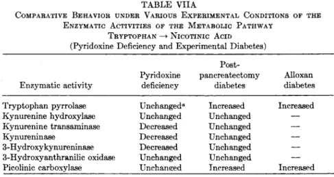
On the basis of the above review, it is possible to determine rather accurately the enzyme patterns occurring in certain physiopathological experimental conditions. Table VIIA presents the patterns observed in pyridoxine deficiency and in experimental diabetes. It is immediately ap- parent that, in these two experimental conditions, distinct variations exist, which appear to be most probably specific. One of the arguments in favor
TABLE VIIA
COMPARATIVE BEHAVIOR UNDER VARIOUS EXPERIMENTAL CONDITIONS OF THE ENZYMATIC ACTIVITIES OF THE METABOLIC PATHWAY
TRYPTOPHAN —> NICOTINIC ACID
(Pyridoxine Deficiency and Experimental Diabetes) Post-
Pyridoxine pancreatectomy Alloxan Enzymatic activity deficiency diabetes diabetes Tryptophan pyrrolase Unchanged" Increased Increased Kynurenine hydroxylase Unchanged Unchanged — Kynurenine transaminase Decreased Unchanged —
Kynureninase Decreased Unchanged —
3-Hydroxykynureninase Decreased Unchanged — 3-Hydroxyanthranilic oxidase Unchanged Unchanged — Picolinic carboxylase Unchanged Increased Increased
a In pyridoxine-deficient animals, tryptophan pyrrolase after tryptophan load is lower than in normally fed animals submitted to the same load.
of the specificity of the aforementioned enzymatic alterations follows from a comparison with the patterns obtained under other experimental conditions. This is shown in Table VIIB, which also records the re
sponses obtained in the intact rat after administration of tryptophan and cortisone.
In parallel investigations on certain enzymatic activities of rat liver and kidney after hypophysectomy or adrenalectomy, we have also noted some differences which reveal the specificity of observed variations. For example, it was found that kynurenine hydroxylase is increased in the
T A B L E V I I B
COMPARATIVE BEHAVIOR UNDER VARIOUS EXPERIMENTAL CONDITIONS OF THE ENZYMATIC ACTIVITIES OF THE METABOLIC PATHWAY
TRYPTOPHAN NICOTINIC ACID
(Under Other Experimental Conditions) Cortisone
Hypophy Adrenal administra Tryptophan Enzymatic activity sectomy ectomy tion load Tryptophan pyrrolase Unchanged" Decreased Increased Increased Kynurenine hydroxylase Increased Increased — Unchanged Kynurenine transaminase Unchanged Unchanged — Unchanged Kynureninase Decreased Decreased — Unchanged 3-Hydroxyanthranilic oxidase Unchanged Unchanged Unchanged Decreased Ficolinic carboxylase Unchanged Unchanged Unchanged Unchanged
" In hypophysectomized animals, tryptophan pyrrolase after tryptophan load is lower than in intact animals submitted to the same load.
6. ENZYMES OF TRYPTOPHAN - > NICOTINIC ACID PATHWAY 263 liver of hypophysectomized or adrenalectomized rats, but that in the former this increment is greater either without, or after, tryptophan load. This enzymatic activity, moreover, is also strongly increased in the kidneys of hypophysectomized rats, while it remains practically unchanged in the kidneys of adrenalectomized animals (38-40). Appar- ently, specific and primary enzymatic activities occur which depend directly upon the experimentally induced pathological conditions.
The patterns of these enzymatic lesions correspond frequently to changes in urinary metabolites. In comparing these two patterns, it is relevant to point out that because methods for determining urinary metabolites were evolved before those for determining enzymatic activi- ties, the two parameters were not studied simultaneously. However, most of the results were obtained by the same group of investigators under basically comparable experimental conditions. It must be emphasized also that the enzymatic activities, studied for the most part in the liver, may represent, at least in certain cases, and mainly if not exclusively, the function of this organ, whereas the variations in the urinary excretion of metabolites express, so to speak, the tryptophan metabolism function of the body as a whole.
On the other hand, the quantitative variations of a metabolite in the urine do not necessarily correspond to like variations in the quantity of a metabolite formed in the body. For purposes of comparison, however, it is generally assumed that decreased (or increased) urinary excretion is an index of a reduced (or increased) formation of the metabolite.
Despite the above qualifications, it is still interesting to compare the variations in enzymatic activity and urinary metabolites because of the suggestions afforded for researches in human pathology. The pat- terns obtained, respectively, in rats fed a pyridoxine-free diet, after a load of 500 mg/kg L-tryptophan per osy and in diabetic rats 6 months after pancreatectomy (80) are shown in Figs. 3 and 4.
The differences between the enzymatic activities and the urinary metabolites are apparent from a comparison of the two patterns. In pyridoxine deficiency, the variations in enzymatic activities and in the corresponding urinary metabolites enable us to note a kynurenine in- crease in the urine. TPy, which regulates the metabolic step from which this metabolite is derived, remains unaltered in the liver of rats not receiving a tryptophan load; it is decreased in normal animals, after an amino acid load, but this is always associated with an elevated kynure- nine formation. Of the enzymatic activities which act on kynurenine, it is kynurenine transaminase and kynureninase which cause a decrease.
An increase in urinary kynurenine, therefore, is due both to an increased production of this substance (after a tryptophan load) and to a reduced
(INDOLES)- - TRYPTOPHAN- - (5-OH-DERIVATIVES)
— Tryptophan pyrrolase FORMYL KYNURENINE
KYNURENIC ACID
Kynurenine transaminase
J
-Formylase
KYNURENINE
Kynureninase
ANTHRANILIC ACID
XANTHURENIC ACID
Kynurenine transaminase t
- Kynurenine hydroxylase
3-OH-KYNURENINE
— 3-OH-Kynureninase
3-OH-ANTHRANILIC ACID 3-OH-Anthranilic oxidase -
QUINOLINIC ACID - (INTERMEDIATE)
Picolinic carboxylase
PICOLINIC ACID
NICOTINIC ACID (AND DERIVATIVES)
DPN ΤΡΝ
— unchanged
— decrease
= i n c r e a s e FIG. 3. A simplified scheme of the tryptophan metabolism (via kynurenine).
Pyridoxine-deficient rat, after tryptophan load.
production of kynurenic acid (kynurenine transaminase activity) and anthranilic acid (kynureninase activity).
As to the transformation of kynurenine to 3-hydroxykynurenine, kynurenine hydroxylase remains unchanged: The excretion of 3-hydroxy
kynurenine increases in relation to a decrease in the 3-hydroxykynure-
6. ENZYMES OF TRYPTOPHAN - » NICOTINIC ACID PATHWAY 265
(INDOLES) - TRYPTOPHAN- -(5-OH-DERIVATIVES)
• Tryptophan pyrrolase FORMYLfcYNURENINE
KYNURENIC ACID
Kynurenine transaminase
-Formylase Kynureninase
KYNURENINE ^ *~ ANTHRANILIC ACID
XANTHURENIC ACID
Kynurenine transaminase
f
• Kynurenine hydroxylase
3-OH-KYNURENINE
— 3 - OH - Kynur eninase
3-OH-ANTHRANILIC ACID ( β )
3-OH-Anthranilic oxidase * H
(
Picolinic carboxylasei
- ^ QUTNOLINIC ACID-* (INTERMEDIATE) 1 ^-PICOLINIC ACID
unchanged decrease
= increase (·) after 3-OH-
anthranilic acid load FIG. 4. A simplified scheme of the tryptophan metabolism (via kynurenine).
Diabetic rat (pancreatectomy).
ninase activity, which governs the conversion of this substance to 3-hydroxyanthranilic acid. The increased urinary elirhination of both 3-hydroxykynurenine and its alternate metabolite, xanthurenic acid, can therefore be explained in terms of a reduced conversion of 3-hydroxy
kynurenine in this path. However, the formation of xanthurenic acid
NICOTINIC ACID (AND DERIVATIVES)
\
DPN TPN
ι
depends on kynurenine transaminase (or, better, 3-hydroxykynurenine transaminase) whose activity is depressed.
Interpretation of this fact remains obscure, especially so since there are no doubts as to the increase of urinary output of xanthurenic acid in pyridoxine deficiency, however induced, in experimental animals and in humans. It is perhaps worthy of mention that the data relative to kynurenine transaminase derive from determinations using kynure
nine and not 3-hydroxykynurenine as substrate. In this light, the hy
pothesis might be advanced that the characteristics of transamination may vary with the substrate. It would be interesting, therefore, to investigate whether the results of experiments of Wiss and Knox on kynureninase would also apply to kynurenine transaminase. Wiss (81) showed that the degradation curves of kynurenine and 3-hydroxykynure
nine follow the same course, but that the curve for the latter is distinctly higher, while Knox (82) observed that DL-3-hydroxykynurenine is broken down by kynureninase more rapidly than DL-kynurenine.
In the patterns related to post-pancreatectomy diabetes, an increase of TPy in the liver and an increase of kynurenine in the urine is noted.
These variations are coincident, moderate, and present only in the ad
vanced phase of this type of diabetes.
Other variations are observed in the metabolic pathway 3-hydroxy- anthranilic acid —»nicotinic acid and its derivatives. The elimination of nicotinic acid and its derivatives is appreciably and rapidly reduced both before and after a tryptophan, nicotinic acid, or 3-hydroxyan- thranilic acid loading. In the latter test the urinary elimination of 3-hydroxyanthranilic acid itself, and of quinolinic acid, is also decreased;
in no case was it possible to reveal, even qualitatively, the presence of picolinic acid. Among the enzymatic activities which operate in these metabolic steps, 3-hydroxyanthranilic oxidase remains constant; and picolinic carboxylase is markedly increased in the liver, but not sig
nificantly so in the kidney. The metabolite which is formed with the intervention of 3-hydroxyanthranilic oxidase is practically undetectable in the urine. On the other hand, the fact that 3-hydroxyanthranilic oxidase is unchanged shows no contrast with the reduced excretion of 3-hydroxyanthranilic acid after a load. It is not possible to interpret the picolinic carboxylase increase, which accompanies the absence of the corresponding metabolite in the urine.
Of definite interest is the observation of a biochemical lesion starting at the level of 3-hydroxyanthranilic acid. This lesion is an early one, and of such prevalence as to appear characteristic of this pathological condition. It is clearly different from the hitherto known patterns both for the variations of enzymatic activities and of urinary metabolites.
6. ENZYMES OF TRYPTOPHAN - » NICOTINIC ACID PATHWAY 267 V . GENERAL CONSIDERATIONS
The many references to results obtained after tryptophan administra- tion prompt some consideration of the value and meaning of load tests
carried out with this amino acid, which are now widely used also in human physiopathology. It should be noted that a number of investigators have employed DL-tryptophan in these tests. We have always used L-tryptophan (83, 84). Since the organism metabolizes the L-form of the amino acid naturally, it is obviously preferable to administer this form in tests, in order also to reduce the incidence of untoward reac- tions. On the basis of personal experience over many years, and of the extensive literature on the subject, it is felt that these load tests should be regarded as "tests of function" and accorded the same consideration as the other load tests used in clinical investigations. In these tests the organism—and in particular the liver—is forced to overmetabolize the compound used, a situation which brings into evidence even latent alterations in the metabolism of the substance. Obviously, a distinction must be made between a metabolic alteration found without the chal- lenge of a massive dose of the amino acid, or of one of its metabolites, and an alteration observed after a load. The former is an expression of an already established damage, whilst the latter permits the early dis- covery of a borderline situation or of latent damage; frequently, how- ever, both are steps on the same ladder.
Load tests can be carried out either with the amino acid or with its metabolites, as we did in 1935, administering 3 gm of tryptophan or 1.5 gm of kynurenine per os in humans (85). Load tests with a metabolite can elicit a response for a particular step in the metabolic path. This may be highly important in some instances, but never allows a total examination of the in vivo metabolism of the test amino acid.
For example, experiments done in our laboratory on post-pancreatec- tomy diabetic rats, with a tryptophan load, pointed to a lesion at the level of step 3—hydroxyanthranilic acid -> nicotinic acid and its deriva- tives. Subsequent load tests with 3-hydroxyanthranilic and nicotinic acid were carried out, and these confirmed our deductions. Hence, investiga- tions on certain levels of a metabolic path can be expected to make a useful contribution to the study of the evolution of syndromes, with lesions already ascertained for the total pathway. However, the necessity of studying the entire metabolic path is shown by the patterns given in Tables VIIA and VIIB, for the enzymatic activities, and in Figs. 3 and 4, for the urinary metabolites. These patterns make it possible to distin- guish with sufficient accuracy the various metabolic aberrations in the tryptophan —» nicotinic acid pathway.
Until it becomes possible to carry out enzymatic determinations on samples obtained from humans, the problem of selection between determinations of enzymatic activities or of urinary metabolites can be explored only in laboratory animals. Since it is not always possible to estimate the enzymatic situation from variations in urinary metabo
lites, we consider it necessary to study not only the enzymatic activities, directly, but also the corresponding urinary metabolites. The ability of a given enzyme to operate on two different substrates which yield different metabolites, and the influence on the metabolic process of extraneous factors capable of interfering with the excretion of the metab
olites (for instance, at the level of the renal excretion threshold), clearly point to the need for parallel measurements of enzymatic activities and urinary metabolites. This need for investigation of the entire metabolic path as completely as possible applies in general to all studies of the in vivo conversions of amino acids. In fact, it is reason
able to assume, as indeed some experiments have already indicated, that there is interdependence between these metabolic paths which may arise from biochemical compensations, adjustments, and interactions. It is exceedingly unlikely that one could gain knowledge of them by determinations of single, or a limited number of, enzymes and metabo
lites. In this connection, one needs only to recall that, for example, pyridoxal phosphate-dependent enzymes operate both on the nicotinic acid pathway and on the 5-hydroxyindoles pathway (86).
Such a relationship has been demonstrated by our observations on pyridoxine-deficient rats (87) and those of Boulet et al. (86) in a case of carcinoid syndrome in man. In pyridoxine deficiency the excre
tion of 5-hydroxyindoleacetic acid by the rat was unchanged, and that of xanthurenic acid was increased. This leads to the deduction that the pyridoxal phosphate-dependent processes are definitely damaged insofar as the transamination occurring on the nicotinic acid pathway is con
cerned, whereas no alteration of the decarboxylation on the 5-hydroxy- indole pathway was disclosed. In the case of the carcinoid syndrome, tryptophan metabolism was apparently diverted towards serotonin, as if pyridoxal phosphate had been utilized for 5-hydroxtryptophan decar
boxylase. At an advanced stage of the disease, however, signs of pyri
doxine and niacin deficiency also appeared. These observations reveal that preferential metabolic pathways may disclose their interrelation
ship when the normal metabolic balance is broken.
Thus, stresses on enzymatic balance may provide evidence for the existence of conditioning factors. An understanding of these factors, their mechanisms, and points of weakness may in some instances aid in the determination of the primary cause of a metabolic lesion. To wit, a
6. ENZYMES OF TRYPTOPHAN —» NICOTINIC ACID PATHWAY 269 lesion at the level of pyridoxal phosphate-dependent enzymes, for ex- ample, may occur not only in pyridoxal phosphate deficiency, but also when one of the factors or one of the mechanisms of the interenzymatic regulation is altered. Since the patterns of metabolic organization are biochemically coordinated among themselves, and with all other physio- logical activities of the body, it seems not unreasonable, therefore, to assume a coordination between different tissues and organs which take part in the same biochemical function. For instance, the metabolic scheme (Fig. 2) shows that kynurenine hydroxylase activity is present in the kidneys and in the liver. Hence, close coordination may exist between these two organs with regard to this enzyme. Experiments with hypophy- sectomized or adrenalectomized rats disclosed that its activity was modified in both liver and kidneys in the former case, and in the liver only in the latter.
In addition to considerations of enzymatic correlations, there are the problems of self-regulation of enzymatic activities. Previously men- tioned metabolic schemes proposed by Feigelson and Greengard (88) indicate the existence of equilibrium and regulation systems for trypto- phan pyrrolase. These include humoral agents in maternal blood or in the placenta which are capable of suppressing this enzymatic activity in the fetus. Tissue factors and conditions which stimulate, inhibit, or stabilize the enzyme and metal ions which influence the kinetics of the individual reactions are also implicated, as well as many other factors
(89).
VI. SUMMARY
Quantitative variations in the activities of enzymes operating at the individual steps of the tryptophan metabolism "via kynurenine" have been found both in health and in experimental disease.
Tryptophan pyrrolase activity (which appears at different stages of neonatal development according to animal species) increases under the action of various physical and chemical agents, remains unchanged in experimental toxic liver diseases, decreases in tumoral tissues, and varies in different ways as a result of unbalanced diets, vitamin deficiencies, removal of endocrine glands, and hormone administration.
Kynurenine hydroxylase is increased in the rat after hypophysectomy or adrenalectomy; it markedly decreased in riboflavin deficiency and after administration of dimethylaminobenzene.
Kynurenine transaminase and kynureninase are not increased in any of the pathologic situations so far studied; they are markedly de- creased in pyridoxine deficiency and even absent from tumoral tissues.
Kynureninase is unchanged in regenerating liver and reduced in the rat
after hypophysectomy or adrenalectomy; kynurenine transaminase is unchanged after adrenalectomy or hypophysectomy.
3-Hydroxyanthranilic oxidase and picolinic carboxylase remain un
changed in the rat after hypophysectomy or adrenalectomy; in alloxan diabetes the former is unchanged and the latter increased.
The pattern of the enzyme variations is compared with that of the urinary metabolites of tryptophan in certain experimental conditions
(hypophysectomy, adrenalectomy, alloxan diabetes).
Under the heading of general considerations, the value of loading tests with tryptophan or its metabolites and the choice between deter
minations of enzyme activities and of urinary metabolites are briefly discussed. Exhaustive research into all metabolic paths is recommended;
some aspects of the problem of interenzymatic correlations and their conditioning and self-regulating factors are touched on. In the latter con
nection the importance of the interplay of the endocrine glands is emphasized.
V I I . ANALYTICAL METHODS A. Tryptophan Pyrrolase
Tryptophan pyrrolase is the liver enzyme which catalyzes the trans
formation of tryptophan to formylkynurenine, which is in turn rapidly hydrolyzed to kynurenine by formylase; the latter is present in the liver at a concentration 600 times greater than that of tryptophan pyrrolase.
The determination of liver tryptophan pyrrolase activity is carried out by estimating the kynurenine formed per unit time in an incubation mixture containing tryptophan and liver homogenate or its fractions
(22, 90).
1. Determination on the Homogenate a. Reagents. (1) KC1, 0.14M, in 0.0025Ν NaOH.
(2) L-Tryptophan, 0.03 M: Dissolve 613 mg in 20 ml of 1% N a H C 03, neutralize to pH 7, and dilute to 100 ml with distilled water.
(3) Phosphate buffer, 0.2 ikf, pH 7.
(4) Zinc acetate, 5%.
(5) Sodium hydrate, 0.2 N.
b. Preparation of Homogenate (Liver). The liver, removed immedi
ately after sacrifice and exsanguination of the animal, is homogenized in a Potter-Elvehj em glass homogenizer with Teflon pestle, with 9 volumes of ice-cold 0.14 Μ KC1, then centrifuged for 1 minute at 600 X g. The supernatant is used in the enzyme assay; an aliquot is used for de
termining the total Ν with Markham's method (91).
6. ENZYMES OF TRYPTOPHAN - » NICOTINIC ACID PATHWAY 271 c. Incubation Mixture. (1) Phosphate buffer, 0.2 M: 0.5 ml = 0.1 mmole.
(2) Liver homogenate, 2.0 ml, corresponding to 200 mg of wet liver.
(3) L-Tryptophan, 0.03 M: 0.3 ml = 9 /xmoles.
(4) KC1, 0.14M: 2.0 ml.
A blank is obtained by adding the L-tryptophan solution at the end of incubation, immediately before deproteinization.
d. Procedure. The incubation mixture is poured into a fermentation flask and shaken in an atmosphere of air at 38°C for 1 hour. At the end of incubation, the mixture is deproteinized by adding 2 ml of 5% zinc acetate and, after mixing, 2 ml of 0.2 Ν sodium hydrate. The protein precipitate is removed by centrifuging for 2 minutes at 600 X g and filtering on paper. The same procedure is followed for the blank.
The extinction of the filtrate is read in the spectrophotometer at 365 mju,, which corresponds to the maximum absorption of kynurenine.
e. Calculation. The extinction (E) owing to the formed kynurenine is obtained by subtracting the blank (2?biank) from the sample extinction
(•^sample) ·
Ε = Es&mple — Ebl&nk
By multiplying the value Ε by a constant1 (K — 0.227), the concentration (C) of kynurenine as /xmoles/ml in the deproteinized incubation mixture may be obtained.
C = EK
The enzymatic activity is expressed as //.moles of kynurenine formed in 1 hour by an amount of homogenate corresponding to 100 mg total Ν by applying the formula
/maoles kynurenine/hr/100 mg Ν = C pr—; ^ — /8 8 Ί u — 100
J 1 1 & 2 X mg Ν in 1 ml homogenate where 8.8 is the final volume (in milliliters) of the deproteinized in
cubation mixture and 2 is the quantity of homogenate (in milliliters) used for the test.
2. Determination on the Homogenate Fractions
The assay of liver tryptophan pyrrolase can be carried out on par
ticle-free supernatant, in which the enzymatic activity is located, with or without addition of microsomes. These particles are per se devoid of
1 Constant Κ refers to a path of 1 cm in length, in this as well as in the methods adopted for the other enzymes.
enzymatic activity, but then have the property of activating the super
natant enzyme (21, 90).
a. Reagents. (1) KC1, 0.14 M, containing 0.0025 Ν sodium hydrate.
(2) L-Tryptophan, 0.03 M: Dissolve 613 mg in 20 ml of 1% N a H C 03, neutralize to pH 7, and dilute to 100 ml with distilled water.
(3) Phosphate buffer, 0.2 M, pH 7.
(4) Metaphosphoric acid, 15%.
(5) Sodium hydrate, 10 N.
b. Preparation of the Fractions. The liver, removed immediately after sacrifice and exsanguination of the animal, is homogenized in a Waring blendor with 7 volumes of ice-cold 0.14 Μ KC1 containing 0.0025 Ν sodium hydrate. All the operations are carried out at 2°C.
Tissue debris is discarded by centrifugation for 30 minutes at 600 X g. The supernatant is centrifuged for 40 minutes at 13,000 X g to remove mitochondria. The supernatant is centrifuged in a Spinco ultra- centrifuge at 105,000 X g for 50 minutes, to obtain the microsomal fraction and the particle-free supernatant. The microsomes are pre
pared by suspending the sediment in a glass Potter-Elvehjem homog- enizer with Teflon pestle in a 0.14 Μ KC1 solution containing 0.0025 Ν sodium hydrate, centrifuging at 105,000 X g for 50 minutes, and re- suspending in a volume of the same solution less than that of the original 13,000 X g supernatant. It is advisable, in fact, for a better evaluation of the activation by microsomes, to add the microsomes to the incubation mixture in an amount greater than that of the corresponding particle- free supernatant.
An aliquot of the supernatant fraction and of the microsome fraction is used for the assay of the total Ν according to Markham's method (91).
c. Incubation Mixture. (1) Phosphate buffer, 0.2 M, pH 7: 2.5 ml
— 0.5 mmole.
(2) L-Tryptophan solution, 0.03 M: 3.0 ml = 90 /mioles.
(3) H20 : 3.0 ml.
(4) Particle-free supernatant: 2.5 ml.
(5) Suspension of microsomes in 0.14 Μ KC1 containing 0.0025 Ν sodium hydrate (or KC1 solution without microsomes): 2.5 ml.
d. Procedure. The incubation mixture is placed in a fermentation flask and incubated in air with shaking at 37°C.
Every 30 minutes, 2-ml samples are taken from the incubation mix
ture, deproteinized with 1 ml metaphosphoric acid, and centrifuged for 2 minutes at 600 X g. The clear liquid is decanted and neutralized with 0.11 ml of sodium hydrate; for each sample taken, a total volume of 3.1 ml is obtained. For each of these samples, the extinction is read at 365 m/x in a spectrophotometer.
6. ENZYMES OF TRYPTOPHAN - » NICOTINIC ACID PATHWAY 273 e. Calculation. The values of the readings are plotted against time.
From the resulting curve, only the values on its straight portion are considered, which generally correspond to the readings taken after 30, 60, 90, and 120 minutes of incubation. From these values, the extinction variation (Δ^) per minute is calculated as
A E / m i n = g * - f ' » to — t\
where Ε is the extinction, and t2 and tt are the times (in minutes) cor
responding to the upper and lower extremes of the straight part of the curve.
Tryptophan pyrrolase activity is expressed as m/xmoles of kynurenine produced in 1 minute, by applying the formula
i i · / · Λ Ρ / · v 6-75 X 100 nnzmoles kynurenine/min = Δ^/min X — ^ —
where 6.75 is the factor for referring the extinction to the total volume of incubated mixture, 0.141 is the extinction of 0.1 ^mole kynurenine in 3.1 ml of the solution, and 100 is the factor for expressing the amount of kynurenine in m/xmoles.
B. Formylase
The enzyme is not determined separately, for the reasons discussed in Section ΙΙ,Β.
C. Kynurenine Hydroxylase
Kynurenine hydroxylase catalyzes the transformation of kynurenine to 3-hydroxykynurenine.
The activity determination is based on the assay of 3-hydroxykynure
nine formed during the incubation of mitochondria with kynurenine in the presence of TPNH (41, 92).
1. Reagents a. Saccharose, 0.25 M.
b. Saccharose, 0.25 i k f , with EDTA [ (ethylenedinitrilo)tetraacetic acid], 0.001 i k f .
c. Sodium cholate, 2%, pH 7.8.
d. Trichloroacetic acid, 16%.
e. Tris [tris(hydroxymethyl)aminomethane] buffer, 0.3 i k f , pH 8.3.
/. KC1, 0.1 i k f .
g. Nicotinamide, 0.2 ikf.
h. KCN, 0.1 M.
i. Cysteine, 0.05 M.
j . TPNH, 0.005 M.
k. L-Kynurenine sulfate, 0.01 Μ.
I HC1, N.
m. Sodium nitrite, 0.25%.
n. Ammonium sulfamate, 10%.
2. Preparation of Mitochondria (Liver and Kidney)
The tissue, removed immediately after sacrifice and exsanguination of the animal, is homogenized in a Potter-Elvehjem glass homogenizer with 9 volumes of ice-cold saccharose-EDTA solution. All operations are carried out at 2°C. The homogenate is centrifuged for 10 minutes at 600 X g, the supernatant is collected and centrifuged at 13,000 X g for 10 minutes. The sediment (mitochondria) is washed by suspending it by homogenization in a volume of saccharose-EDTA solution 9 times the volume of the tissue used for the preparation, and centrifuging for 10 minutes at 20,000 X g; the washing is repeated a second time; the super
natant is decanted and the mitochondria are suspended in a 0.25 Μ saccharose solution, at a volume corresponding to one-half that of the original tissue. An equal volume of 2% sodium cholate at pH 7.8 is added; the mixture, left at 2°C for 90 minutes, is stirred frequently with a glass rod. On a sample of the mitochondria suspension, the total pro
teins are measured as total Ν according to Markham's method (91).
3. Incubation Mixture a. Tris buffer, 0.3 Μ: 0.5 ml = 150 /xmoles.
b. KC1, 0.1 M: 0.3 ml = 30 /unoles.
c. Nicotinamide, 0.2 Μ: 0.3 ml = 60 jumoles.
d. KCN, 0.1 M: 0.3 ml = 30 /unoles.
e. Cysteine, 0.05 M: 0.1 ml = 5 /mioles.
/. TPNH, 0.005 Μ: 0.1 ml = 0.5 /anole.
g. L-Kynurenine sulfate, 0.01 Μ: 0.1 ml = 1 /rniole.
h. Liver mitochondria suspension: 0.3 ml, or kidney mitochondria suspension: 0.6 ml.
i. Twice-distilled water up to a total volume of 3.0 ml.
4. Procedure
The TPNH-free mixture is preincubated with shaking at 30°C for 5 minutes; TPNH is then added and after 10 minutes the mixture is deproteinized with 1 ml of 16% trichloroacetic acid and centrifuged. On
6. ENZYMES OF TRYPTOPHAN - » NICOTINIC ACID PATHWAY 275 the supernatant, 3-hydroxykynurenine is measured according to the spectrophotometric method with nitrous acid (35).
For the blank, the TPNH-free mixture is prepared and deproteinized at once. TPNH is then added; the mixture is centrifuged and the same procedure is followed as for the incubated mixture.
The colorimetric reaction is carried out by keeping the tubes in the dark. To 1 ml of trichloroacetic filtrate, 0.1 ml water, 1 ml Ν HC1, and 0.2 ml sodium nitrite are added; the mixture is shaken frequently and 0.2 ml of 10% ammonium sulfamate are added 3 minutes after the addition of sodium nitrite; the mixture is thoroughly shaken to eliminate the excess N2 and, 10 minutes after the addition of sodium nitrite, the reading at 390 τημ is taken against a blank made by substituting sodium nitrite solution with an equal volume of water.
5. Calculation
The extinction value of the blank (EhiSLnk) is subtracted from the extinction value of the incubated sample (2£8 amPie), thus obtaining the extinction value (E) corresponding to the concentration of the 3-hydroxy
kynurenine formed.
Ε = ^sample — Eh\ank
Ε is multiplied by a constant Κ (Κ = 0.768), thus obtaining the con
centration in jumoles/ml of the 3-hydroxykynurenine in the deproteinized incubation mixture.
The enzymatic activity is expressed in /maoles of 3-hydroxykynure
nine formed by 1 mg of mitochondrial proteins in 1 hour, by applying the formula
μπιοΐββ of 3-hydroxykynurenine/hr/mg proteins
= Ε Κ χ 4 X 6 X 3 . 3
mg proteins in 1 ml mitochondrial suspension where 4 is the volume (in milliliters) of the deproteinized mixture, 6 is the factor for referring the value to 1 hour of incubation, and 3.3 is the factor for referring the value to 1 ml of mitochondrial suspension.
D. Kynurenine Transaminase
Kynurenine transaminase is the enzyme which catalyzes the formation of kynurenic acid from kynurenine.
The determination of kynurenine transaminase level in liver and kidney is carried out by evaluating the amount of kynurenic acid
formed in the incubation mixture by the enzyme (44).
1. Reagents a. Phosphate buffer, 0.066 AT, pH 6.8.
b. Sodium a-ketoglutarate, 0.06 Μ in phosphate buffer, 0.1 M, pH 6.8.
c. L-Kynurenine sulfate, 0.0125 Μ in phosphate buffer, 0.066 M, pH 6.8.
d. Boric acid, 1%, in absolute ethanol.
2. Preparation of Homogenate (Liver or Kidney)
The organ, removed immediately after sacrifice and exsanguination of the animal, is homogenized in a glass Potter-Elvehjem homogenizer with 2 volumes of ice-cold water and then centrifuged for 10 minutes at 600 X g. The supernatant is diluted with 2 volumes of cold 0.06 Μ sodium α-ketoglutarate solution in 0.1 Μ phosphate buffer and thereafter used in the incubation mixture.
The total Ν is estimated with Markham's method on an aliquot of tissue homogenate (91).
3. Incubation Mixture a. Phosphate buffer, 0.066 Μ: 0.6 ml = 40 /mioles.
b. Tissue homogenate: 0.5 ml corresponding to 55 mg wet tissue + 20 /mioles «-ketoglutarate + 33.3 //.moles phosphate buffer.
c. L-Kynurenine sulfate, 0.0125 M: 0.4 ml = 5 /mioles + 26.7 ^moles phosphate buffer (final content of phosphate buffer 100 /mioles).
4. Procedure
The incubation mixture is incubated at 37°C in a stoppered tube;
after 30 minutes; 1 ml is removed, deproteinized with 7 ml of 1% boric acid in absolute ethanol, and centrifuged. On the supernatant, diluted 1:3 with 1% boric acid in absolute ethanol, spectrophotometric readings are carried out at wavelengths corresponding to the maxima of absorp
tion of kynurenine and kynurenic acid (respectively, 365 and 330 m/x).
At the same time, a blank is prepared in which L-kynurenine is added at the end of incubation after deproteinization.
5. Calculation
With # ι and E2 are indicated the values of the readings taken, re
spectively, at Λ330 and λ365 ηΐμ after subtraction of the values cor
responding to the blank.
The extinction values read at each of these two wavelengths are the sum of the extinctions of kynurenic acid (KA) and kynurenine (Ky), since each one of the compounds presents some absorption even at the